
Give me detailed information on cardiovascular system explain every minute thing, every structure but keep it in bullet points and display every bit of information digramatically
Reading File
Searching sources...
Finding Sources
Finding Sources
Finding Sources
Finding Sources
Finding Sources
Good - I can see cardiology and anatomy textbooks. Let me search the TOC of key books.
Finding Sources
Finding Sources
Finding Sources
Excellent! Gray's Anatomy for Students has a perfect "CARDIOVASCULAR SYSTEM" section. Let me read these key sections now in parallel.
Reading File
Reading File
Reading File
Reading File
Reading File
Reading File
Now let me fetch the key diagrams and also read the pericardium and valves sections:
Loading Image
Loading Image
Loading Image
Loading Image
Reading File
Loading Image
Loading Image
Loading Image
Reading File
Loading Image
I now have all the information and diagrams needed. Let me update the todos and compile the comprehensive response.Here is your complete, detailed reference on the cardiovascular system - sourced directly from Gray's Anatomy for Students and Braunwald's Heart Disease.
The Cardiovascular System - Complete Reference
1. OVERVIEW
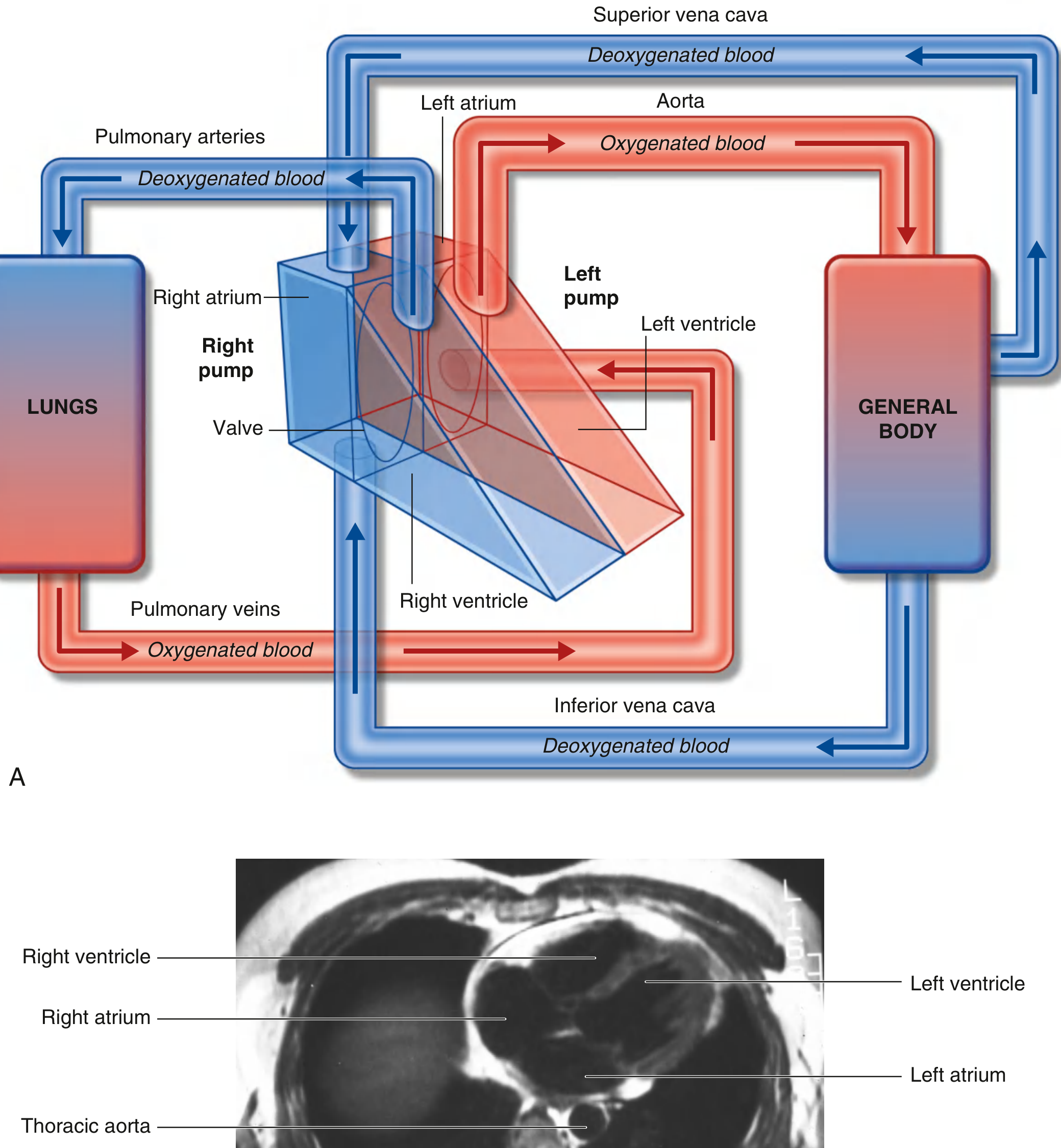
- The cardiovascular system = heart + blood vessels (a closed network of tubes)
- Primary function: Pump blood to deliver oxygen, nutrients, hormones, and immune cells throughout the body while removing carbon dioxide and metabolic waste
- The system operates as two parallel circuits:
PULMONARY CIRCUIT SYSTEMIC CIRCUIT
Right Heart → Lungs → Left Heart Left Heart → Body → Right Heart
(deoxygenated blood pumped (oxygenated blood pumped
to lungs for gas exchange) to all body tissues)
2. THE HEART
A. General Shape and Position
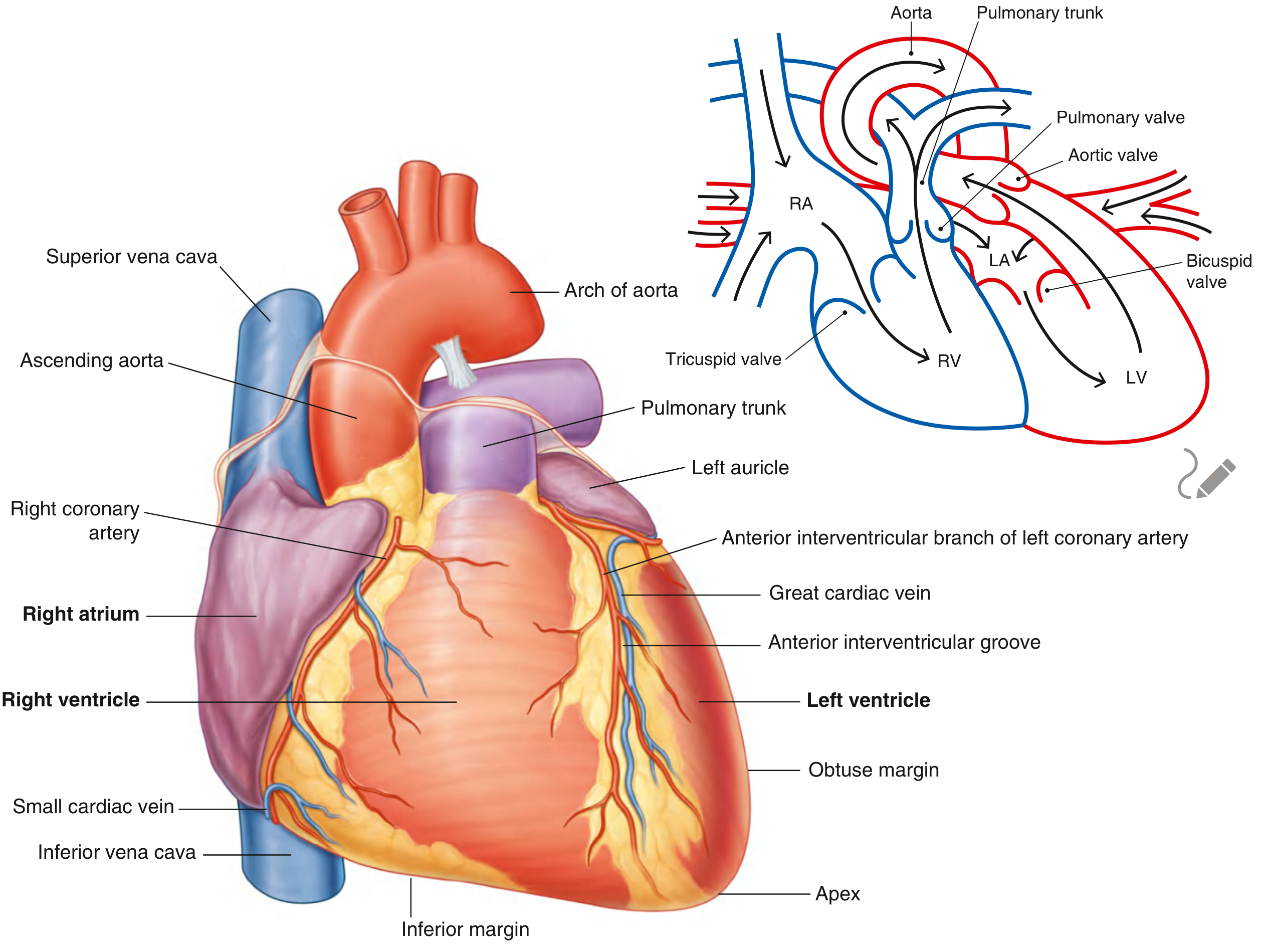
- Shape: Pyramid fallen on its side - three surfaces and an apex
- Located in the mediastinum of the thoracic cavity, within the pericardial sac
- Apex - formed by the inferolateral part of the left ventricle; points forward, downward, and to the left
- Located deep to the left 5th intercostal space, 8-9 cm from the midsternal line
- Produces the apex beat (point of maximal impulse)
- Base (posterior surface) - quadrilateral, directed posteriorly; formed mainly by the left atrium + small portion of right atrium + proximal parts of great veins (superior/inferior venae cavae + pulmonary veins)
- Fixed posteriorly at the level of vertebrae TV to TVIII (standing: TVI to TIX)
- Esophagus lies immediately posterior to the base
B. Surfaces
| Surface | Also Called | Formed By |
|---|---|---|
| Anterior | Sternocostal | Mainly right ventricle |
| Inferior | Diaphragmatic | Left + right ventricle |
| Right | Right pulmonary | Right atrium |
| Left | Left pulmonary | Left ventricle + small part of left atrium |
C. The Pericardium
- Fibrous pericardium - tough outer sac of dense connective tissue; attached below to the central tendon of the diaphragm; prevents overdistension
- Serous pericardium - two-layered membrane:
- Parietal layer - lines the inner surface of the fibrous pericardium
- Visceral layer (epicardium) - directly covers the outer heart surface
- Pericardial cavity - space between the two layers containing a thin film of serous fluid (15-50 mL); acts as lubricant to reduce friction
- Pericardial sinuses:
- Transverse pericardial sinus - posterior to the great arteries (aorta + pulmonary trunk) and anterior to the superior vena cava; clinically important for cardiac surgery
- Oblique pericardial sinus - posterior to the left atrium, bounded by pulmonary veins and inferior vena cava
3. CARDIAC CHAMBERS
A. The Four Chambers
- The heart = two pumps in series, separated by a muscular partition (septa)
- Right pump: receives deoxygenated blood from body → sends to lungs
- Left pump: receives oxygenated blood from lungs → sends to body
RIGHT ATRIUM
- Forms the right border of the heart in anatomical position
- Receives blood from:
- Superior vena cava (upper posterior portion)
- Inferior vena cava (lower posterior portion)
- Coronary sinus (returns blood from heart wall itself)
- Internal anatomy:
- Divided into two continuous spaces by the crista terminalis (smooth muscular ridge)
- Sinus of venae cavae - posterior part, smooth walls; where both venae cavae empty
- Anterior part (atrium proper) - contains musculi pectinati (parallel ridges of muscle)
- Fossa ovalis - oval depression on the interatrial septum; remnant of the fetal foramen ovale (allows fetal blood to bypass the lungs)
- Limbus of fossa ovalis - elevated margin around fossa ovalis
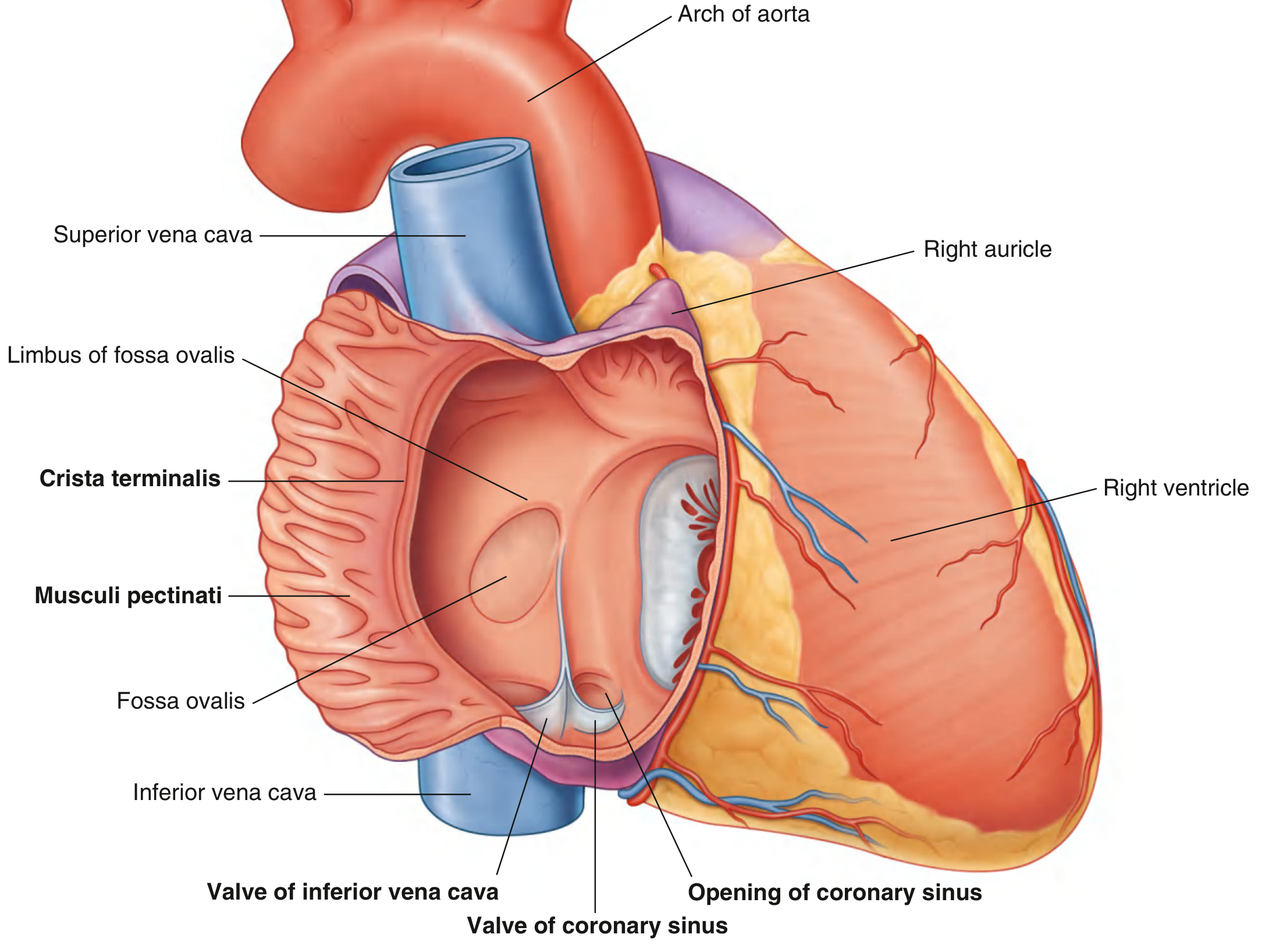
- Tricuspid valve (right atrioventricular valve) - closes the right AV orifice during ventricular contraction
RIGHT VENTRICLE
- Located anterior to the right atrium and left ventricle
- Inflow: right AV orifice (from right atrium)
- Outflow: pulmonary trunk (via the pulmonary valve)
- Internal anatomy:
- Trabeculae carneae - irregular muscular ridges/columns on the inner wall
- Papillary muscles (3 in RV): anterior, posterior, septal - project into the cavity
- Chordae tendineae - fibrous strings connecting papillary muscles to valve cusps; prevent valve eversion during systole
- Septomarginal trabecula (moderator band) - muscular band crossing from interventricular septum to base of anterior papillary muscle; carries part of the right bundle branch
- Conus arteriosus (infundibulum) - smooth-walled outflow portion leading to pulmonary valve
LEFT ATRIUM
- Located posteriorly; forms most of the base of the heart
- Receives blood from: 4 pulmonary veins (2 right, 2 left) entering posterior wall
- Internal anatomy:
- Smooth posterior wall (developed from pulmonary veins)
- Musculi pectinati - only in the left auricle
- Bicuspid/mitral valve guards the left AV orifice
- Wall slightly thicker than right atrium
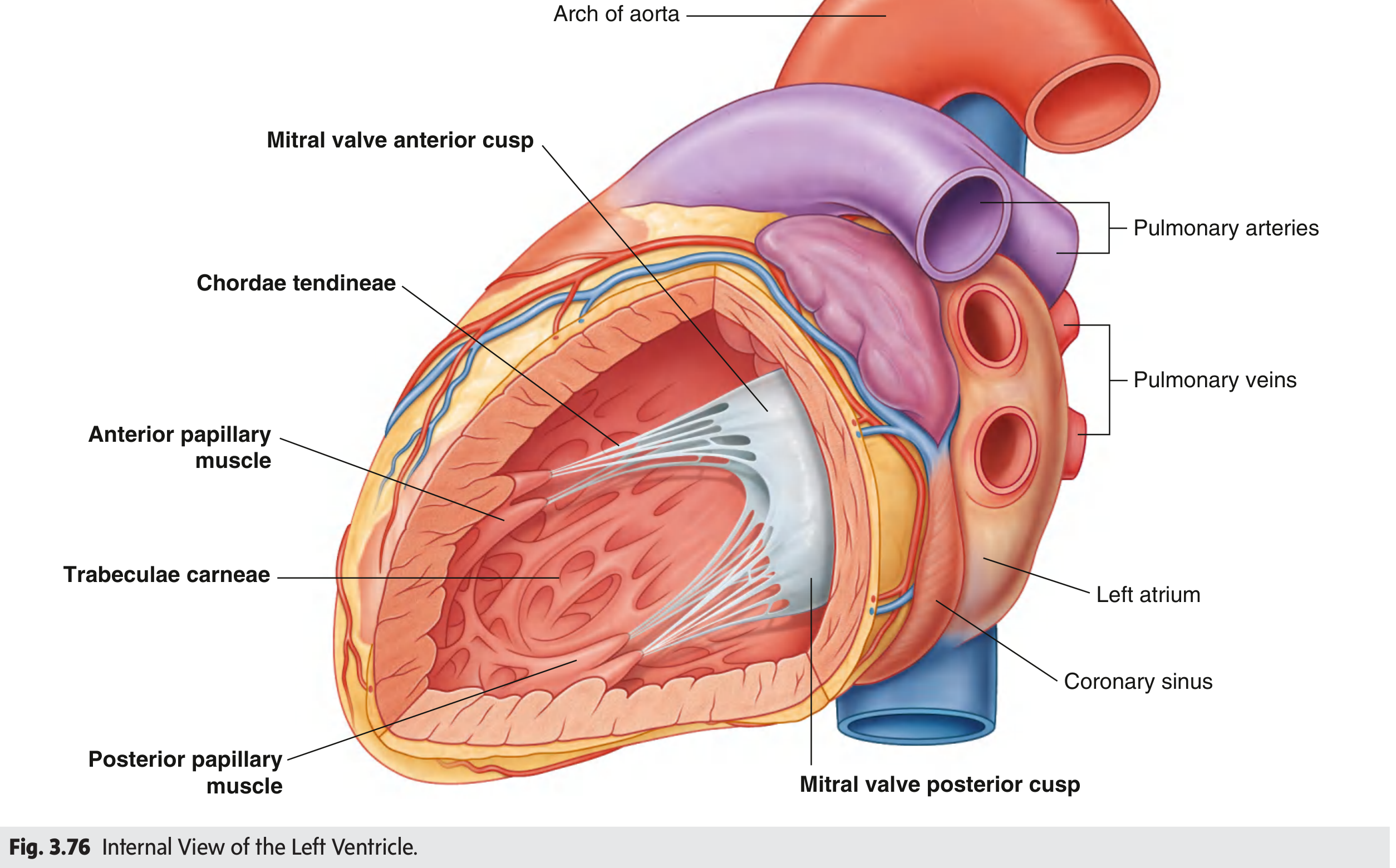
LEFT VENTRICLE
- Located posterior and to the left of the right ventricle
- Thicker wall than RV (approximately 3x thicker) because it must pump blood against higher systemic pressure (vs. pulmonary pressure)
- Inflow: left AV orifice (from left atrium)
- Outflow: aorta (via aortic valve) through the aortic vestibule
- Internal anatomy:
- Trabeculae carneae - more numerous and finer than right ventricle
- Two papillary muscles: anterior and posterior
- Chordae tendineae - same function as right side
- Interventricular septum - two parts:
- Muscular part - thick, forms the major portion
- Membranous part - thin, upper portion; separated into interventricular and atrioventricular parts
4. CARDIAC VALVES
A. Atrioventricular (AV) Valves
| Feature | Tricuspid (Right AV) | Mitral / Bicuspid (Left AV) |
|---|---|---|
| Cusps | 3 (anterior, posterior, septal) | 2 (anterior, posterior) |
| Location | Between RA and RV | Between LA and LV |
| Function | Prevents backflow to RA during systole | Prevents backflow to LA during systole |
| Papillary muscles | 3 (anterior, posterior, septal) | 2 (anterior, posterior) |
- Both AV valves are anchored via chordae tendineae to papillary muscles
- Papillary muscles contract BEFORE the ventricular walls → they tense the chordae tendineae to prevent cusp eversion into the atria
B. Semilunar Valves
| Feature | Pulmonary Valve | Aortic Valve |
|---|---|---|
| Cusps | 3 semilunar (anterior, left, right) | 3 semilunar (right, left, posterior/non-coronary) |
| Location | RV to pulmonary trunk | LV to ascending aorta |
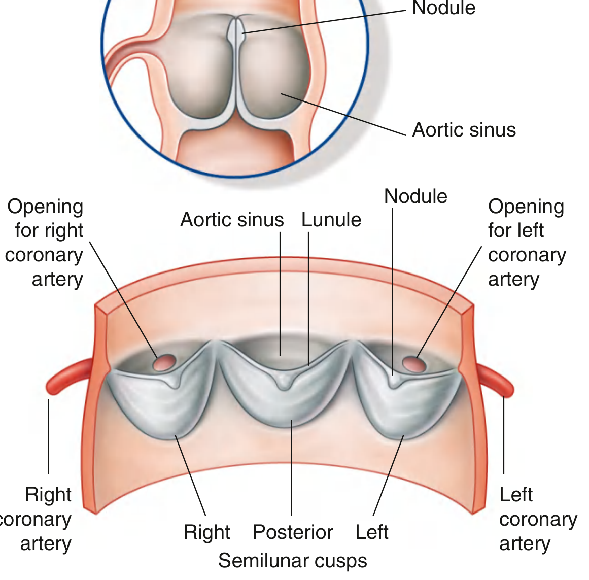
| Sinuses | Pulmonary sinuses | Aortic sinuses (R, L, posterior) |
- Aortic sinuses are pocket-like spaces between each semilunar cusp and the aortic wall
- Right and left coronary arteries originate from the right and left aortic sinuses - blood recoils after systole into these sinuses and automatically perfuses coronary arteries
- Nodule - thickening at the midpoint of each cusp's free edge; ensures complete valve closure
C. Auscultation Areas
| Valve | Auscultation Point |
|---|---|
| Mitral | Left 5th intercostal space, midclavicular line (apex) |
| Tricuspid | Left 4th/5th intercostal space, lower sternal border |
| Aortic | Right 2nd intercostal space, sternal border |
| Pulmonary | Left 2nd intercostal space, sternal border |
5. CARDIAC SKELETON
-
A collection of dense fibrous connective tissue forming four rings between atria and ventricles
-
Components:
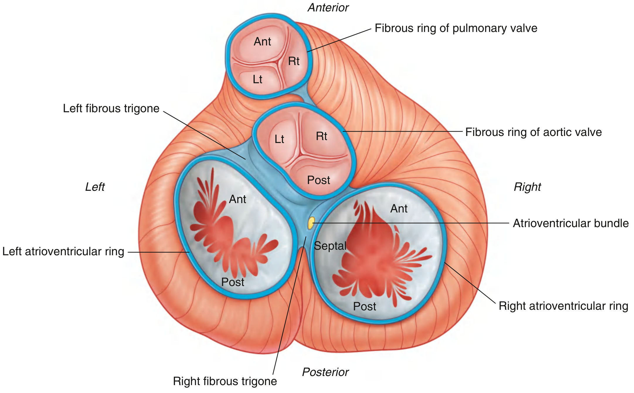
- 4 annuli fibrosi (fibrous rings) - surround both AV orifices, the aortic orifice, and pulmonary trunk opening
- Right fibrous trigone - thickened connective tissue between aortic ring and right AV ring
- Left fibrous trigone - between aortic ring and left AV ring
-
Functions of the cardiac skeleton:
- Maintains integrity of the valve openings
- Provides attachment points for valve cusps
- Provides origin for atrial and ventricular myocardium
- Electrically isolates atria from ventricles (the AV bundle is the single conducting connection)
6. CORONARY CIRCULATION
A. Coronary Arteries
- Arise from the aortic sinuses of the ascending aorta
- Circle the heart in the coronary sulcus; branches run in the interventricular sulci
RIGHT CORONARY ARTERY (RCA)
- Originates from the right aortic sinus
- Runs in the coronary sulcus between right atrium and right ventricle
- Branches:
- Sinu-atrial nodal branch - supplies the SA node (in ~60% of people from RCA)
- Right marginal branch - runs along the inferior (acute) margin toward apex
- AV nodal branch - supplies the AV node (in ~80% of people from RCA)
- Posterior interventricular branch - in the posterior interventricular sulcus
- Supplies: right atrium, right ventricle, SA node, AV node, posterior third of interventricular septum
LEFT CORONARY ARTERY (LCA)
- Originates from the left aortic sinus
- Passes between the pulmonary trunk and left auricle before dividing into:
- Anterior interventricular (left anterior descending, LAD) branch - runs in the anterior interventricular sulcus to the apex; supplies anterior 2/3 of interventricular septum and anterior walls of both ventricles
- Circumflex branch - continues in the coronary sulcus; gives off the left marginal branch; supplies left atrium and posterior/lateral wall of left ventricle
B. Coronary Veins
- Great cardiac vein - runs with the anterior interventricular artery; becomes the coronary sinus
- Middle cardiac vein - runs with the posterior interventricular artery
- Small cardiac vein - runs with the right marginal artery
- Coronary sinus - lies in the coronary sulcus on the posterior surface; drains into the right atrium between the IVC opening and right AV orifice
7. CARDIAC CONDUCTION SYSTEM
- The musculature of both atria and ventricles can contract spontaneously
- The conduction system initiates and coordinates contraction in a specific sequence
Components (in order of impulse transmission):
SA Node → Atrial Myocardium → AV Node → AV Bundle (Bundle of His)
→ Right & Left Bundle Branches → Purkinje Fiber Network → Ventricular Myocardium
SINU-ATRIAL (SA) NODE - "The Pacemaker"
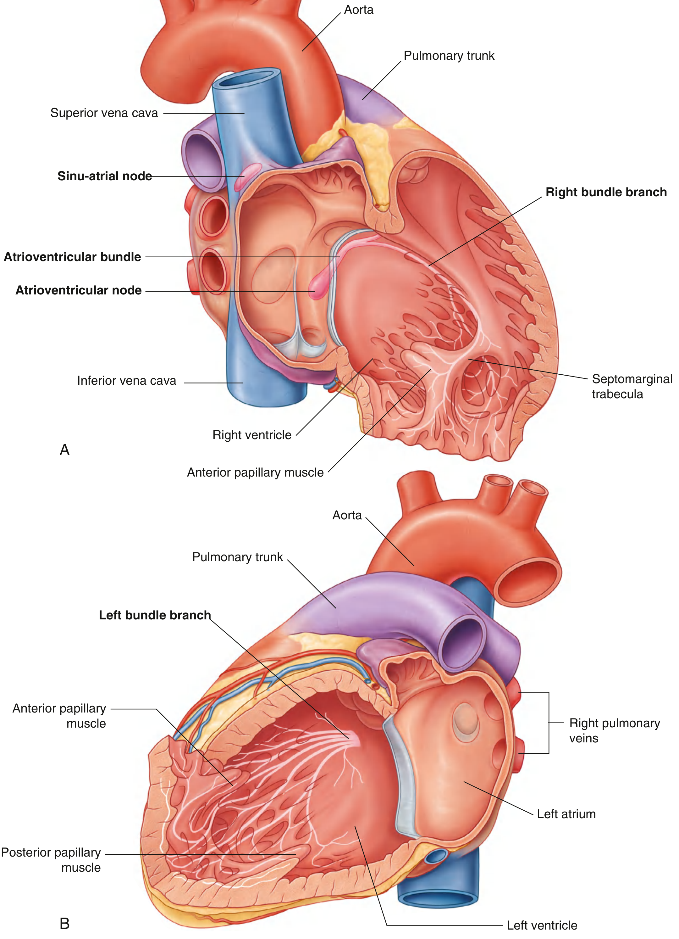
- Located at the superior end of the crista terminalis at the junction of the superior vena cava and right atrium
- Spontaneous firing rate: 60-100 beats/min at rest
- Sets the cardiac rhythm (hence "pacemaker")
- Excitation spreads across both atria → atrial contraction
ATRIOVENTRICULAR (AV) NODE
- Located near the opening of the coronary sinus, close to the attachment of the septal cusp of the tricuspid valve, within the AV septum
- Delays impulse transmission (~0.1 sec) → allows atria to finish contracting before ventricles begin
- The only electrical connection between atria and ventricles through the cardiac skeleton
ATRIOVENTRICULAR (AV) BUNDLE (Bundle of His)
- Direct continuation of the AV node
- Runs along the lower border of the membranous part of the interventricular septum
- Then splits into:
- Right bundle branch - descends on right side of interventricular septum → enters the septomarginal trabecula → reaches the base of the anterior papillary muscle → spreads as Purkinje fibers
- Left bundle branch - passes to left side of muscular interventricular septum → descends to apex of left ventricle → branches to Purkinje fibers
PURKINJE FIBER NETWORK (Subendocardial Plexus)
- Terminal network of specialized conduction cells spreading throughout ventricular walls
- Extremely rapid conduction (3-4 m/sec vs. 0.3-0.5 m/sec in normal myocardium)
- Produces a wave of excitation moving from apex to outflow tracts → ensures efficient blood ejection
8. CARDIAC INNERVATION
- The autonomic nervous system regulates heart rate, force of contraction, and cardiac output
- Both sympathetic and parasympathetic fibers converge to form the cardiac plexus:
- Superficial part - inferior to aortic arch, between it and the pulmonary trunk
- Deep part - between the aortic arch and tracheal bifurcation
| System | Effect on Heart Rate | Effect on Contractility | Effect on Coronary Arteries |
|---|---|---|---|
| Parasympathetic (vagus n.) | Decreases (bradycardia) | Reduces | Constricts |
| Sympathetic (T1-T5) | Increases (tachycardia) | Increases | Dilates |
- Parasympathetic: preganglionic fibers travel as cardiac branches of vagus nerves → synapse in ganglia within cardiac plexus or atrial walls
- Sympathetic: postganglionic fibers from superior, middle, and inferior cervical ganglia + upper thoracic ganglia (T1-T5) → enter cardiac plexus
9. THE CARDIAC CYCLE
SYSTOLE (Contraction) DIASTOLE (Relaxation/Filling)
─────────────────────────────────────────────────────────────────────
1. Isovolumic Contraction 4. Isovolumic Relaxation
(mitral closes, aortic not yet open) (aortic closes, mitral not yet open)
2. Rapid Ejection 5. Rapid Filling (E wave)
3. Reduced Ejection 6. Diastasis (slow filling)
7. Atrial Systole (A wave/"atrial kick")
- Isovolumic contraction - LV pressure rises after QRS (mitral valve closes → S1) before aortic valve opens; volume unchanged
- Rapid ejection - aortic valve opens when LV pressure exceeds aortic pressure (~80 mmHg)
- Reduced ejection - LV pressure peaks then falls; ejection continues by aortic recoil (Windkessel effect)
- Isovolumic relaxation - aortic valve closes (→ S2) to mitral valve opening; volume unchanged; Ca²⁺ re-sequestered into SR
- Rapid filling (E wave) - mitral valve opens; most of diastolic LV filling occurs here
- Diastasis - pressures equalize; LV filling virtually stops
- Atrial systole ("atrial kick") - atrial contraction forces remaining ~20-30% blood into LV; produces A wave; responsible for S4 if pathological
Normal Pressures:
| Chamber/Vessel | Pressure |
|---|---|
| Right atrium | 0-8 mmHg |
| Right ventricle | 15-30 / 0-8 mmHg |
| Pulmonary artery | 15-30 / 4-12 mmHg |
| Left atrium | 4-12 mmHg |
| Left ventricle | 100-140 / 3-12 mmHg |
| Aorta | 100-140 / 60-90 mmHg |
10. BLOOD VESSELS
A. General Wall Structure (Three Tunics)
LUMEN
│
├── TUNICA INTIMA (innermost)
│ └── Endothelial lining (single cell layer); basement membrane
│
├── TUNICA MEDIA (middle)
│ └── Smooth muscle + elastic fibers (variable amounts by vessel type)
│
└── TUNICA EXTERNA / ADVENTITIA (outermost)
└── Connective tissue; anchors vessel to surroundings
B. Arteries (carry blood AWAY from heart)
| Type | Tunica Media | Examples | Function |
|---|---|---|---|
| Large elastic arteries | Abundant elastic fibers | Aorta, brachiocephalic trunk, pulmonary trunk | Expand during systole, recoil during diastole to maintain flow (Windkessel) |
| Medium muscular arteries | Mostly smooth muscle | Femoral, axillary, radial arteries | Regulate diameter; control regional blood flow |
| Small arteries & arterioles | Thin smooth muscle | Terminal arteries | Control capillary filling; major determinant of peripheral vascular resistance |
C. Capillaries (exchange vessels)
- Smallest vessels; walls = single layer of endothelium + basement membrane
- Site of gas, nutrient, and waste exchange between blood and tissues
- Types:
- Continuous capillaries - tight junctions; found in muscle, brain (blood-brain barrier), skin
- Fenestrated capillaries - pores in endothelium; found in kidneys, small intestine, endocrine glands
- Sinusoidal (discontinuous) capillaries - wide, irregular lumen with gaps; found in liver, spleen, bone marrow
D. Veins (carry blood TOWARD heart)
| Type | Key Features | Examples |
|---|---|---|
| Large veins | Thickest layer = tunica externa; some smooth muscle in media | Superior vena cava, inferior vena cava, hepatic portal vein |
| Small/medium veins | Thin walls; large lumens; valves present | Superficial limb veins, deep leg veins |
| Venules | Smallest veins; drain capillaries | Post-capillary venules |
- Key differences between arteries and veins:
- Veins have thinner tunica media
- Veins have larger luminal diameter relative to wall thickness
- Veins have valves (paired cusps, especially in peripheral veins below heart level) - prevent backflow; essential for venous return against gravity
- Multiple veins (venae comitantes) often run alongside arteries in peripheral regions
E. Major Vessels
Aorta and its Segments:
Heart (LV) → Ascending Aorta → Aortic Arch → Descending Thoracic Aorta
→ Abdominal Aorta → Common Iliac Arteries (R & L)
- Aortic arch branches: brachiocephalic trunk (→ right subclavian + right common carotid), left common carotid artery, left subclavian artery
Pulmonary Circulation Vessels:
- Pulmonary trunk → right and left pulmonary arteries → lungs
- Pulmonary veins (4 total: 2 right, 2 left) → return oxygenated blood to left atrium
Venae Cavae:
- Superior vena cava (SVC) - drains upper body (head, neck, upper limbs) → right atrium
- Inferior vena cava (IVC) - drains lower body (abdomen, pelvis, lower limbs) → right atrium
11. CARDIAC HISTOLOGY (Microscopic Structure)
A. Layers of the Heart Wall
EPICARDIUM (outer) = visceral layer of serous pericardium; contains coronary vessels and fat
│
MYOCARDIUM (middle) = cardiac muscle; the bulk of the heart wall
│
ENDOCARDIUM (inner) = thin layer of endothelium; continuous with vessel endothelium
B. Cardiac Muscle Cells (Cardiomyocytes)
- Striated muscle but involuntary (unlike skeletal muscle)
- Branched cells that interconnect
- Intercalated discs - specialized junctions between cells containing:
- Desmosomes - provide mechanical strength
- Gap junctions (connexons) - allow ion flow → electrical coupling → coordinated contraction
- T-tubules, sarcoplasmic reticulum (SR) for Ca²⁺ storage
- Rich in mitochondria (~30% of cell volume) - rely almost entirely on aerobic metabolism
- Cannot undergo significant cell division after birth (minimal regenerative capacity)
12. CARDIAC PHYSIOLOGY SUMMARY
A. Starling's Law of the Heart
- "The more the heart is filled during diastole (preload), the more forcefully it contracts and the more blood it ejects"
- Increased venous return → increased end-diastolic volume → increased stroke volume
- Achieved by increased overlap between actin-myosin filaments
B. Key Definitions
| Term | Definition | Normal Value |
|---|---|---|
| Stroke Volume (SV) | Volume ejected per beat | ~70 mL |
| Heart Rate (HR) | Beats per minute | 60-100/min |
| Cardiac Output (CO) | CO = HR × SV | ~5 L/min at rest |
| Ejection Fraction (EF) | SV / End-Diastolic Volume × 100 | ≥55% (normal) |
| Preload | Ventricular filling pressure (end-diastolic volume) | - |
| Afterload | Resistance against which ventricle pumps (aortic pressure/SVR) | - |
| Contractility (Inotropy) | Intrinsic force of contraction independent of preload/afterload | - |
C. Heart Sounds
| Sound | Timing | Cause | Best Heard |
|---|---|---|---|
| S1 ("lub") | Start of systole | Closure of mitral + tricuspid valves | Apex (mitral area) |
| S2 ("dub") | End of systole | Closure of aortic + pulmonary valves | Aortic/pulmonary areas |
| S3 | Early diastole | Rapid filling of non-compliant ventricle | Apex; pathological in adults >40 (heart failure) |
| S4 | Late diastole (pre-systole) | Atrial contraction against stiff ventricle | Apex; always pathological |
13. BLOOD PRESSURE REGULATION
- Systolic BP - peak pressure during ventricular contraction (~120 mmHg)
- Diastolic BP - pressure during relaxation (~80 mmHg)
- Mean Arterial Pressure (MAP) = Diastolic BP + 1/3 (Pulse Pressure) ≈ 93 mmHg
- Pulse pressure = Systolic - Diastolic = ~40 mmHg
Mechanisms of regulation:
- Short-term: Baroreceptor reflex (carotid sinus, aortic arch) → alters heart rate and vasomotor tone
- Medium-term: Renin-angiotensin-aldosterone system (RAAS), ADH
- Long-term: Renal fluid regulation; vascular remodeling
14. PULMONARY vs. SYSTEMIC CIRCULATION AT A GLANCE
PULMONARY SYSTEMIC
─────────────────────────────────────────────────────────
Pump: Right ventricle Left ventricle
Resistance: Low High
Pressure: 15-30 / 4-12 mmHg 100-140 / 60-90 mmHg
Vessels: Short, wide Long, branched
Function: Gas exchange Nutrient/O₂ delivery
*Sources: Gray's Anatomy for Students (9780323934237); Braunwald's Heart Disease (9780323722193); Fuster and Hurst's The Heart 15e (9781264257560)*That is everything you need on the cardiovascular system, compiled directly from Gray's Anatomy for Students, Braunwald's Heart Disease, and Fuster & Hurst's The Heart. Here's a quick summary of what was covered:
What was covered (14 major sections):
- Overview - Two circuits (pulmonary + systemic), core function
- Heart position and surfaces - Pyramid shape, apex, base, 4 surfaces
- Pericardium - Fibrous, parietal serous, visceral (epicardium), transverse/oblique sinuses
- Four chambers - Right atrium (crista terminalis, fossa ovalis), Right ventricle (trabeculae, moderator band), Left atrium, Left ventricle (thick wall, papillary muscles)
- Cardiac valves - Tricuspid, mitral (AV valves); pulmonary, aortic (semilunar valves); auscultation sites
- Cardiac skeleton - Four fibrous rings, right + left trigones; electrical isolation function
- Coronary circulation - RCA (SA node, AV node branches), LCA (LAD + circumflex); cardiac veins + coronary sinus
- Conduction system - SA node → AV node → Bundle of His → Bundle branches → Purkinje fibers
- Cardiac innervation - Sympathetic (increases HR/contractility) vs. parasympathetic (decreases HR/contractility)
- Cardiac cycle - All 7 phases (isovolumic contraction through atrial kick); normal pressures table
- Blood vessels - Tunica layers; artery types; capillary types; vein types; major vessel anatomy
- Cardiac histology - Epicardium/myocardium/endocardium; cardiomyocyte features (intercalated discs, gap junctions)
- Cardiac physiology - Starling's law, stroke volume, cardiac output, ejection fraction, heart sounds (S1-S4)
- Blood pressure regulation - Short/medium/long-term mechanisms
This is a shared conversation. Sign in to Orris to start your own chat.