
Write black eye for exam purpose and make it for 3 or 5 marks
The medical-library skill must be invoked first for this medical topic. Let me read it and search simultaneously.
Reading File
Searching the Web
black eye periorbital hematoma causes treatment exam notes
Now let me search the medical library for black eye content:
Finding Sources
Finding Sources
Excellent! Found great content from Forensic Medicine and Kanski's Ophthalmology. Let me read the key sections:
Reading File
Reading File
Now let me also get the Forensic Medicine content on "raccoon eyes" / bilateral black eyes and the medicolegal significance, plus fetch the clinical images:
Finding Sources
Loading Image
I now have everything needed for a thorough exam answer. Here is the complete write-up:
BLACK EYE (Periorbital Hematoma)
P C Dikshit Textbook of Forensic Medicine and Toxicology | Kanski's Clinical Ophthalmology 10th ed.
Definition
A black eye (also called periorbital hematoma or periorbital ecchymosis) is bruising and/or a focal collection of blood (haematoma) in the tissues surrounding the eye, accompanied by oedema. It is the most common blunt injury to the eyelid or forehead.
Causes / Mechanisms (3-Mark Level)
A black eye may result from three distinct mechanisms:
1. Direct Violence
- A direct blow to the eye, cheek, eyebrow, or nose
- Often associated with abrasion or laceration of the adjacent skin (upper cheek, eyebrow, nose)
2. Gravitational Seepage
- An injury to the front of the scalp or above the eyebrow causes blood to seep downward beneath the scalp into the eyelids under gravity
- No direct injury to the eye itself
3. Percolation of Blood (Most Forensically Important)
- Results from a fracture of the anterior cranial fossa of the skull
- Often a contrecoup injury caused by a fall on the back of the head
- Leads to secondary fracture of the paper-thin orbital roof bone
- Blood tracks forward and collects around the eye
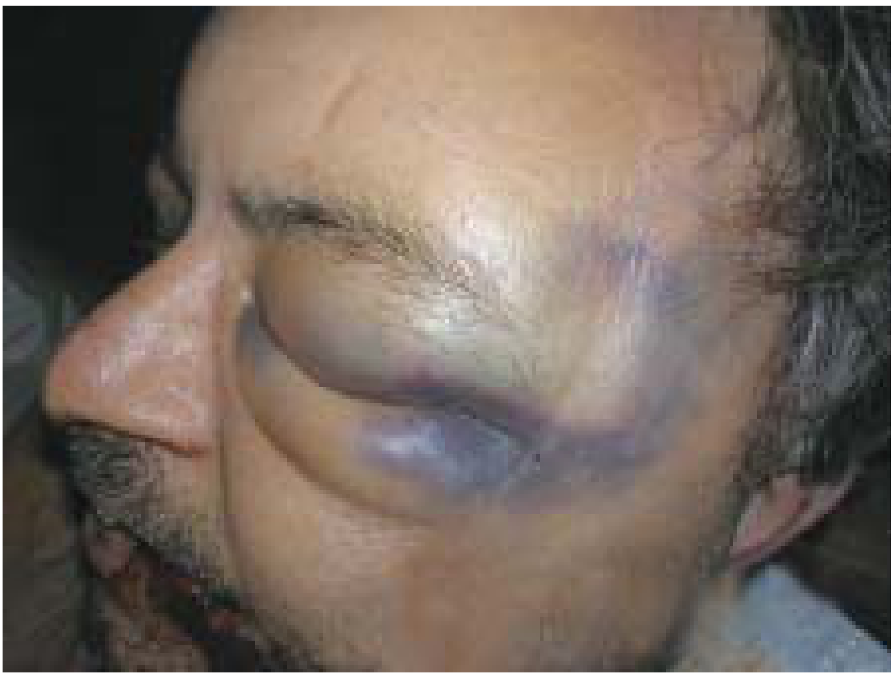
Additional Serious Conditions to Exclude (5-Mark Addition)
A black eye is generally innocuous, but the following serious associated conditions must always be excluded:
| Condition | Key Feature |
|---|---|
| Trauma to the globe/orbit | Examine the globe before lids become oedematous; do NOT apply pressure on globe until integrity confirmed |
| Orbital roof fracture | Black eye + subconjunctival haemorrhage without visible posterior limit |
| Basal skull fracture | Bilateral periorbital haematoma = "Panda eyes" / "Raccoon eyes" |
TIP (Exam Favourite): Bilateral periorbital haematoma ("panda eyes" / "raccoon eyes") is a classic sign of basilar skull fracture. It typically develops over several hours after injury (not immediately).
Medicolegal Importance
- A unilateral black eye with no other facial injury should raise suspicion of percolation from anterior cranial fossa fracture
- Bilateral black eyes without local trauma = strong indicator of skull base fracture
- Battle's sign (mastoid ecchymosis) + raccoon eyes together are pathognomonic of basilar skull fracture
- Important in cases of assault, road traffic accidents, and child abuse
Investigations
- CT scan of orbit and skull if orbital roof or skull fracture is suspected
- MRI or bedside ultrasonography if globe injury is suspected (avoid pressure on globe)
- Ophthalmologic consultation for vision changes, pupil abnormalities, or subconjunctival haemorrhage without posterior limit
Treatment
| Phase | Management |
|---|---|
| First 24-48 hours | Cold compress (ice pack wrapped in cloth) for 10-20 min intervals - reduces swelling and bleeding |
| After 48 hours | Warm compress to promote reabsorption |
| Pain relief | Paracetamol/acetaminophen (avoid aspirin and NSAIDs - increase bleeding) |
| Underlying injury | Treat fracture, globe injury, or skull base fracture accordingly |
Summary Box (Quick Revision)
- Synonym: Periorbital hematoma, Periorbital ecchymosis, "Shiner"
- 3 mechanisms: Direct violence / Gravitational seepage / Percolation (anterior cranial fossa fracture)
- Bilateral black eye = Panda/Raccoon eyes = Skull base fracture (exam favourite)
- Subconjunctival haemorrhage without posterior limit = orbital roof fracture
- Contrecoup mechanism - fall on back of head causing bilateral periorbital bruising
- Do NOT press on the globe before ruling out globe injury
Sources: P C Dikshit Textbook of Forensic Medicine and Toxicology (pp. 192-193); Kanski's Clinical Ophthalmology: A Systematic Approach, 10th ed. (p. 22.1)
For 1st year mbbs only
For 1st year MBBS, black eye is covered under Anatomy (orbital region) and Physiology (not really) - mainly it's an anatomy topic. Let me search the library for the anatomical basis.
Searching sources...
Finding Sources
Finding Sources
Now let me get the anatomy-focused content - specifically the anatomical basis explaining WHY a black eye forms, which is the 1st year MBBS angle:
Finding Sources
Finding Sources
The key phrase is found: "extravasation of blood in the loose areolar tissue around the eye globe." Now let me read the full Essentials of FMT section for the complete 1st-year-level content, and also look for anatomy of eyelid layers:
Reading File
Finding Sources
I now have all the anatomical basis needed. Let me also quickly fetch the black eye image from Essentials of FMT:
Loading Image
Here is the rewritten answer tailored specifically for 1st Year MBBS - the focus is on Anatomy (the structural/anatomical basis), which is what 1st year examiners test:
BLACK EYE (Periorbital Hematoma)
For 1st Year MBBS | Anatomy
Definition
Black eye (periorbital hematoma / periorbital ecchymosis) is extravasation or seepage of blood into the loose areolar connective tissue around the eye globe, giving the appearance of a spectacle-shaped bruise.
Also called: Spectacle haematoma | Periorbital bruising | Shiner
Anatomical Basis - WHY Blood Collects Here (Most Important for 1st Year)
The key lies in the layers of the eyelid and the nature of the surrounding tissue:
| Layer | Structure |
|---|---|
| 1st | Skin (very thin over eyelid) |
| 2nd | Subcutaneous tissue - contains loose areolar tissue (NO fat) |
| 3rd | Orbicularis oculi muscle |
| 4th | Orbital septum (fibrous layer) |
| 5th | Tarsus + conjunctiva |
The subcutaneous layer of the eyelid contains loose areolar tissue with no fat - this tissue:
- Offers very little resistance
- Has large potential space
- Allows blood to collect rapidly and in large amounts
- Blood tracks easily in all directions within this space
This is why even a small amount of bleeding causes dramatic swelling and discolouration around the eye.
Causes (3 Mechanisms - Must Know)
1. Direct blow to the orbit
- Punch, kick, or blunt trauma directly over the eye/cheek/nose
- Blood seeps into the loose areolar tissue of the eyelids immediately
2. Injury to the forehead / scalp (Gravitational seepage)
- Blow on the forehead or above the eyebrow
- Blood tracks downward under the scalp by gravity into the eyelids
- No direct injury to the eye itself
- There is NO local injury sign around the orbit
3. Fracture of the anterior cranial fossa (Most important - Exam favourite)
- Fall on the back of the head (contrecoup injury)
- Fractures the paper-thin orbital roof (roof of orbit)
- Blood percolates forward through cracked orbital plates and collects around the eye
- This produces bilateral black eyes = "Raccoon eyes" / "Panda eyes"
Anatomical Boundaries that Limit Blood Spread
Blood collecting in a black eye is limited by:
- Above - eyebrow (fibrous attachment of skin to bone)
- Below - orbital margin (skin attached firmly to periosteum)
- Medially/Laterally - orbital rim attachments
This explains the classic spectacle-shaped outline of the bruise.
Key Anatomical Point: Why NO fat in eyelid subcutaneous tissue?
The absence of fat in the eyelid subcutaneous layer is unique to this region (unlike rest of face). This:
- Allows eyelids to move freely
- But also means blood and fluid can collect with almost no resistance - hence dramatic swelling
Important Exam Points (3 & 5 Mark)
| Point | Fact |
|---|---|
| Tissue responsible | Loose areolar tissue under the eyelid skin |
| Why so dramatic | Loose areolar tissue offers no resistance to blood collection |
| Unilateral black eye (with no local injury) | Suspect fracture anterior cranial fossa or scalp injury |
| Bilateral black eye | Classic sign of basal skull fracture (raccoon/panda eyes) |
| Contrecoup mechanism | Fall on back of head → fracture orbital roof → bilateral black eyes |
| Associated sign | Battle's sign (bruising behind ear) = middle cranial fossa fracture |
3-Mark Answer (Write This)
Black eye is bruising around the eye due to extravasation of blood into the loose areolar tissue of the eyelids. It may result from: (1) direct blow to the orbit, (2) gravitational seepage of blood from a forehead injury tracking downward, or (3) percolation of blood from a fracture of the anterior cranial fossa (contrecoup injury), which fractures the thin orbital roof. Bilateral black eyes ("raccoon eyes") indicate a basal skull fracture.
5-Mark Answer - Add These Points
- Anatomy of eyelid layers and why loose areolar tissue allows blood accumulation
- Anatomical boundaries limiting the spread of blood (spectacle shape)
- Contrecoup mechanism in detail - fall on occiput, fracture of orbital roof
- Bilateral vs unilateral: differentiate cause
- Medicolegal importance: bilateral black eye without local injury = skull base fracture
Sources: The Essentials of Forensic Medicine and Toxicology 36th ed. (2026); P C Dikshit Textbook of FMT; Kanski's Clinical Ophthalmology 10th ed.; Gray's Anatomy for Students
This is a shared conversation. Sign in to Orris to start your own chat.