


Give all the details Abt the parasitology stool examination specimens answers to asked questions for microbiology mbbs practical examination
Enterobius vermicularis pinworm adult worm morphology
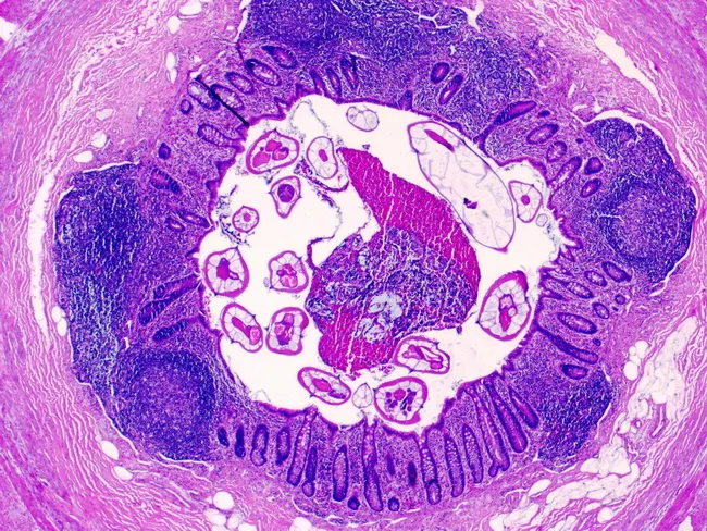
A high‑quality light microscopic image of a resected vermiform appendix demonstrates Enterobius vermicularis (pinworm) infection. The specimen was prepared as a paraffin section and stained with Hematoxylin and Eosin, viewed at high magnification (approximately 400x). The transverse cross‑sections reveal a circular arrangement of degenerative luminal parasites embedded in mucosa and luminal debris, with multiple adult pinworms characterized by a thick eosinophilic cuticle and distinctive lateral alae. The parasite bodies display a smooth, tapered morphology and a cylindrical outline; internal structures are limited by the staining. Surrounding appendiceal mucosa shows mild chronic inflammatory changes with eosinophilic infiltration, consistent with helminthic infestation rather than primary bacterial appendicitis. The focal luminal obstruction by parasites may account for symptoms or overflow. The image emphasizes gross features such as the worm's external cuticle and the lateral alae, as well as microanatomical context including mucosa‑submucosa interface and muscularis propria. Clinically, identifying pinworms in the appendix supports a diagnosis of enterobiosis and prompts anti‑helminthic therapy (e.g., albendazole or mebendazole) and screening of close contacts. This histopathology image is educational for parasitology, helminthic infections, surgical pathology, and differential diagnosis of appendiceal irritation in pediatric and adult patients.
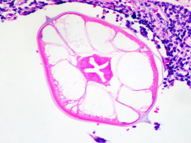
This brightfield light microscopy image shows a histologic cross-section of Enterobius vermicularis (pinworm) within a hysterectomy specimen. The nematode appears as a rounded, straw-colored structure with a thick, eosinophilic cuticle. The most distinctive feature is the lateral blade-like ridges along the worm circumference, diagnostic for pinworms. Internal anatomy includes a central digestive tract and surrounding musculature; the surrounding colonic mucosa/submucosa demonstrates mild inflammatory infiltrate with eosinophils, lymphocytes, and occasional plasma cells. The orientation is transverse, revealing the layered circular muscle and cuticular ridges within cross-section. Clinically, this incidental finding has limited impact on gynecologic cancer management but confirms Enterobius vermicularis infestation in the gastrointestinal tract. Differential considerations include other nematodes such as Ascaris lumbricoides and Trichuris trichiura, which have distinct cuticular patterns. The diagnostic significance lies in recognizing a common intestinal parasite in an extraneous surgical specimen; this image serves educational and parasitology morphology purposes for pathologists, residents, and medical students, highlighting characteristic transverse cross-sections and spined cuticle morphology.
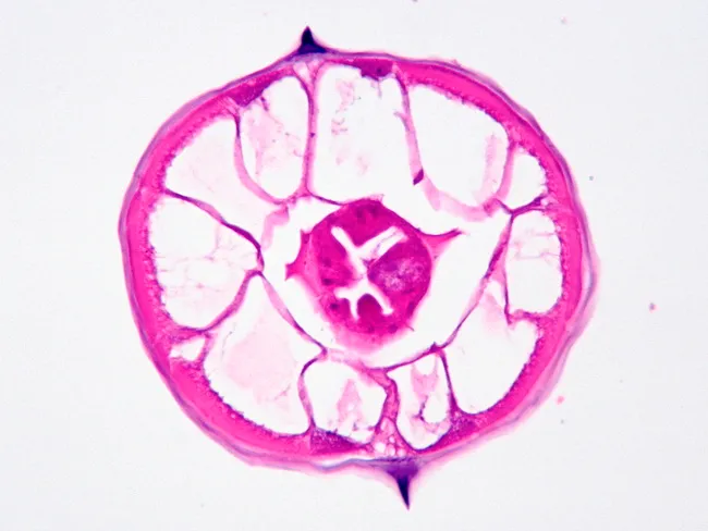
This image illustrates a transverse cross-section of Enterobius vermicularis (pinworm) captured by bright-field light microscopy after routine histological staining. The specimen is a nematode cuticle with a thick, pink-stained epidermal syncytial layer and underlying hypodermis, surrounding a central pseudocoel containing the alimentary canal. The outer surface displays characteristic longitudinal ridges (alae) along the body, which are highly diagnostic for E. vermicularis and help distinguish it from other intestinal nematodes. In this transverse section, the circular muscle layer and surrounding tissues are visible as concentric bands; the central intestinal cavity may appear as a variably stained lumen. The cuticle shows a defined border with folds that contribute to the worm’s distinctive morphology. The staining highlights the structural organization of nematode body wall, sensory and digestive components, and the internal organ arrangement, including dorsal and ventral nerve cords and radial musculature. Clinically, identification of these lateral alae in parasite sections supports a diagnosis of enterobiasis, particularly when correlated with patient symptoms such as perianal itching and epidemiologic context. This image is valuable for parasitology education, histopathology teaching, and differential diagnosis of intestinal nematodes in biopsy or stool preparation. Additional clinical relevance includes parasite burden assessment and educational demonstrations for students in pathology.
Taenia solium scolex proglottid tapeworm morphology
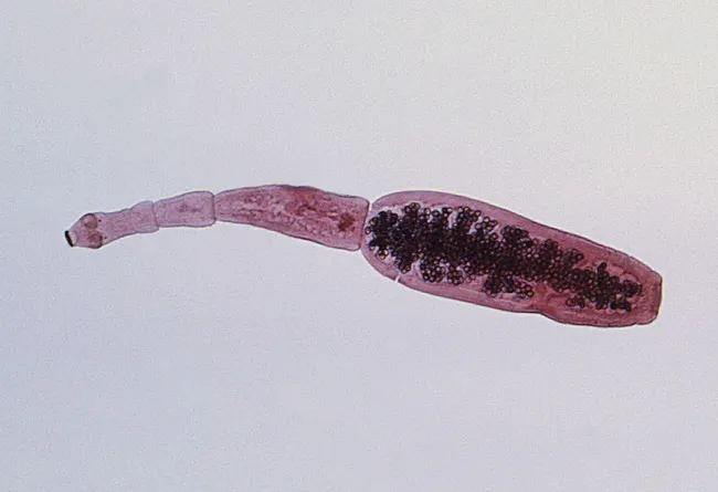
This image is a macroscopic photomicrograph of an adult cestode segment consistent with Echinococcus granulosus, the dog tapeworm. The specimen length is about 3–6 mm and shows a defined scolex with four suckers and a rostellum bearing 25–50 hooks, a classic genus-defining feature. Behind the scolex, three proglottids are visible, with the terminal proglottid gravid and longer than wide, indicating mature sexual production. The gravid segment contains densely packed eggs, visible as numerous dark structures within the proglottid. Morphology supports genus-level identification and differentiation from Taenia species in educational and diagnostic settings. Echinococcus spp. have a life cycle requiring dogs or other canids as definitive hosts and intermediate hosts such as sheep or cattle; humans become accidental intermediate hosts after ingesting eggs, leading to hydatid disease via larval cyst formation mostly in the liver, lungs, or CNS. In veterinary and medical contexts, this image aids teaching parasitology, veterinary parasitology, zoonotic risk assessment, and public health strategies—dog deworming, hygiene, and prevention of fecal contamination. Image courtesy of CDC/Dr. Peter M. Schantz.
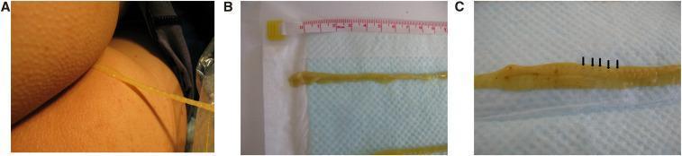
A series of clinical photographs documenting the extraction and gross morphology of a tapeworm (Cestoda). Figure A shows a long, yellowish, ribbon-like structure being carefully extracted from a patient's anal region into a clear plastic bag to maintain specimen integrity. Figure B displays the parasite post-extraction against a measuring tape and blue absorbent pad, highlighting its significant length, flat morphology, and a spoon-shaped scolex (head). Figure C provides a close-up view of the parasite's strobila, revealing distinct proglottids (segments). Black arrows point to central dark spots within the segments, which represent the gravid uterus. This image serves as a clinical reference for parasitic helminth infections, specifically diphyllobothriasis associated with the consumption of raw fish. Key educational features include the visualization of the scolex, proglottid segmentation, and macroscopic diagnostic characteristics used in identifying cestode species.
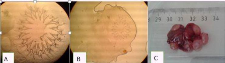
This composite image illustrates the morphology and clinical manifestation of Cysticercus fasciolaris, the larval stage of the tapeworm Taenia taeniaeformis. Panes A and B are diagnostic microscopic images showing the parasite's scolex. Pane A provides a high-magnification view of the double row of large and small chitinous hooks arranged in a radial pattern, characteristic of the cestode's rostellum. Pane B shows the complete scolex structure with the hooks centrally located. Pane C is a gross clinical photograph of an infected liver specimen, likely from a rodent model used in parasitological research. The liver appears dark reddish-brown and is heavily distorted by multiple white-to-pinkish, smooth, spherical cysts measuring approximately 0.5 to 1.0 cm in diameter. A ruler in the background provides a scale showing the total mass of the infected tissue spanning about 5 cm. This visual serves as an educational tool for identifying helminthic infections and understanding the macro- and microscopic pathology of larval tapeworm infestation in organ tissue.
Taenia solium egg embryophore oncosphere tapeworm
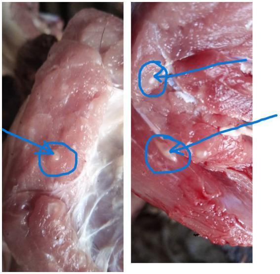
This clinical photograph shows a gross specimen of raw pork muscle tissue infested with Taenia solium metacestodes, a condition known as porcine cysticercosis. The image displays the pinkish-red, fibrous texture of skeletal muscle containing multiple embedded, small, white to translucent, oval-shaped structures, commonly referred to as 'bladder worms' or cysticercus cellulosae. Blue circles and arrows highlight specific cysts, which are typically 5 to 10 mm in length and represent the intermediate larval stage of the human tapeworm T. solium. This image is medically significant in the context of zoonotic disease transmission and public health, illustrating the physical manifestations of cysticercosis that meat inspectors identify to prevent the human consumption of infected meat, which can lead to taeniasis or the more severe neurocysticercosis if eggs are ingested. The presentation is a classic example used in parasitology and food safety education to demonstrate parasitic contamination in livestock.
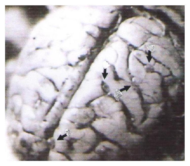
This clinical photograph displays a gross pathological view of a brain (macaque model) illustrating neurocysticercosis. The anatomical focus is on the cerebral cortex, where the characteristic gyri and sulci are visible. Multiple small, discrete, circular to oval-shaped cystic lesions are distributed within the subarachnoid space and along the cortical sulci. These lighter-colored, fluid-filled cysts (indicated by black arrows) represent the larval stage (cysticerci) of the Taenia solium tapeworm. The image demonstrates the typical multifocal distribution of the parasites in a heavy infection scenario, which clinically correlates with symptoms of increased intracranial pressure, seizures, and neurological deficits. This material serves as a translational model for human neurocysticercosis, a major cause of acquired epilepsy globally. The visible pathology highlights the relationship between parasitic localization in the subarachnoid spaces and the resulting parenchymal compression or inflammatory response.
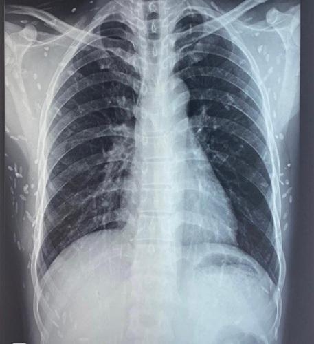
This diagnostic image is an anteroposterior (AP) chest X-ray demonstrating a classic presentation of disseminated cysticercosis. The radiograph reveals numerous small, ovoid, radiopaque calcifications—often described as 'rice-grain' calcifications—distributed throughout the soft tissues of the thoracic wall, axillae, and neck. These lesions represent the calcified larval cysts (cysticerci) of the Taenia solium tapeworm within the skeletal muscles. The lung parenchyma appears clear, without evidence of active infiltrates, consolidation, or pleural effusion. The cardiac silhouette is within normal limits for size and shape, and the visualized bony structures of the rib cage and clavicles appear intact. This radiological finding is pathognomonic for the chronic, calcified stage of muscular cysticercosis and is a key clinical indicator for investigating further neurological involvement, such as neurocysticercosis, in patients presenting with seizures.
Taenia saginata beef tapeworm morphology proglottid
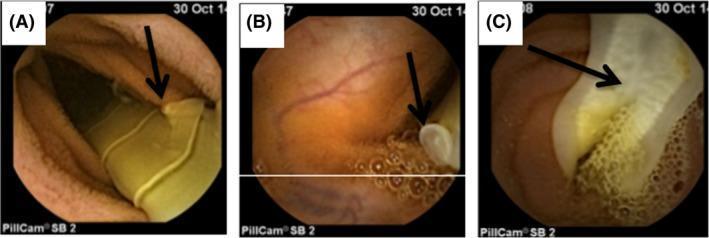
Diagnostic imaging via capsule endoscopy (PillCam SB 2) capturing Taenia saginata (beef tapeworm) infestation within the human small intestine. Panel A displays the yellowish, translucent strobila with a prominent arrow indicating a lateral genital pore on a proglottid. Panel B shows a close-up of a genital pore against the backdrop of the intestinal mucosa and intraluminal air bubbles. Panel C demonstrates a gravid proglottid with a visible internal uterine system, appearing more opaque and textured compared to immature segments. These clinical images highlight the utility of video capsule endoscopy in detecting intestinal helminths and differentiating species based on morphological features, such as the number and pattern of uterine branches. Taenia saginata typically presents with 15-20 uterine branches, a key diagnostic feature distinguishing it from the rosette-shaped uterus of Diphyllobothrium latum. The presence of these parasites is often a consideration in the differential diagnosis of unexplained abdominal symptoms or iron deficiency anemia when standard upper and lower endoscopies are normal.
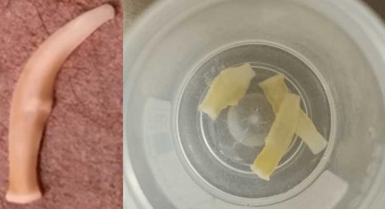
This clinical photograph displays macroscopic specimens of ivory-colored, ribbon-like worm segments isolated from a human stool sample. The image is divided into two panels: the left shows a single, elongated, and slightly curved proglottid with a smooth, translucent texture and ivory hue, exhibiting a tapered end and a central constriction. The right panel shows several similar segments collected in a clear specimen container; these fragments are flattened, rectangular to irregular in shape, and pale yellow-white, consistent with Taenia saginata (beef tapeworm) proglottids. This visual illustrates the macroscopic presentation of taeniasis, a parasitic infection typically acquired through the consumption of undercooked beef containing cysticerci. The presence of motile or stationary segments in feces is a pathognomonic clinical sign, often accompanied by perianal itching or mild abdominal discomfort. The image serves as an educational reference for medical parasitology and gastrointestinal infectious diseases, highlighting the importance of macroscopic stool examination when microscopic wet mounts for eggs are negative.
Parasitology Stool Examination - MBBS Practical Viva Answers
SPECIMEN 1: Enterobius vermicularis (Pin Worm / Thread Worm)
1. Identification
2. Brief Morphology
- Small, creamy-white/yellow worms
- Characteristic cervical alae (fin-like ridges) present on both sides of the head - this is the key diagnostic feature
- Buccal capsule absent; esophagus has a distinctive posterior bulb
- Length: ~10 mm (some sources say up to 13 mm)
- Tail: Long, sharply pointed and straight - hence the name "pinworm"
- Gravid females are distended with eggs
- Length: ~3-4 mm, diameter ~150 µm
- Posterior extremity: curved/coiled ventrally
- Single spicule (~70 µm), brown, hook-like
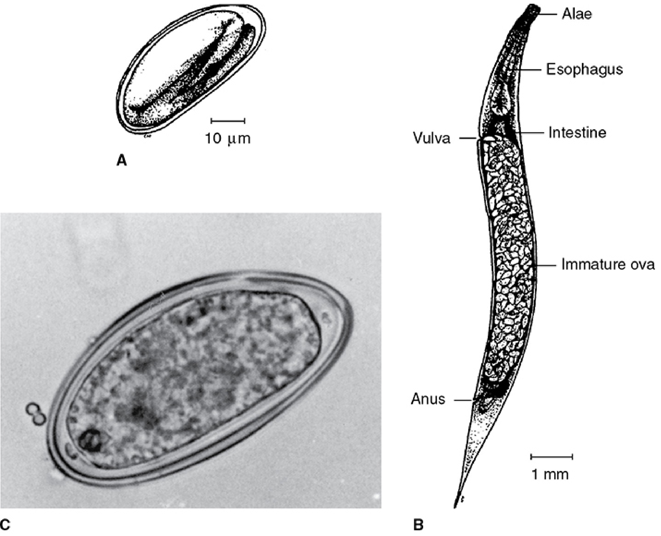
- Size: 25 x 50 µm
- Shape: Ovoid/plano-convex (flattened on one side) - asymmetrical - UNIQUE feature
- Shell: Clear, thin, double-layered
- Contains a fully developed larva (infective immediately or within 4-6 hours)
- NOT found in stool - collected from perianal skin using cellophane tape (Scotch tape test)
3. Life Cycle
| Stage | Location | Details |
|---|---|---|
| Adult | Caecum & proximal colon | Definitive host = human only |
| Gravid female | Migrates at night | Down to perianal area to deposit eggs |
| Eggs deposited | Perianal skin | Become infective in 4-6 hours |
| Infection | Oral-fecal | Auto-infection via scratching perianal area → hand → mouth |
| Larvae hatch | Small intestine | After ingestion of eggs |
| Adults establish | Colon/caecum | Cycle repeats in ~1 month |
- Retroinfection - larvae can migrate from anal skin back into the rectum
- Autoinfection is the main reason infections persist in families
- No intermediate host - direct life cycle
- Eggs may become airborne, be inhaled, swallowed
4. Pathogenesis & Clinical Features
- Female worm migrates nocturnally to perianal region to deposit eggs
- This migration causes intense irritation and scratching
- Eggs under fingernails lead to auto-reinfection and spread to contacts
- Perianal itching (pruritus ani) - the cardinal symptom, worse at night
- Sleep disturbance, restlessness, irritability (especially in children)
- Secondary bacterial infection of scratch marks
- Vulvovaginitis in girls (worms may migrate to vagina/fallopian tubes)
- Rarely: appendicitis if worms enter the appendix
- Eosinophilia is mild or absent
- Most infections are asymptomatic
- Sellotape/Cellophane tape test (Graham's method) - applied perianally in early morning before bathing - NOT stool examination
- Stool examination is not the preferred method (eggs rarely found in stool)
SPECIMEN 2: Taenia solium (Pork Tapeworm)
1. Identification
2. Brief Morphology
- Length: 2-4 meters
- Flat, ribbon-like body (strobila) divided into segments (proglottids)
- Number of proglottids: 800-1,000
| Part | Features |
|---|---|
| Scolex (head) | Small & globular (~1 mm); 4 suckers + ROSTELLUM with 2 rows of hooks (22-32 hooks) → "ARMED tapeworm" |
| Neck | Short, unsegmented - site of new proglottid formation |
| Immature proglottids | Near neck, small, undifferentiated |
| Mature proglottids | Middle region, contain developed reproductive organs |
| Gravid proglottids | Terminal, packed with eggs; uterus has 7-13 lateral branches each side |
- Spherical, thick-walled, brown
- Contains oncosphere (hexacanth embryo with 6 hooks)
- Outer shell is radially striated (embryophore)
- Size: ~35 µm
- Non-acid fast
- Taenia eggs of T. solium and T. saginata are morphologically identical
3. Life Cycle
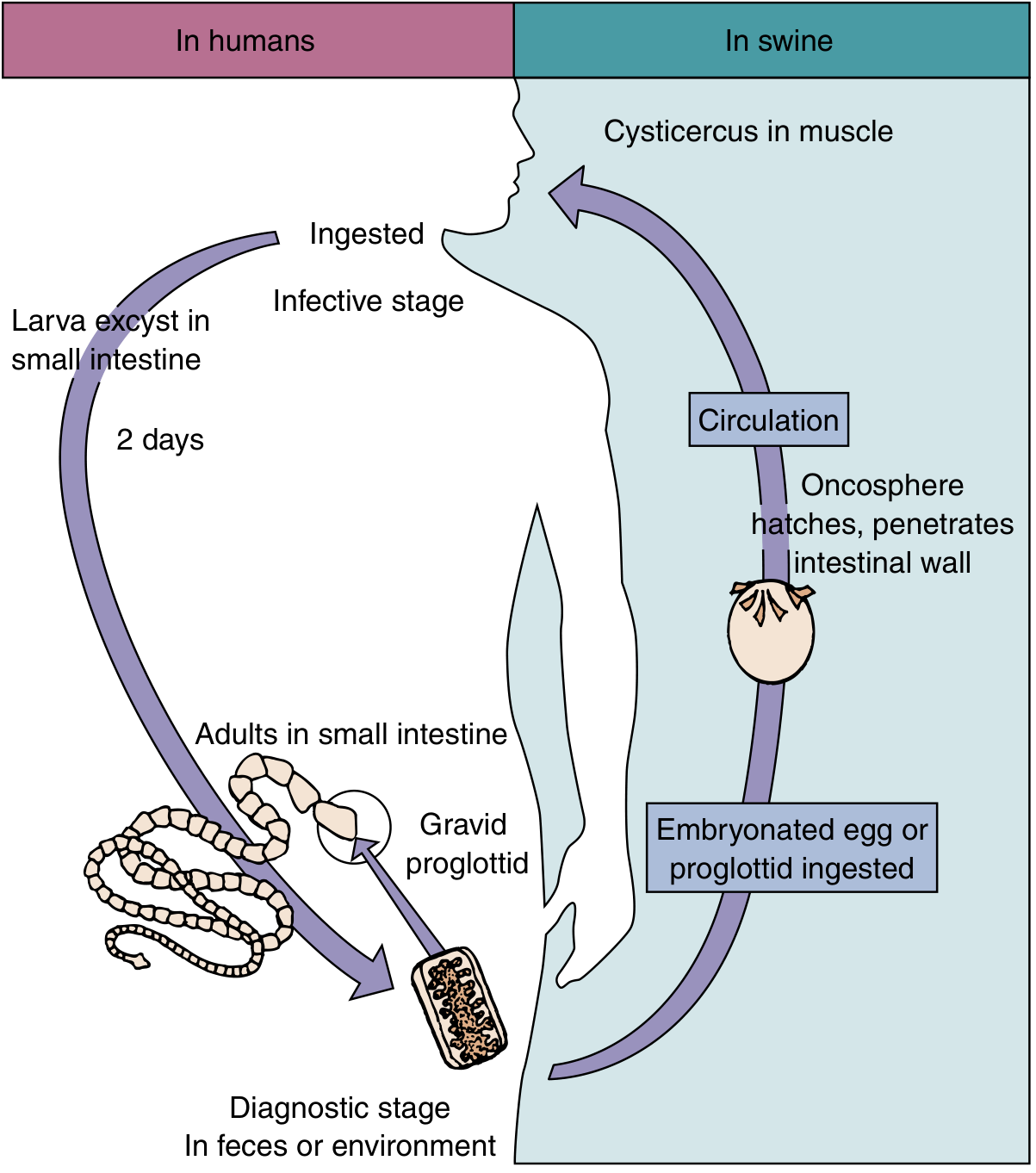
| Stage | Event |
|---|---|
| Gravid proglottids | Pass in human feces → embryonated eggs released |
| Pig ingests eggs | Oncosphere hatches, penetrates intestinal wall |
| Via circulation | Oncosphere travels to muscles/tissues |
| Forms cysticercus | Cysticercus cellulosae - bladder worm (~1 cm) in pork muscle ("measly pork") |
| Human eats raw/undercooked pork | Cysticercus excysts in small intestine |
| Adult worm | Develops in human intestine in 2-3 months |
4. Mode of Transmission, Pathogenesis & Clinical Features
- Taeniasis (intestinal): Eating raw/undercooked pork containing cysticerci
- Cysticercosis: Ingesting T. solium eggs via contaminated food, water, or auto-infection (feco-oral)
- Usually asymptomatic
- Mild: nausea, abdominal discomfort, diarrhea, weight loss
- Passage of proglottid segments in stool
- Vague hunger sensations
-
Neurocysticercosis (NCC) - most serious:
- Seizures (most common presentation)
- Headache, raised intracranial pressure
- Mental disturbances, hydrocephalus
- Leading cause of acquired epilepsy in endemic areas
-
Ophthalmocysticercosis:
- Diminution of visual acuity, blindness
-
Subcutaneous cysticercosis:
- Palpable nodules under skin
- Taenia saginata (beef tapeworm)
- Taenia asiatica (Asian tapeworm)
- Echinococcus granulosus (hydatid cyst)
- Echinococcus multilocularis (alveolar hydatid)
- Hymenolepis nana (dwarf tapeworm)
- Diphyllobothrium latum (fish tapeworm)
SPECIMEN 3: Taenia saginata (Beef Tapeworm)
1. Identification
2. Diagram & Description
- Length: 4-10 meters (much longer than T. solium)
- Flat, ribbon-like (strobila)
- Number of proglottids: 1,000-2,000
| Part | Features |
|---|---|
| Scolex (head) | Large & quadrate (square-shaped); 4 prominent suckers; NO rostellum, NO hooks → "UNARMED tapeworm" |
| Neck | Longer than T. solium |
| Gravid proglottids | Uterus has 15-20 lateral branches each side (more than T. solium → key differentiation) |
| Proglottids motility | Active - can crawl out of anus independently |
3. Uses of the Specimen (Stool examination)
- Diagnose taeniasis by macroscopic identification of proglottid segments
- Differentiate T. saginata from T. solium by counting uterine branches (T. saginata: 15-20; T. solium: 7-13)
- Inject ink into the uterus of proglottid to demonstrate branching pattern
- Stool examination for eggs (though eggs are indistinguishable from T. solium microscopically)
- Epidemiological studies - confirms exposure to beef-borne infection
4. Ovum Morphology (collected by specimen/stool exam)
- Shape: Spherical/sub-spherical
- Size: 31-43 µm
- Color: Brown/yellowish-brown
- Shell: Thick, radially striated embryophore (outer shell)
- Contents: Oncosphere (hexacanth embryo) - 6 hooklets visible inside
- Acid-fast: T. saginata eggs are acid-fast (unlike T. solium which is non-acid-fast) - key practical difference
- Outer membrane: Often lost by the time eggs reach stool
[Outer striated embryophore shell]
        ↓
[Thick radially-striated wall]
        ↓
[Inner oncosphere with 3 pairs of hooklets = 6 hooks total]
- Perianal swab method - Scotch tape applied perianally
- Stool examination - gravid proglottids disintegrate and release eggs
- Sieving stool through gauze to collect proglottid segments
Key Comparison Table (T. solium vs T. saginata)
| Feature | T. solium (Pork) | T. saginata (Beef) |
|---|---|---|
| Length | 2-4 m | 4-10 m |
| Scolex | Small, globular | Large, quadrate |
| Rostellum | Present (armed) | Absent (unarmed) |
| Hooks | 2 rows (22-32) | None |
| Proglottids | 800-1,000 | 1,000-2,000 |
| Uterine branches | 7-13 each side | 15-20 each side |
| Neck | Short | Long |
| Intermediate host | Pig | Cattle |
| Larva name | Cysticercus cellulosae | Cysticercus bovis |
| Human as int. host | Yes (Cysticercosis) | No |
| Egg acid-fast | Non-acid fast | Acid fast |
| Diseases caused | Taeniasis + Cysticercosis | Taeniasis only |
| Proglottid motility | Passive (inactive) | Active (crawls) |
Quick Viva Points to Remember
- Pinworm diagnosis = Sellotape test (NOT stool exam) - eggs are found perianally, not in stool
- T. solium = ARMED (has rostellum + hooks); T. saginata = UNARMED (no rostellum, no hooks)
- Cysticercosis only with T. solium, NOT with T. saginata
- Neurocysticercosis = most common cause of acquired epilepsy in developing countries
- Larva of T. solium = Cysticercus cellulosae; Larva of T. saginata = Cysticercus bovis
- E. vermicularis - only nematode diagnosed by perianal egg collection, not stool
- Characteristic features of E. vermicularis egg = plano-convex (flattened on one side), embryonated, asymmetric oval
- Gravid proglottid ink injection method is used to count uterine branches and differentiate Taenia species
- Sherris & Ryan's Medical Microbiology, 8th ed., pp. 1823-1830 (Intestinal Nematodes)
- Jawetz, Melnick & Adelberg's Medical Microbiology, 28th ed., pp. 754-756 (Taenia)
- Medical Microbiology 9e, Fig. 76.3 (T. solium life cycle)