
Upper extremity muscles origin insertion innervation and muscle action
Reading File
Now let me search the medical library for upper extremity muscle anatomy in parallel:
Finding Sources
Finding Sources
Finding Sources
Finding Sources
Finding Sources
Excellent! Found the THIEME Atlas of General Anatomy and Gray's Anatomy. Let me read the key sections in parallel:
Reading File
Reading File
Finding Sources
Now let me read the detailed origin/insertion/innervation/action tables from the THIEME Atlas in parallel:
Reading File
Reading File
Reading File
Now let me search for the detailed tables with origin, insertion, innervation, and action in the THIEME Atlas:
Finding Sources
Finding Sources
Excellent! Miller's Orthopaedics has a detailed table. Let me read it:
Reading File
I have an excellent detailed table. Let me read the continuation to get forearm and hand muscles:
Reading File
Loading Image
I now have everything I need. Let me read the full detailed tables from Miller's Orthopaedics:
Reading File
I have all the core data I need. Now let me compile the comprehensive reference table for all upper extremity muscles. I have the full table from Miller's Orthopaedics (lines 9707) which I already read. Let me now write the final answer:
Here is a complete reference for the upper extremity muscles - origin, insertion, innervation, and action - organized by region.
Source: Miller's Review of Orthopaedics, 9th Edition (Table 2.8) and THIEME Atlas of General Anatomy and Musculoskeletal System
Muscle Attachments - Shoulder & Arm (Anterior and Posterior Views)
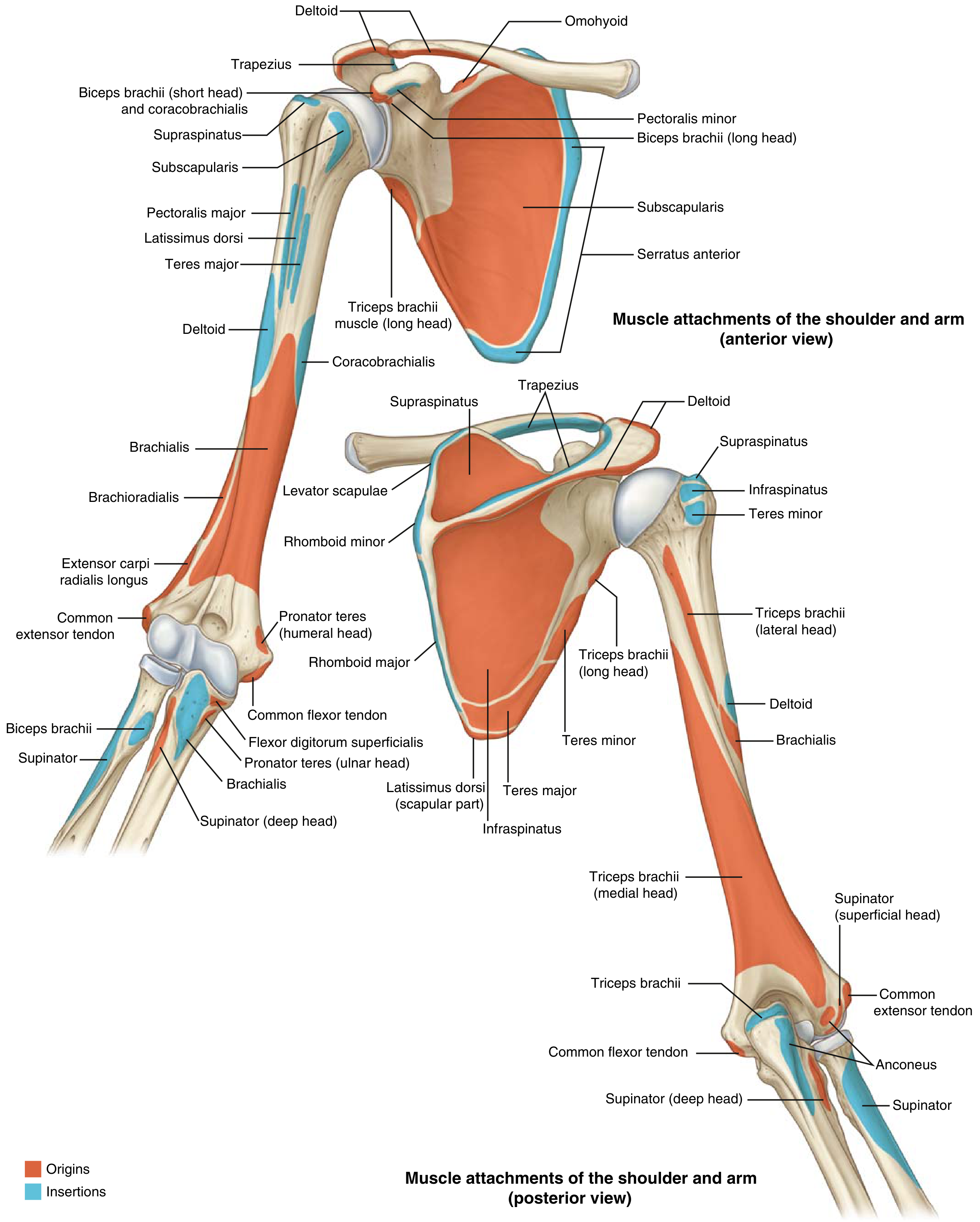
1. Muscles Connecting Trunk to Scapula / Shoulder Girdle
| Muscle | Origin | Insertion | Action | Innervation |
|---|---|---|---|---|
| Trapezius | Spinous processes C7-T12, external occipital protuberance, nuchal ligament | Clavicle (lateral 1/3), scapula (acromion, spine) | Rotating scapula (upper fibers elevate, lower depress, middle retract) | Cranial nerve XI (spinal accessory) + C3, C4 |
| Latissimus dorsi | Spinous processes T6-S5, iliac crest, ribs 9-12 | Humerus (intertubercular groove - floor) | Extension, adduction, internal rotation of humerus | Thoracodorsal nerve (C6, C7, C8) |
| Rhomboid major | Spinous processes T2-T5 | Scapula (medial border) | Adducting/retracting scapula, rotating glenoid downward | Dorsal scapular nerve (C5) |
| Rhomboid minor | Spinous processes C7-T1, nuchal ligament | Scapula (medial border at root of spine) | Adducting/retracting scapula | Dorsal scapular nerve (C5) |
| Levator scapulae | Transverse processes C1-C4 | Scapula (superior medial angle) | Elevating and rotating scapula (glenoid downward) | C3, C4; dorsal scapular nerve (C5) |
| Serratus anterior | Ribs 1-9 (outer surface) | Scapula (ventral/costal medial border) | Protracting scapula, preventing winging, rotating glenoid upward | Long thoracic nerve (C5, C6, C7) |
| Pectoralis minor | Ribs 3-5 (anterior surface) | Scapula (coracoid process) | Protracting and depressing scapula | Medial pectoral nerve (C8, T1) |
| Subclavius | Rib 1 (first costochondral junction) | Inferior clavicle | Depressing clavicle, stabilizing sternoclavicular joint | Nerve to subclavius (C5, C6) |
2. Muscles of the Shoulder Joint (Acting on Humerus)
| Muscle | Origin | Insertion | Action | Innervation |
|---|---|---|---|---|
| Deltoid | Lateral clavicle (1/3), acromion, scapular spine | Humerus (deltoid tuberosity) | Abduction (middle), flexion (anterior), extension (posterior); all fibers assist abduction | Axillary nerve (C5, C6) |
| Pectoralis major | Sternum, costal cartilages ribs 1-6, clavicle (medial 1/2) | Humerus (lateral lip of intertubercular groove) | Adduction, internal rotation, flexion (clavicular head) of arm | Medial and lateral pectoral nerves (C5-T1) |
| Teres major | Inferior angle of scapula (dorsal surface) | Humerus (medial lip of intertubercular groove) | Adduction, internal rotation, extension of arm | Lower subscapular nerve (C6, C7) |
| Coracobrachialis | Coracoid process of scapula | Humerus (medial surface, mid-shaft) | Flexion and adduction of arm | Musculocutaneous nerve (C6, C7) |
3. Rotator Cuff Muscles (SITS)
| Muscle | Origin | Insertion | Action | Innervation |
|---|---|---|---|---|
| Supraspinatus | Supraspinous fossa of scapula | Humerus (greater tuberosity - superior facet) | Initiates abduction (first 15°), stabilizes glenohumeral joint | Suprascapular nerve (C5, C6) |
| Infraspinatus | Infraspinous fossa of scapula | Humerus (greater tuberosity - middle facet) | External rotation of arm, stabilizes glenohumeral joint | Suprascapular nerve (C5, C6) |
| Teres minor | Lateral border of scapula (dorsal) | Humerus (greater tuberosity - inferior facet) | External rotation and adduction of arm | Axillary nerve (C5, C6) |
| Subscapularis | Subscapular fossa (ventral scapula) | Humerus (lesser tuberosity) | Internal rotation of arm, anterior glenohumeral stability | Upper and lower subscapular nerves (C5, C6) |
4. Muscles of the Arm
| Muscle | Origin | Insertion | Action | Innervation |
|---|---|---|---|---|
| Biceps brachii (long head) | Supraglenoid tubercle of scapula | Radial tuberosity; bicipital aponeurosis | Flexion of elbow, supination of forearm, weak shoulder flexion | Musculocutaneous nerve (C5, C6) |
| Biceps brachii (short head) | Coracoid process of scapula | Same | Same | Musculocutaneous nerve (C5, C6) |
| Brachialis | Anterior humerus (distal 1/2) | Coronoid process and tuberosity of ulna | Flexion of elbow (primary flexor regardless of pronation/supination) | Musculocutaneous nerve (C5, C6); small branch from radial nerve (C7) |
| Triceps brachii (long head) | Infraglenoid tubercle of scapula | Olecranon of ulna (posterior) | Extension of elbow; long head also extends/adducts arm | Radial nerve (C7, C8) |
| Triceps brachii (lateral head) | Posterior humerus (above radial groove) | Olecranon | Extension of elbow | Radial nerve (C7, C8) |
| Triceps brachii (medial head) | Posterior humerus (below radial groove) | Olecranon | Extension of elbow (most active component) | Radial nerve (C7, C8) |
| Anconeus | Lateral epicondyle of humerus (posterior) | Lateral olecranon and posterior ulna | Assists elbow extension; abducts ulna in pronation | Radial nerve (C7, C8) |
5. Anterior (Flexor) Forearm Muscles
Superficial Layer
| Muscle | Origin | Insertion | Action | Innervation |
|---|---|---|---|---|
| Pronator teres | Medial epicondyle (humeral head); coronoid process of ulna (ulnar head) | Lateral surface of radius (mid-shaft) | Pronation of forearm; weak elbow flexion | Median nerve (C6, C7) |
| Flexor carpi radialis | Medial epicondyle (common flexor tendon) | Base of 2nd (and 3rd) metacarpal | Wrist flexion and radial deviation | Median nerve (C6, C7) |
| Palmaris longus | Medial epicondyle (common flexor tendon) | Palmar aponeurosis | Wrist flexion; tenses palmar fascia | Median nerve (C7, C8) |
| Flexor carpi ulnaris | Medial epicondyle (humeral head); olecranon and posterior ulna (ulnar head) | Pisiform, hook of hamate, 5th metacarpal | Wrist flexion and ulnar deviation | Ulnar nerve (C7, C8) |
| Flexor digitorum superficialis (FDS) | Medial epicondyle, coronoid process, oblique line of radius | Middle phalanges of fingers 2-5 (splits to allow FDP through) | Flexion of PIP joints; assists MCP and wrist flexion | Median nerve (C7, C8, T1) |
Deep Layer
| Muscle | Origin | Insertion | Action | Innervation |
|---|---|---|---|---|
| Flexor digitorum profundus (FDP) | Anterior/medial ulna and interosseous membrane | Distal phalanges of fingers 2-5 | Flexion of DIP joints; assists PIP and MCP flexion | Median nerve (AIN) - index/middle (C8, T1); Ulnar nerve - ring/little (C8, T1) |
| Flexor pollicis longus (FPL) | Anterior radius and adjacent interosseous membrane | Distal phalanx of thumb | Flexion of IP joint of thumb | Anterior interosseous nerve (branch of median; C8, T1) |
| Pronator quadratus | Anterior distal ulna | Anterior distal radius | Pronation of forearm | Anterior interosseous nerve (C8, T1) |
6. Posterior (Extensor) Forearm Muscles
Superficial Layer (all arise from common extensor tendon at lateral epicondyle)
| Muscle | Origin | Insertion | Action | Innervation |
|---|---|---|---|---|
| Extensor carpi radialis longus (ECRL) | Lateral supracondylar ridge of humerus | Base of 2nd metacarpal | Wrist extension and radial deviation | Radial nerve (C6, C7) |
| Extensor carpi radialis brevis (ECRB) | Lateral epicondyle (common extensor tendon) | Base of 3rd metacarpal | Wrist extension (primary extensor) | Posterior interosseous nerve (deep radial; C7, C8) |
| Extensor digitorum (ED) | Lateral epicondyle (common extensor tendon) | Extensor hood - middle and distal phalanges of fingers 2-5 | Extension of fingers 2-5 at MCP, PIP, DIP | Posterior interosseous nerve (C7, C8) |
| Extensor digiti minimi (EDM) | Lateral epicondyle (common extensor tendon) | Extensor hood of little finger | Extension of little finger | Posterior interosseous nerve (C7, C8) |
| Extensor carpi ulnaris (ECU) | Lateral epicondyle + posterior border of ulna | Base of 5th metacarpal | Wrist extension and ulnar deviation | Posterior interosseous nerve (C7, C8) |
Deep Layer
| Muscle | Origin | Insertion | Action | Innervation |
|---|---|---|---|---|
| Supinator | Lateral epicondyle, radial collateral ligament, supinator crest of ulna | Anterior and lateral radius (proximal 1/3) | Supination of forearm | Posterior interosseous nerve (C6, C7) |
| Abductor pollicis longus (APL) | Posterior ulna, radius, and interosseous membrane | Base of 1st metacarpal | Abduction and extension of thumb; radial deviation of wrist | Posterior interosseous nerve (C7, C8) |
| Extensor pollicis brevis (EPB) | Posterior radius and interosseous membrane | Base of proximal phalanx of thumb | Extension of MCP joint of thumb; abduction of thumb | Posterior interosseous nerve (C7, C8) |
| Extensor pollicis longus (EPL) | Posterior ulna and interosseous membrane | Base of distal phalanx of thumb | Extension of IP joint of thumb; retropulsion (adduction) of thumb | Posterior interosseous nerve (C7, C8) |
| Extensor indicis (EI) | Posterior ulna and interosseous membrane | Extensor hood of index finger | Independent extension of index finger | Posterior interosseous nerve (C7, C8) |
Radial Group (Mobile Wad)
| Muscle | Origin | Insertion | Action | Innervation |
|---|---|---|---|---|
| Brachioradialis | Lateral supracondylar ridge (proximal 2/3) | Styloid process of radius | Flexion of elbow (especially in neutral/semi-pronated position) | Radial nerve (C5, C6) |
7. Intrinsic Muscles of the Hand
Thenar Muscles (Thumb)
| Muscle | Origin | Insertion | Action | Innervation |
|---|---|---|---|---|
| Abductor pollicis brevis (APB) | Flexor retinaculum, scaphoid, trapezium | Radial base of proximal phalanx of thumb | Abduction of thumb (perpendicular to palm) | Recurrent branch of median nerve (C8, T1) |
| Opponens pollicis | Flexor retinaculum and trapezium | Radial shaft of 1st metacarpal | Opposition of thumb | Recurrent branch of median nerve (C8, T1) |
| Flexor pollicis brevis | Flexor retinaculum, trapezium (superficial head); 1st metacarpal (deep head) | Radial base of proximal phalanx of thumb | Flexion of MCP joint of thumb | Superficial head: Median nerve; Deep head: Ulnar nerve (C8, T1) |
| Adductor pollicis | Oblique head: capitate, bases of 2nd/3rd metacarpals; Transverse head: 3rd metacarpal shaft | Ulnar base of proximal phalanx of thumb | Adduction of thumb toward palm | Deep branch of ulnar nerve (C8, T1) |
Hypothenar Muscles (Little Finger)
| Muscle | Origin | Insertion | Action | Innervation |
|---|---|---|---|---|
| Abductor digiti minimi (ADM) | Pisiform and pisohamate ligament | Ulnar base of proximal phalanx of little finger | Abduction of little finger | Deep branch of ulnar nerve (C8, T1) |
| Flexor digiti minimi brevis | Hook of hamate and flexor retinaculum | Ulnar base of proximal phalanx of little finger | Flexion of MCP joint of little finger | Deep branch of ulnar nerve (C8, T1) |
| Opponens digiti minimi | Hook of hamate and flexor retinaculum | Ulnar shaft of 5th metacarpal | Opposition of little finger | Deep branch of ulnar nerve (C8, T1) |
| Palmaris brevis | Flexor retinaculum/palmar aponeurosis | Dermis of hypothenar skin | Wrinkling skin of hypothenar eminence; deepens palm | Superficial branch of ulnar nerve (C8, T1) |
Metacarpal Muscles
| Muscle | Origin | Insertion | Action | Innervation |
|---|---|---|---|---|
| Lumbricals 1 & 2 | Radial side of FDP tendons to index and middle fingers | Radial lateral bands of extensor hood (fingers 2, 3) | MCP flexion + IP extension | Median nerve (C8, T1) |
| Lumbricals 3 & 4 | Adjacent sides of FDP tendons to ring and little fingers | Radial lateral bands of extensor hood (fingers 4, 5) | MCP flexion + IP extension | Deep branch of ulnar nerve (C8, T1) |
| Dorsal interossei (4) | Adjacent sides of two metacarpals (bipennate) | Bases of proximal phalanges and extensor hoods | Abduction of fingers (DAB); MCP flexion, IP extension | Deep branch of ulnar nerve (C8, T1) |
| Palmar interossei (3) | Ulnar side of 2nd metacarpal; radial sides of 4th and 5th metacarpals | Bases of proximal phalanges and extensor hoods | Adduction of fingers (PAD); MCP flexion, IP extension | Deep branch of ulnar nerve (C8, T1) |
Key Memory Aids
- Rotator cuff = SITS: Supraspinatus, Infraspinatus, Teres minor, Subscapularis
- "DAB" vs "PAD": Dorsal interossei Abduct; Palmar interossei Adduct
- All thenar muscles = median nerve EXCEPT adductor pollicis (ulnar) and deep head of FPB (ulnar)
- All hypothenar muscles = ulnar nerve
- "LOAF" muscles are median nerve: Lumbricals 1&2, Opponens pollicis, Abductor pollicis brevis, Flexor pollicis brevis
- Common flexor origin = medial epicondyle (pronator teres, FCR, palmaris longus, FCU, FDS)
- Common extensor origin = lateral epicondyle (ECRB, ED, EDM, ECU)
- AIN (anterior interosseous nerve) innervates FDP to index/middle, FPL, pronator quadratus - a median nerve branch
- PIN (posterior interosseous nerve) = deep branch of radial nerve - innervates all deep extensors
Sources: Miller's Review of Orthopaedics, 9th Ed., Table 2.8; THIEME Atlas of General Anatomy and Musculoskeletal System
This is a shared conversation. Sign in to Orris to start your own chat.