
Striatum
Reading File
The striatum is the largest component of the basal ganglia, receiving input from the cortex and relaying it through the direct and indirect pathways to regulate voluntary movement. Let me search the medical library for detailed content.
Searching sources...
Finding Sources
Finding Sources
Good results. Now let me read the key sections from multiple authoritative sources in parallel.
Reading File
Reading File
Reading File
Reading File
Excellent content from four textbooks. Let me also fetch the key diagram and read the procedural memory section.
Loading Image
Reading File
Reading File
I now have rich, comprehensive content from four major textbooks. Here is the full synthesis:
The Striatum
Anatomy and Nomenclature
The striatum is the largest nucleus of the basal ganglia. The term derives from the Latin corpus striatum ("striped body"), named for the striated appearance created by bands of white matter interconnecting its components through the anterior limb of the internal capsule.
The striatum consists of two subdivisions:
- Dorsal striatum: caudate nucleus + putamen
- Ventral striatum: nucleus accumbens + olfactory tubercle
The caudate nucleus is a large C-shaped structure (head, body, tail) that closely follows the contour of the lateral ventricle. The putamen and globus pallidus together form the lentiform nucleus, lying lateral to the internal capsule. The caudate and putamen are separated by the internal capsule but remain interconnected by gray matter bridges - the source of the "striated" appearance.
- Gray's Anatomy for Students, p. 1337
Functional Subdivisions
The striatum is subdivided along a ventromedial-dorsolateral continuum based on its cortical input:
| Region | Primary Input | Function |
|---|---|---|
| Ventral striatum (nucleus accumbens) | Limbic cortex, amygdala, hippocampus | Reward, motivation, emotion |
| Caudate nucleus | Prefrontal/association cortex | Cognition, executive function |
| Putamen | Sensorimotor cortex | Motor control |
Classic teaching divides a "neurologists' striatum" (dorsal, motor) from a "psychiatrists' striatum" (ventral, emotional) - though neuroimaging data show the dorsal striatum also participates in emotional regulation.
- Stahl's Essential Psychopharmacology, p. [block 1]
Cellular Architecture
More than 90% of striatal neurons are medium spiny neurons (MSNs) - GABAergic projection neurons. They are segregated into two populations:
| Population | Peptide co-transmitter | Dopamine receptor | Pathway |
|---|---|---|---|
| MSN type 1 | Substance P + Dynorphin | D1 (excitatory cAMP) | Direct pathway |
| MSN type 2 | Enkephalin | D2 (inhibitory cAMP) | Indirect pathway |
The remaining 5-10% are interneurons - both GABAergic and cholinergic - that provide local circuit modulation. This architecture is strikingly uniform across all functional territories, suggesting the striatum applies the same computational operations on diverse inputs.
- Eric Kandel - Principles of Neural Science, 6th ed.
Inputs to the Striatum
The striatum is the principal input nucleus of the basal ganglia. It receives:
- Corticostriatal projections (glutamatergic, excitatory) - from virtually the entire cerebral cortex; topographically organized. Frontal cortex projects to the head of the caudate and rostral putamen.
- Thalamostriatal projections - particularly from the intralaminar nuclei (centromedian nucleus).
- Nigrostriatal dopaminergic projections - from substantia nigra pars compacta (SNpc); modulates signal-to-noise in striatum, enhancing strong inputs while suppressing weak ones.
- Raphe nuclei - serotonergic inputs.
- Pedunculopontine nucleus - cholinergic inputs.
Signals arriving at the striatum represent competing behavioral options being generated in parallel across the brain; the basal ganglia circuit acts as a selection mechanism among them.
- Localization in Clinical Neurology, 8e, p. 1098
- Kandel Principles of Neural Science, 6th ed.
Direct and Indirect Pathways
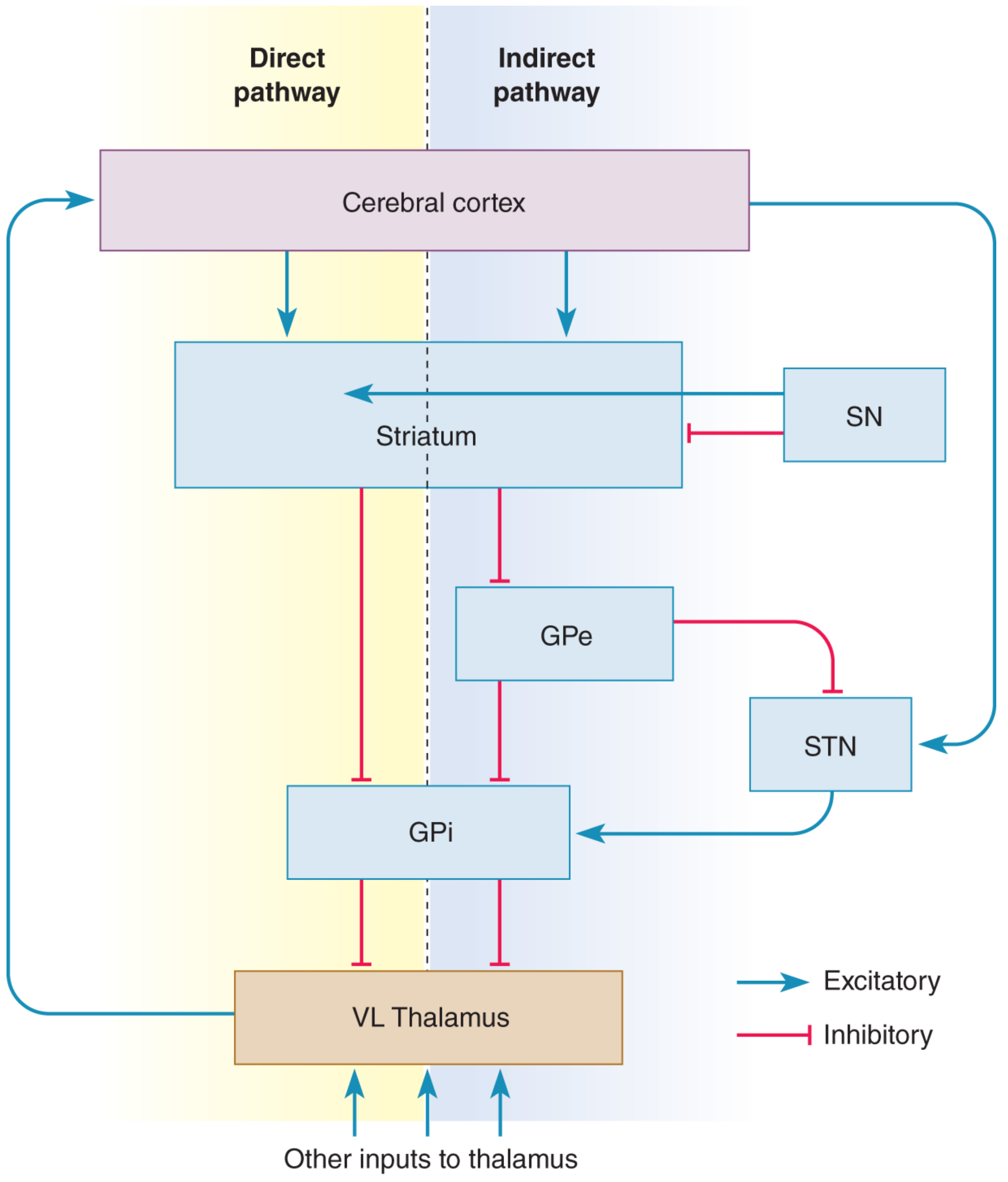
Direct Pathway (facilitates movement)
Cortex → Striatum (D1 MSNs) → GPi → VL Thalamus → Motor Cortex (SMA)
At rest, GPi neurons are tonically active and inhibit the thalamus. Cortical activation excites D1 MSNs → they inhibit GPi → GPi is released from tonic firing → VL thalamus is disinhibited → SMA activation is boosted. Net result: movement facilitation.
Indirect Pathway (suppresses movement)
Cortex → Striatum (D2 MSNs) → GPe → STN → GPi → VL Thalamus
Cortical activation excites D2 MSNs → inhibits GPe → GPe releases STN from inhibition → STN excites GPi → GPi more strongly inhibits VL thalamus → movement suppression.
The cortex also drives the STN directly via the "hyperdirect" pathway, providing fast inhibitory control that can rapidly suppress ongoing movements.
Dopamine's role: D1 receptor stimulation facilitates the direct pathway; D2 receptor stimulation inhibits the indirect pathway. Dopamine therefore promotes movement initiation via both routes.
- Neuroscience: Exploring the Brain, 5th ed., p. 1354-1356
Dopaminergic Modulation
Dopamine from SNpc has nuanced effects on medium spiny neurons:
-
Increases signal-to-noise in the striatum - amplifies responses to strong cortical inputs, suppresses weak ones
-
Long-term potentiation (LTP) and long-term depression (LTD) of glutamatergic synapses on MSNs require dopamine - this is the cellular basis for reinforcement learning in the striatum
-
Kandel Principles of Neural Science, 6th ed.
Role in Memory: Procedural Learning
The striatum is a key node in the procedural memory system (habit learning), distinct from the hippocampal declarative memory system. Classic double-dissociation experiments in rodents (radial arm maze) showed:
- Hippocampal lesions impair declarative/place memory but spare stimulus-response habit learning
- Striatal lesions impair habit learning but spare declarative memory
In humans, the same dissociation is seen in patients like H.M. (who had hippocampal damage) - they can form new procedural habits but cannot encode new declarative memories.
- Neuroscience: Exploring the Brain, 5th ed., p. 2197-2198
Clinical Correlations
| Condition | Mechanism | Dominant Features |
|---|---|---|
| Parkinson's disease | Loss of dopaminergic SNpc neurons → reduced D1/D2 stimulation → indirect pathway dominates → excess GPi inhibition of thalamus | Bradykinesia, akinesia, rigidity, resting tremor |
| Huntington's disease | Loss of D2 MSNs in indirect pathway first → indirect pathway underactivated → thalamus releases from inhibition | Chorea, hyperkinesia |
| Hemiballismus | Subthalamic nucleus lesion → reduces excitation of GPi → thalamus disinhibited | Violent flinging movements of contralateral limbs |
| Schizophrenia | Dopamine overactivity in ventral striatum | Positive symptoms (hallucinations, delusions) |
- Neuroscience: Exploring the Brain, 5th ed., p. 1356
- Localization in Clinical Neurology, 8e, p. 1099
This is a shared conversation. Sign in to Orris to start your own chat.