
VT management step by step
I'll start by reading the medical library skill, then search for VT management content in parallel.
Searching sources...
Reading File
Now searching the library and PubMed in parallel:
Finding Sources
Finding Sources
Searching PubMed
"ventricular tachycardia"[MeSH Terms] AND management
Now reading the three most relevant textbook sections in parallel:
Reading File
Reading File
Reading File
Excellent content retrieved. Now reading more from Braunwald's for long-term management and getting a relevant VT algorithm image:
Reading File
Searching Images
ventricular tachycardia management algorithm ACLS
I now have comprehensive content from three major textbooks. Let me also read the long-term management section further and get the ICD/ablation data:
Reading File
I now have comprehensive data from multiple authoritative sources. Here is the complete step-by-step VT management guide:
Ventricular Tachycardia (VT) - Step-by-Step Management
Step 1: Immediate Assessment - Is There a Pulse?
The single most important first question divides management into two completely different pathways.
No Pulse (Pulseless VT) = Cardiac Arrest Protocol
- Start CPR immediately
- Defibrillate (unsynchronized shock) as soon as the defibrillator is available
- Follow ACLS guidelines: epinephrine 1 mg IV every 3-5 min, amiodarone 300 mg IV push (then 150 mg if needed)
- Identify and treat reversible causes (H's and T's)
Pulse Present → Proceed to Step 2
Step 2: Assess Hemodynamic Stability
| Feature | Unstable | Stable |
|---|---|---|
| Blood pressure | Hypotension | Maintained |
| Mental status | Altered/unconscious | Alert |
| Chest pain / pulmonary edema | Present | Absent |
Step 3: Hemodynamically UNSTABLE VT with Pulse
Synchronized electrical cardioversion immediately. Do not delay for pharmacology.
- If conscious, give sedation/analgesia (procedural sedation) before shocking if clinical status allows - do not delay if severely compromised
- Deliver synchronized DC cardioversion (biphasic 100-200 J; escalate if unsuccessful)
- After restoration of sinus rhythm, proceed to identify and reverse precipitating causes (Step 6)
Step 4: Hemodynamically STABLE VT - Monomorphic
First, try to clarify the diagnosis if uncertain:
- Obtain a 12-lead ECG
- IV adenosine can be used diagnostically - if it terminates the arrhythmia or reveals an underlying rhythm, it was likely SVT with aberrancy, not VT
Pharmacologic Options (first-line):
1. Procainamide (preferred for stable VT)
- More effective than amiodarone or lidocaine for acute termination
- Dose: 10 mg/kg IV (up to 1000 mg) over 20 minutes
- Caution: vasodilation, hypotension in ~30% of patients
- Contraindicated in end-stage renal disease (metabolite NAPA accumulates → QT prolongation → polymorphic VT)
2. Amiodarone
- Dose: 150 mg IV over 10 minutes, then infusion (1 mg/min x 6 hours, then 0.5 mg/min)
- More effective at slower heart rates; better for preventing recurrence after termination
- Also vasodilator; hypotension in ~30%
3. Lidocaine
- Less effective than amiodarone overall
- More efficacious at faster heart rates; effective for termination but NOT for preventing recurrence
- Useful in acute ischemia setting
- Dose: 50 mg IV bolus, then 2-4 mg/min infusion
- Alternative to amiodarone in patients where amiodarone is contraindicated (e.g., severe iodine allergy)
Key rule: If the patient deteriorates during pharmacologic treatment → immediate synchronized cardioversion.
Special case - Idiopathic VT (no structural heart disease):
- IV beta-blockers - often effective
- Fascicular reentrant VT (idiopathic LV tachycardia): IV verapamil or diltiazem will usually terminate it
- Warning: Calcium channel blockers are absolutely contraindicated for VT in structural heart disease (risk of hemodynamic collapse)
Step 5: Hemodynamically STABLE VT - Polymorphic VT
Polymorphic VT requires identifying the QT interval:
Polymorphic VT with prolonged QT = Torsades de Pointes (TdP)
- Stop all QT-prolonging drugs
- IV Magnesium 1-2 g over 5-10 min (even if Mg levels are normal)
- Correct electrolyte imbalances (K⁺, Mg²⁺)
- Correct bradycardia - overdrive pacing at rate sufficient to suppress TdP bursts is definitive treatment
- Acquired long QT (drug-induced) with bradycardia: isoproterenol to increase rate temporarily
- Congenital long QT: beta-blockers + overdrive pacing; avoid beta-agonists
Polymorphic VT with normal QT
- Suspect acute ischemia/MI - check ECG and cardiac biomarkers
- Treat underlying ischemia urgently (reperfusion)
- If Brugada or early repolarization syndrome: isoproterenol IV, quinidine IV, overdrive pacing
Step 6: Identify and Reverse Precipitating Causes
This is critical for preventing early recurrence after any VT episode:
| Cause | Action |
|---|---|
| Hypokalemia | Correct K⁺ aggressively |
| Hypomagnesemia | IV Mg replacement |
| Hypoxia | Supplemental O₂, intubate if needed |
| Acidosis | Correct pH |
| Acute MI / ischemia | Emergent reperfusion |
| Heart failure | Treat pulmonary edema (diuresis, afterload reduction) |
| Drug toxicity (Na channel blockers) | Hypertonic NaHCO₃ |
| Hyperkalemia (sinusoidal VT) | Calcium, insulin/glucose, kayexalate |
| Sympathomimetic excess (milrinone, dobutamine) | Reduce or stop infusion |
Step 7: Electrical Storm
Definition: VT/VF recurring ≥3 times in 24 hours, requiring repeated shocks.
This is a life-threatening emergency. Management strategy (from Braunwald's eTable 67.1):
- ACLS protocol
- IV amiodarone infusion
- Reduce sympathetic tone - this is the cornerstone:
- Sedation, escalating to general anesthesia if needed
- IV beta-blocker - propranolol (non-selective) more effective than metoprolol in one study; esmolol (500 mcg/kg load, then 50-200 mcg/kg/min) if short half-life desired
- If ischemic cause: urgent coronary reperfusion + consider IABP
- For ICD patients: confirm correct device function; consider disabling VT detection if patient is awake and tolerating VT, to allow shocks only when necessary
- Emergent catheter ablation if initial measures fail
- Advanced sympatholysis:
- Stellate ganglion block
- High thoracic epidural anesthesia
- Hemodynamic support if needed:
- IABP, percutaneous VAD (Impella/TandemHeart)
- ECMO for refractory cases
- Bridge to LVAD / transplant in terminal cardiomyopathy
Step 8: Long-Term Management (After Stabilization)
Evaluation
- 12-lead ECG (look for prior MI pattern, LBBB, Brugada, delta waves)
- Echocardiography - assess LV/RV function and structure
- Cardiac MRI - identify scar, infiltrative disease (sarcoid, ARVC, myocarditis)
- Coronary angiography - rule out ischemic substrate
- Genetic testing if non-ischemic cardiomyopathy (~40% are genetic) - important for family screening
ICD Implantation
- Secondary prevention ICD: indicated for virtually all patients who survive sustained VT/VF without reversible cause
- Primary prevention ICD: for patients with structural heart disease and reduced EF (LVEF ≤35%) even without prior VT
Antiarrhythmic Drug Therapy (Long-Term)
- Amiodarone - most effective antiarrhythmic for preventing recurrent VT; use as adjunct to ICD to reduce shock burden
- Sotalol - alternative in patients with structural heart disease
- Beta-blockers - first-line for idiopathic VT; adjunct in structural disease
- Drugs do NOT replace ICD; they reduce recurrence frequency
Catheter Ablation
- Indicated for recurrent VT despite antiarrhythmic drugs
- Effective for scar-related VT (ischemic and non-ischemic cardiomyopathy)
- A 2025 meta-analysis (Santoro et al., Europace 2025, PMID 41316690) comparing catheter ablation vs. antiarrhythmic drugs in ischemic heart disease found catheter ablation superior for reducing VT recurrence
- A 2024 meta-analysis (Askarinejad et al., Sci Rep 2024, PMID 39122752) supports catheter ablation as first-line in structural heart disease with preserved LVEF
- For idiopathic VT (RVOT, fascicular): ablation is highly curative (>90% success) and preferred over lifelong antiarrhythmic therapy
PVCs / Nonsustained VT (No Structural Disease)
- Asymptomatic: no treatment needed (no survival benefit from suppression)
- Symptomatic: beta-blocker (metoprolol 50 mg BID or atenolol 50 mg daily) - safe first choice
- High PVC burden causing cardiomyopathy: aggressive treatment with catheter ablation
Quick Summary Algorithm
VT detected
     │
     ├── No pulse ──► CPR + Defibrillation (ACLS)
     │
     └── Pulse present
              │
              ├── Unstable ──► Sedation → Synchronized Cardioversion
              │
              └── Stable
                       │
                       ├── Monomorphic ──► Procainamide (preferred)
                       │                   Amiodarone
                       │                   Lidocaine
                       │
                       └── Polymorphic
                                │
                                ├── Prolonged QT (TdP) ──► MgSO₄, pacing, stop QT drugs
                                └── Normal QT ──► Treat ischemia, isoproterenol (Brugada)

After stabilization:
  → Correct precipitants → Evaluate substrate → ICD ± ablation ± antiarrhythmics
Sources:
- Tintinalli's Emergency Medicine, 9th ed. - p. 157 (acute management algorithm)
- Braunwald's Heart Disease, 12th ed. - pp. 556-558 (acute and long-term management)
- Goldman-Cecil Medicine, 2 vol. set - p. 590 (electrical storm management)
- Recent evidence: catheter ablation vs. AAD in ischemic VT (Santoro et al., Europace 2025, PMID 41316690) favors ablation for reducing VT recurrence
WCT Mx
Quick answer: Wide Complex Tachycardia (WCT) management hinges on two things - hemodynamic stability and whether you can confidently diagnose the rhythm. If unstable → synchronized cardioversion. If stable → assume VT until proven otherwise, use ECG criteria to differentiate, and treat accordingly. Never give verapamil empirically.
Searching sources...
Finding Sources
Finding Sources
Excellent hits. Now reading the most content-rich sections in parallel:
Reading File
Reading File
Reading File
Excellent content. Now reading the Frameworks monomorphic/polymorphic sections and the Tintinalli undifferentiated WCT treatment section:
Reading File
Reading File
Reading File
Now fetching the key ECG differentiation images and reading the WPW/irregular WCT section:
Loading Image
Reading File
Reading File
I now have all the content needed for a comprehensive, well-structured answer. Here it is:
Wide Complex Tachycardia (WCT) - Step-by-Step Management
Definition: Any tachycardia (rate >100 bpm) with QRS duration ≥ 0.12 seconds (≥120 ms).
Step 1: Rule Out Two Dangerous Mimics First
Before anything else, ask: "Could the wide QRS be caused by something other than a tachyarrhythmia?"
| Cause | Clue | Action |
|---|---|---|
| Hyperkalemia | Sine-wave/sinusoidal QRS, peaked T waves, peaked T, history of CKD | IV Calcium, NaHCO₃, insulin/glucose |
| Sodium channel blocker toxicity | Wide QRS + right axis shift in aVR (tall R in aVR), drug history (TCAs, flecainide, propafenone) | IV NaHCO₃ (hypertonic sodium) |
Both require specific therapy - do not treat these with antiarrhythmics.
Step 2: Assess Hemodynamic Stability
| Sign | Unstable | Stable |
|---|---|---|
| BP | Hypotension | Maintained |
| Consciousness | Altered/unconscious | Alert |
| Pulmonary edema / chest pain | Present | Absent |
Critical rule: Hemodynamic stability does NOT exclude VT. New-onset VT can present with a normal BP. Never use stability as proof of SVT.
Step 3: Unstable WCT → Immediate Synchronized Cardioversion
- Sedate if conscious and clinical status allows - do NOT delay if severely compromised
- Synchronized DC cardioversion (biphasic 100-200 J, escalate if needed)
- Follow with identification and reversal of precipitating causes
- If no pulse → unsynchronized defibrillation + CPR (ACLS protocol)
Step 4: Stable WCT → Classify the Rhythm
Is the rhythm REGULAR or IRREGULAR?
4A. REGULAR Monomorphic WCT
Default assumption: Treat as VT until proven otherwise.
- 80-90% of regular WCTs are VT
- Older age + prior MI makes VT even more likely
ECG Differentiation: Brugada Criteria (sequential - stop when any criterion is YES = VT)
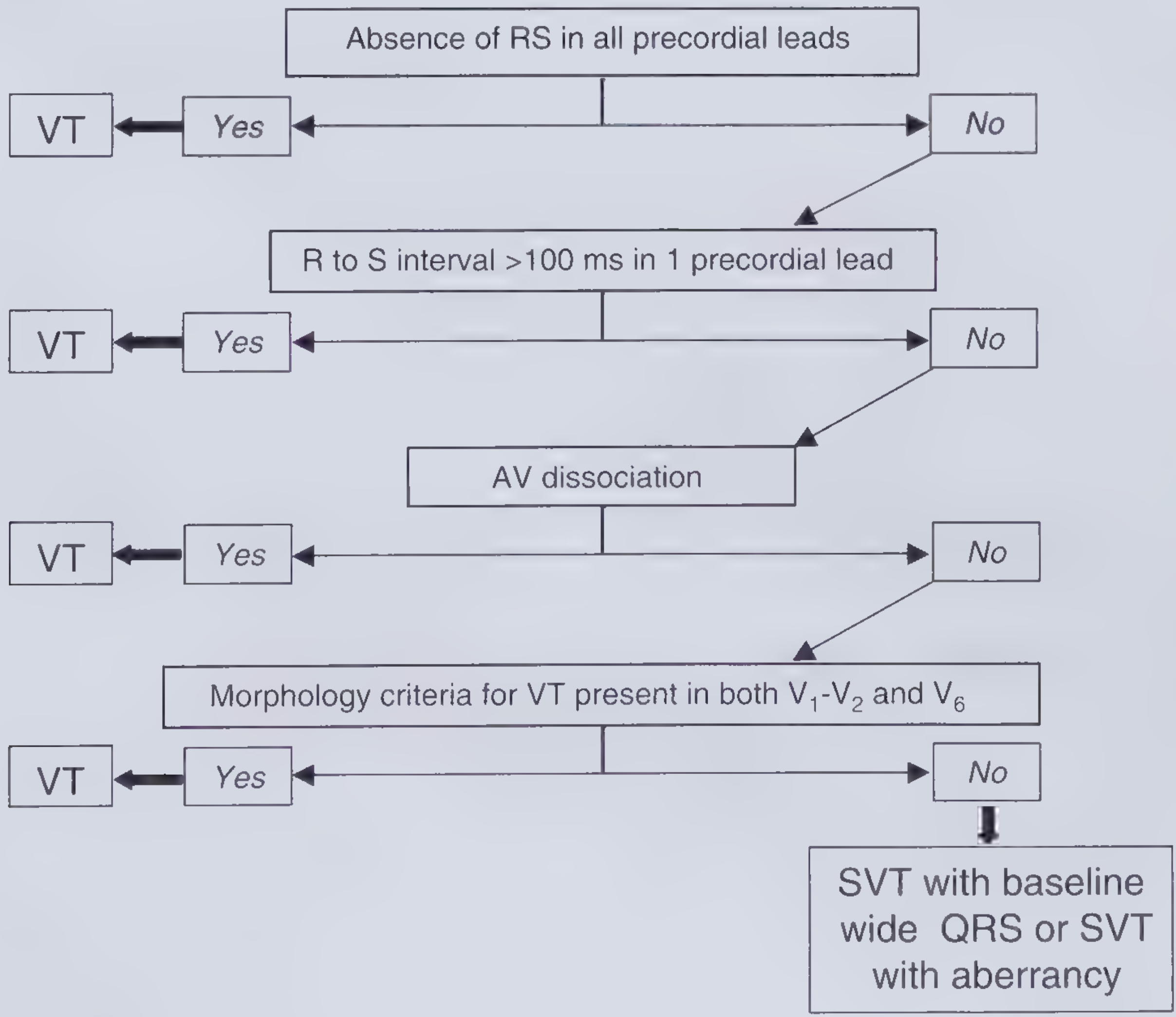
Step through in order - any YES = VT:
- Absence of RS complex in ALL precordial leads (V1-V6)? → Yes = VT
- R-to-S interval >100 ms in any one precordial lead? → Yes = VT
- AV dissociation present? → Yes = VT
- Look for independent P waves in inferior leads and V1-V2
- Seen in only ~10% of VT on surface ECG but pathognomonic when present
- Also look for: capture beats (narrow QRS earlier than expected) and fusion beats (intermediate morphology) - both = VT
- VT morphology criteria in V1-V2 AND V6? → Yes = VT
| Lead | RBBB-pattern | LBBB-pattern |
|---|---|---|
| V1 | Monophasic R, QR, QS, or RS | rS, QS, QR, or S > R |
| V6 | R/S ratio <1, QS, or QR | QR or QS |
If ALL 4 criteria are NO → SVT with aberrancy (baseline BBB or rate-related aberrancy)
Other Criteria (simpler alternatives):
| Criteria | Key Feature |
|---|---|
| Vereckei (aVR) | Initial R wave; initial r/q >40 ms; notch on descending limb; vi/vt ratio <1 → VT |
| Pava | Time to R-wave peak in lead II >50 ms → VT (simplest, fastest) |
Role of Adenosine in Stable Regular WCT:
- IV adenosine (6 mg rapid push, then 12 mg) can be used diagnostically
- If it terminates the tachycardia → was SVT (AVNRT, AVRT) or adenosine-sensitive VT
- If it causes transient AV block revealing underlying flutter/atrial tach → clarifies diagnosis
- If no response → likely VT
- Caution: rare forms of VT are adenosine-sensitive, so termination does NOT absolutely exclude VT
4B. IRREGULAR WCT
Irregular wide-complex tachycardia has a specific differential:
| Cause | Clue | Key Point |
|---|---|---|
| AF with aberrant conduction (BBB) | Irregularly irregular, typical BBB morphology | Treat as AF |
| AF with accessory pathway (pre-excited AF / WPW-AF) | Irregularly irregular, very rapid rate (>200 bpm), variable QRS width and morphology, bizarre QRS | Most dangerous - can degenerate to VF |
| Torsades de Pointes | Polymorphic, sinusoidal "twisting" morphology, prolonged QT in baseline ECG | See Step 5 |
| Atrial flutter with variable block + BBB | P waves, regular flutter waves at ~300 bpm | Treat as flutter |
WPW / Pre-excited AF - CRITICAL RULES:
- NEVER give: AV nodal blockers (adenosine, verapamil, diltiazem, digoxin, metoprolol)
- These block the AV node, forcing all conduction down the accessory pathway → extremely rapid ventricular rate → VF
- Treatment: Synchronized cardioversion (if unstable) or IV procainamide (if stable) - slows conduction in the accessory pathway
Step 5: Polymorphic WCT
First check the QT interval on a prior/baseline ECG (not the tachycardia ECG):
Prolonged QT → Torsades de Pointes (TdP)
- IV Magnesium sulfate 2 g IV over 5-10 min (even if Mg levels normal)
- Stop all QT-prolonging drugs
- Correct K⁺ and Mg²⁺ deficits
- Overdrive pacing at rate >70 bpm (definitive - prevents TdP bursts)
- For acquired TdP with bradycardia: IV isoproterenol temporarily until pacing secured
- Congenital LQTS: beta-blockers + pacing; avoid sympathomimetics
Normal QT → Polymorphic VT
- Suspect acute myocardial ischemia/MI - immediate ECG, troponin, consider angiography
- Defibrillate if unstable
- Treat the ischemia (reperfusion)
- Brugada syndrome / early repolarization: isoproterenol IV, quinidine IV, overdrive pacing
Step 6: Pharmacologic Management of Stable WCT Assumed to Be VT
(When diagnosis is uncertain or confirmed VT - see previous session for full dosing)
| Drug | Role | Notes |
|---|---|---|
| Procainamide | First choice for stable regular WCT / VT | 10 mg/kg IV over 20 min; superior to amiodarone for acute termination; also effective for WPW-AF; avoid in renal failure |
| Amiodarone | Second-line; better for recurrence prevention | 150 mg IV over 10 min; more effective at slower rates |
| Lidocaine | Alternative; good in acute ischemia | 50 mg IV bolus; less effective than others overall |
| Magnesium | TdP, polymorphic VT, hypomagnesemia | 2 g IV over 5-10 min |
| IV beta-blockers | Idiopathic VT, fascicular VT | Esmolol, metoprolol |
| IV verapamil | Fascicular (idiopathic LV) VT ONLY | Contraindicated in all other VTs and in any WCT of uncertain origin |
If clinical status deteriorates during pharmacologic treatment → immediate synchronized cardioversion.
Step 7: When Diagnosis Remains Uncertain in a Stable Patient
- Obtain a cardiology consult before empirical antiarrhythmics
- Unusual forms of WCT may need electrophysiology study
- A prior ECG (if available) is invaluable - compare QRS morphology during tachycardia vs sinus rhythm
- Prior history of structural heart disease, prior MI, or cardiomyopathy → high probability of VT
Quick Summary Algorithm
WCT (QRS ≥120 ms, rate >100)
     │
     ├─ Rule out Hyperkalemia / Na-channel toxicity (treat specifically)
     │
     ├─ NO PULSE → Defibrillate + CPR (ACLS)
     │
     ├─ PULSE + UNSTABLE → Sedation → Synchronized Cardioversion
     │
     └─ PULSE + STABLE
              │
              ├─ IRREGULAR?
              │       ├─ Bizarre/fast/variable → WPW-AF → Procainamide or Cardiovert
              │       ├─ Typical BBB morphology, irregular → AF with aberrancy → treat as AF
              │       └─ Twisting sinusoidal → TdP → MgSO₄, pacing, stop QT drugs
              │
              └─ REGULAR?
                      ├─ ASSUME VT (80-90% of cases)
                      ├─ Apply Brugada criteria → VT confirmed → Procainamide
                      ├─ Adenosine: diagnostic trial (clarifies SVT vs VT)
                      ├─ All Brugada criteria negative → SVT-aberrancy → treat as SVT
                      └─ Uncertain → Cardiology consult; treat as VT empirically
Key Rules to Remember:
- Hemodynamic stability ≠ SVT. Assume VT in any new regular WCT.
- Never give verapamil/diltiazem empirically in WCT - can cause cardiovascular collapse in VT or WPW-AF.
- Irregular WCT with very rapid rate = WPW-AF until proven otherwise - avoid all AV nodal blockers.
- Procainamide is the safest broad-spectrum drug for undifferentiated stable WCT.
- In any doubt, cardiovert.
Sources:
- Rosen's Emergency Medicine, 9th ed. - pp. 1058-1059 (Brugada criteria, WCT differentiation)
- Tintinalli's Emergency Medicine, 9th ed. - pp. 157-159 (treatment table, ECG features, WPW)
- Frameworks for Internal Medicine - pp. 89-91 (classification, algorithms, TdP)
- Braunwald's Heart Disease, 12th ed. - p. 556 (differential diagnosis table)
This is a shared conversation. Sign in to Orris to start your own chat.