
Pulmonary embolism lastest guidelines
pulmonary embolism guidelines 2024 2025 ESC AHA latest recommendations
"pulmonary embolism"[MeSH Terms] AND "guideline"[pt]
https://www.ecccp.org/infographic-aha-acc-2026-guidelines-ac…
PMID: 41712677
pulmonary embolism risk stratification management algorithm CT angiography
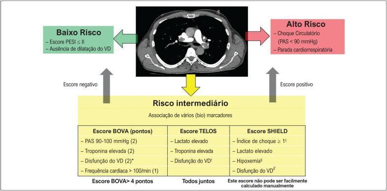
A medical infographic and diagnostic algorithm in Portuguese for risk stratification of acute pulmonary embolism (APE). At the center is a diagnostic image: a transverse/axial CT pulmonary angiogram (CTPA) showing the mediastinum and major hilar vessels, including the pulmonary artery and heart chambers. The algorithm branches into three risk categories: 'Baixo Risco' (Low Risk), defined by a PESI score ≤ II and absence of right ventricular (RV) dilation; 'Alto Risco' (High Risk), characterized by circulatory shock (SBP < 90 mmHg) or cardiorespiratory arrest; and 'Risco intermediário' (Intermediate Risk). The intermediate section details an association of biomarkers across three scoring systems: the BOVA score (incorporating SBP, troponin, RV dysfunction, and heart rate), the TELOS score (lactate, troponin, and RV dysfunction), and the SHIELD score (shock index, lactate, hypoxemia, and RV dysfunction). Arrows indicate the clinical flow based on positive or negative scores, integrating clinical vitals, imaging findings of RV strain, and laboratory markers for triage and management.
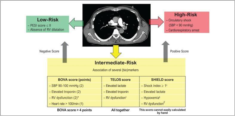
This composite educational graphic combines diagnostic imaging and a clinical decision-making algorithm for the risk stratification of acute pulmonary embolism (PE). At the center is an axial view of a Contrast-Enhanced Computed Tomography (CT) scan of the chest, specifically a CT pulmonary angiogram (CTPA). The image displays the thoracic cage, lungs, mediastinum, and cardiac structures, notably demonstrating filling defects in the pulmonary arteries consistent with PE. The surrounding flowchart categorizes PE severity into three tiers: Low-Risk, Intermediate-Risk, and High-Risk. Low-Risk is defined by a Pulmonary Embolism Severity Index (PESI) score ≤ II and absence of Right Ventricular (RV) dilatation. High-Risk is identified by hemodynamic instability (circulatory shock, SBP < 90 mmHg, or cardiorespiratory arrest). The Intermediate-Risk section utilizes integrated biomarkers and scoring systems, including the BOVA score (focusing on SBP, troponin, RV dysfunction, and heart rate), the TELOS score (lactate, troponin, and RV dysfunction), and the SHIELD score (shock index, lactate, hypoxemia, and RV dysfunction). This educational visual illustrates how imaging findings of RV strain and clinical scores guide patient prognosis and triage.
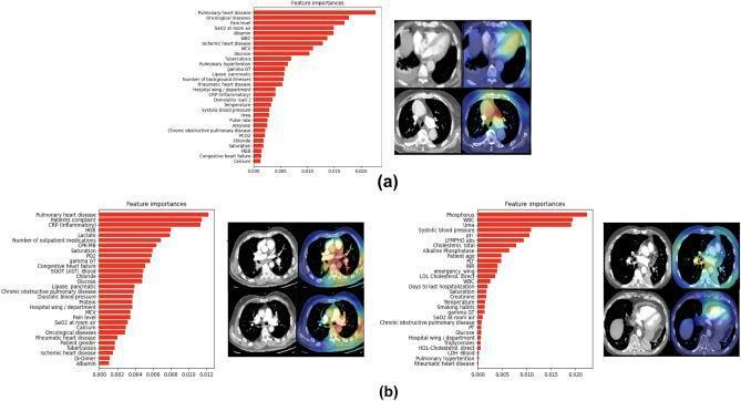
This infographic presents misclassified examples from a multimodal classifier designed for Pulmonary Embolism (PE) risk stratification, comparing Electronic Health Record (EHR) features and 3D CT Pulmonary Angiography (CTPA) imaging. (a) Displays a false negative case featuring a bar chart of EHR feature importance (e.g., Pulmonary heart disease, Patients Complaint, CRP) and corresponding 3D Grad-CAM heatmaps overlaying the left ventricle and pulmonary artery. (b) Shows two false positive examples with their respective feature importance rankings (including Phosphorus, WBC, and Urea) and Grad-CAM visualizations. The heatmaps in these cases highlight the pulmonary trunk, aorta, right ventricle chamber, and areas indicating potential PE clots. This visual provides an educational comparison of how deep learning models utilize diverse data modalities to assess acute PE severity, illustrating anatomical regions of high diagnostic importance such as the heart chambers and major thoracic vessels. It serves as a study of model interpretability and the clinical relevance of multimodal diagnostic fusion.
Pulmonary Embolism: 2026 AHA/ACC Guidelines (Latest)
1. New 5-Tier Clinical Classification System (Categories A-E)
| Category | Description | Key Features |
|---|---|---|
| A | At risk | No acute PE yet; risk factors present |
| B | Beginning/Mild | Hemodynamically stable, no RV strain, low severity score |
| C | Intermediate (3 subcategories) | C1: elevated severity score, normal RV + biomarkers; C2: RV dysfunction OR abnormal biomarkers; C3: both RV dysfunction AND elevated biomarkers |
| D | Deteriorating | Normotensive shock or rapid clinical deterioration; D1-D2 subcategories |
| E | Extremis/High-risk | Overt hemodynamic collapse, cardiac arrest; E1-E2 subcategories |
2. Diagnosis
- CTPA remains the primary imaging modality for confirmed PE diagnosis
- V/Q scanning is preferred in pregnancy and for reducing radiation/contrast exposure
- D-dimer: Use age-adjusted cutoffs (age x 10 mcg/L in patients >50 years); the pregnancy-adapted YEARS algorithm is referenced but with caution
- Wells score / revised Geneva score remain valid pretest probability tools
- Echocardiography: Key for RV assessment to drive C2/C3 vs D/E categorization
- Troponin + BNP/NT-proBNP: Both are now formally incorporated into category assignment (BNP is new vs. ESC 2019, which emphasized only troponin)
3. Anticoagulation
- DOACs are the preferred first-line anticoagulant (Class 1) for most patients without contraindications:
- Rivaroxaban or apixaban (factor Xa inhibitors) - oral from the start, no parenteral bridge needed
- Dabigatran or edoxaban - require 5-10 days of parenteral anticoagulation first
- LMWH/UFH: Preferred in pregnancy, active cancer with GI involvement, and where DOACs are contraindicated
- Duration: Minimum 3 months for provoked PE; longer or indefinite for unprovoked or recurrent VTE with high recurrence risk
4. Advanced (Reperfusion) Therapies - Updated Matrix
| Category | Preferred Advanced Therapy |
|---|---|
| A-C1 | Anticoagulation only (Class 3: do NOT use MT) |
| C2-C3 | Anticoagulation +/- consider catheter-directed therapy if deteriorating |
| D1-D2 | Catheter-directed thrombolysis (CDT) or mechanical thrombectomy (MT) - Class 2b for MT |
| E1 | Systemic thrombolysis OR MT - Class 2a for MT; surgical embolectomy if available |
| E2 (arrest) | Systemic thrombolysis + ECMO; surgical embolectomy |
- Class 2a recommendation for Category E1
- Class 2b for selected D1-D2 patients (especially when bleeding risk is elevated)
- Class 3 (not recommended) for low-risk (A-C1) - guardrail against overuse
- Based partly on the PEERLESS RCT (2025) showing large-bore MT vs. CDT in intermediate-high risk PE
- Reserved for Category E (hemodynamically unstable)
- Contraindicated in low-to-intermediate risk; high bleeding risk (ICH ~2%)
5. Pulmonary Embolism Response Teams (PERTs) - Class 1
6. Outpatient Management
- Category A/B patients with no significant comorbidity, reliable follow-up, and DOAC access can be discharged from the ED
- The Hestia criteria and sPESI score guide safe discharge decisions
7. Anticoagulation Duration & Long-term Follow-up
- 3 months minimum for all PE
- Extended/indefinite therapy: Unprovoked PE with low-moderate bleeding risk, recurrent VTE, active cancer, antiphospholipid syndrome
- Post-PE follow-up clinic at 3-6 months to assess for chronic thromboembolic pulmonary hypertension (CTEPH) - screen with echocardiography; confirm with V/Q scan and right heart catheterization if suspected
- Aspirin is NOT recommended as a substitute for anticoagulation
8. Special Populations
- Pregnancy: LMWH throughout; DOACs contraindicated; CTPA preferred over V/Q if needed; thrombolysis only for life-threatening PE
- Cancer: LMWH or DOACs (edoxaban/rivaroxaban); caution with luminal GI malignancy due to bleeding risk
- Renal impairment: UFH or dose-adjusted LMWH; avoid dabigatran if eGFR < 30
- Right heart thrombus: Consider systemic thrombolysis or surgical removal; anticoagulation alone associated with high mortality
9. Key Differences vs. ESC 2019
| Feature | ESC 2019 | AHA/ACC 2026 |
|---|---|---|
| Risk classification | Low/Intermediate-low/Intermediate-high/High | 5-tier A-E system |
| BNP in risk stratification | Supplementary | Formally incorporated |
| PERTs | Mentioned | Class 1 recommendation |
| MT for intermediate-risk | Not addressed | Class 2b for D1-D2 |
| Normotensive shock | Not explicitly defined | Explicitly categorized (D) |
| Outpatient management | Limited guidance | Explicit ED discharge criteria |
Key Supporting Trials (2025-2026)
- PEERLESS RCT - Large-bore MT vs. CDT in intermediate-high risk PE (informs Class 2a/2b MT recommendations)
- STORM-PE (2026) - MT with anticoagulation vs. anticoagulation alone
- Hi-PEITHO, PEITHO-3, PEERLESS 2 - Ongoing/recently completed; will further refine intermediate-risk therapy
Note: The 2026 guideline has a published erratum (PMID: 42441758); verify the final published version for any corrected recommendations.