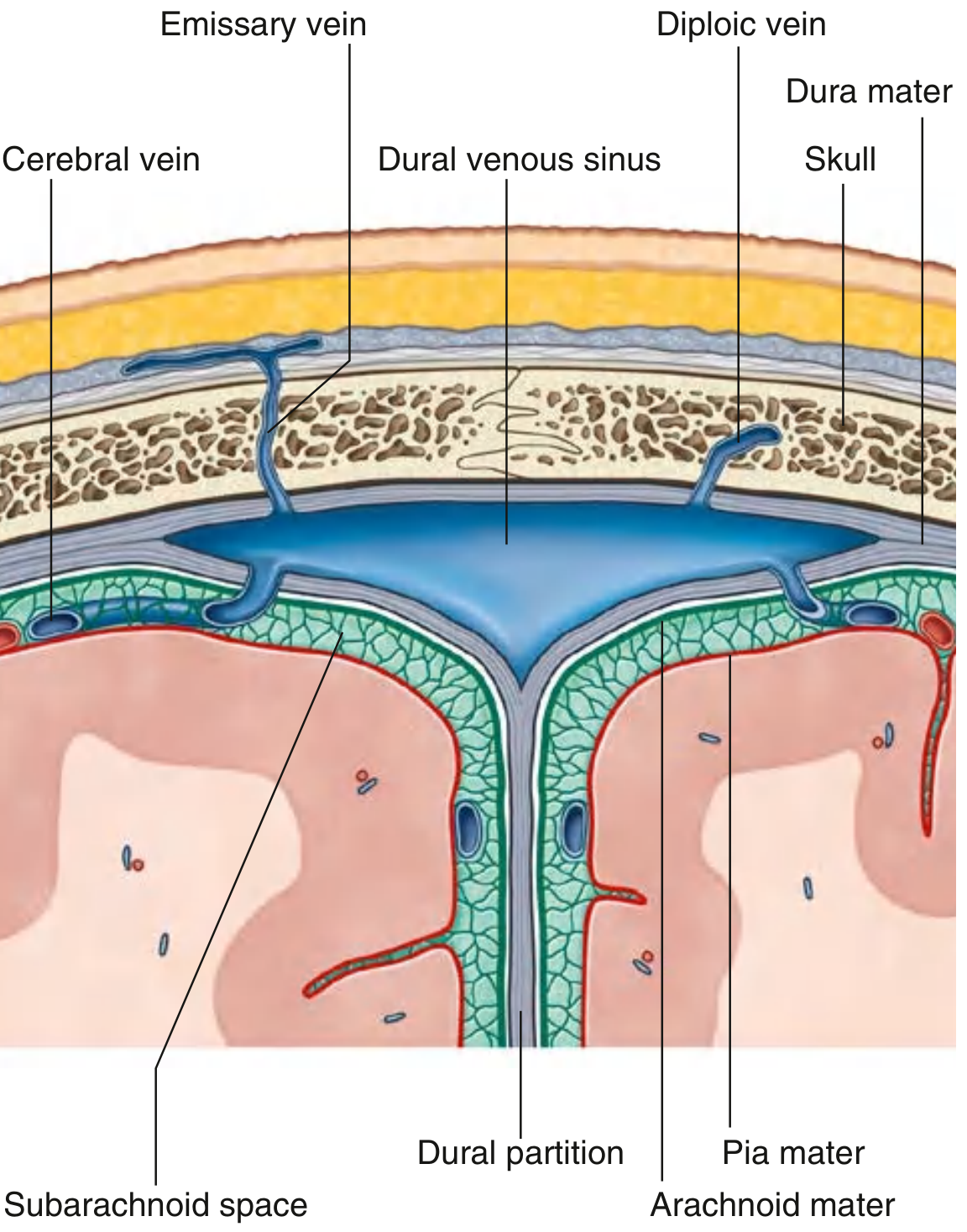
Dural venous sinuses
dural venous sinuses anatomy diagram
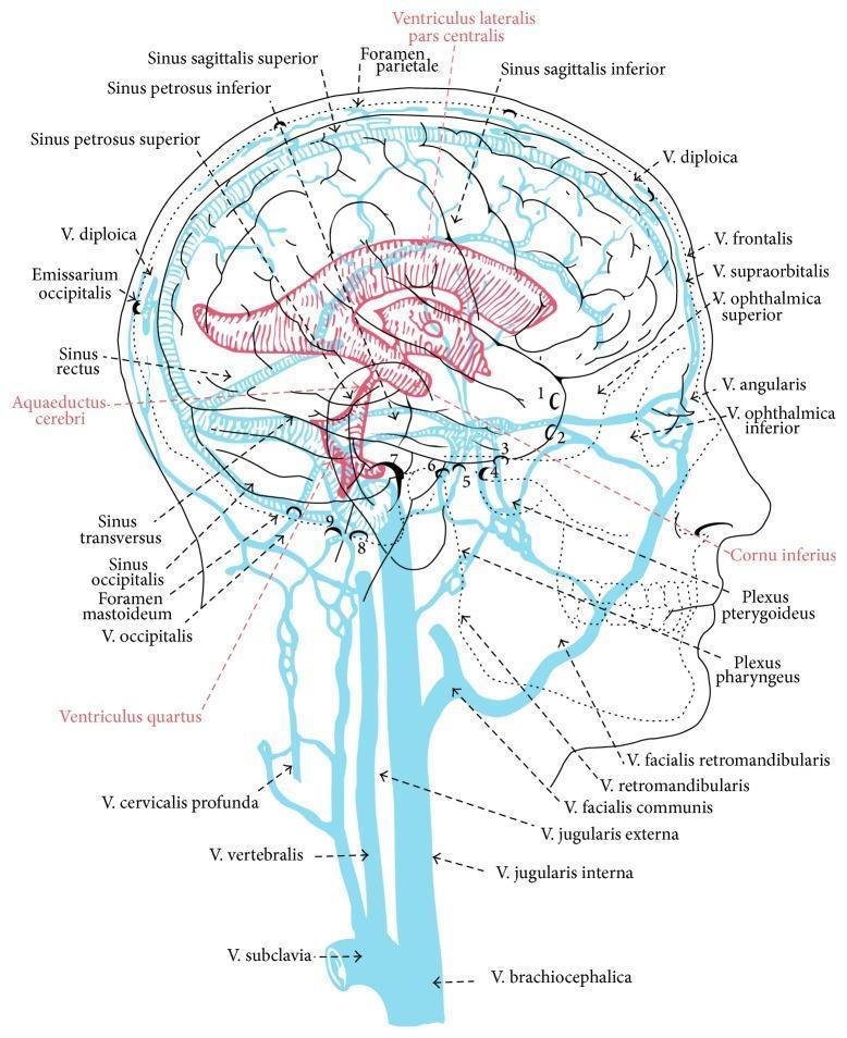
This medical anatomical diagram provides a sagittal view of the human head and neck, illustrating the interconnected dural venous sinus system, the cerebral ventricular system, and the extracranial venous drainage. Key vascular structures highlighted in blue include the sinus sagittalis superior and inferior, sinus rectus, sinus transversus, and sinus occipitalis. The diagram details the transition from intracranial dural sinuses to extracranial pathways, such as the V. jugularis interna, V. vertebralis, and V. brachiocephalica. Additionally, it displays the relationship between the dural sinuses and facial/orbital veins (e.g., V. ophthalmica superior, V. angularis). Overlaid in red is the ventricular system, clearly labeling the ventriculus lateralis, aquaeductus cerebri, and ventriculus quartus, demonstrating the spatial orientation of cerebrospinal fluid pathways relative to the major venous structures. Anatomical landmarks such as the foramen parietale and foramen mastoideum are indicated to show where emissary veins (V. diploica) bridge the intracranial and extracranial compartments. This educational illustration is designed to teach the complex neurovascular anatomy of cranial drainage and ventricular positioning.
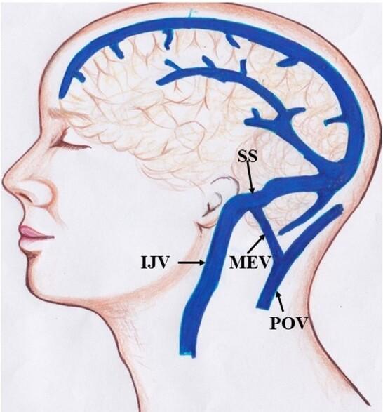
Anatomical diagram depicting the venous drainage of the human head in a lateral view. The illustration highlights the relationship between intracranial dural sinuses and extracranial veins. Key structures identified include the Sigmoid Sinus (SS), which transitions into the Internal Jugular Vein (IJV) as it descends anteriorly. A significant anatomical feature shown is the Mastoid Emissary Vein (MEV), which provides a valveless communication pathway between the intracranial sigmoid sinus and the extracranial Posterior Auricular Vein (POV). The POV is situated posteriorly to the ear and runs roughly parallel to the upper portion of the IJV. The diagram effectively illustrates the concept of emissary veins as collateral drainage routes and their role in intracranial pressure regulation and potential surgical risk during posterior fossa or mastoid procedures. The focus is on neurovascular anatomy, specifically the communication between the dural venous system and superficial scalp veins.
Dural Venous Sinuses
Tributaries
- Cerebral, cerebellar, and brainstem veins
- Diploic veins - run between the inner and outer tables of the skull
- Emissary veins - connect extracranial veins (scalp, face) to the sinuses through skull foramina; because they are valveless, they are a route by which extracranial infection (e.g., scalp cellulitis) can spread intracranially
The Sinuses
| Sinus | Location | Receives |
|---|---|---|
| Superior sagittal | Superior border of falx cerebri | Superior cerebral veins, diploic/emissary veins, CSF (via arachnoid granulations in lateral lacunae) |
| Inferior sagittal | Inferior margin of falx cerebri | Few cerebral veins, veins from falx cerebri |
| Straight | Junction of falx cerebri and tentorium cerebelli | Inferior sagittal sinus + great cerebral vein, posterior cerebral veins, superior cerebellar veins |
| Occipital | In falx cerebelli | Communicates with vertebral venous plexus |
| Confluence of sinuses | Internal occipital protuberance | Superior sagittal, straight, and occipital sinuses |
| Transverse (paired) | Along attachment of tentorium cerebelli | Confluence of sinuses (right usually from superior sagittal, left usually from straight sinus), superior petrosal sinus, diploic/emissary veins |
| Sigmoid (paired) | Continuation of transverse sinus, grooves parietal/temporal/occipital bones | Continues into internal jugular vein |
| Cavernous (paired) | Either side of the sella turcica, against body of sphenoid | Cerebral veins, ophthalmic veins, emissary veins from pterygoid plexus |
| Intercavernous | Anterior/posterior to pituitary stalk | Connects right and left cavernous sinuses |
| Sphenoparietal (paired) | Along inferior surface of lesser wing of sphenoid | Diploic and meningeal veins; drains into cavernous sinus |
| Superior petrosal (paired) | Along petrous temporal bone | Drains cavernous sinus into transverse sinus |
| Inferior petrosal (paired) | Along petro-occipital junction | Drains cavernous sinus into internal jugular vein |
| Basilar | Over the basilar part of occipital bone | Connects inferior petrosal sinuses, communicates with vertebral venous plexus |
Clinical importance: the cavernous sinus
- Through the sinus: internal carotid artery, abducens nerve (CN VI)
- In the lateral wall (superior to inferior): oculomotor nerve (CN III), trochlear nerve (CN IV), ophthalmic nerve (CN V1), maxillary nerve (CN V2)