
Post surgical tachicardia
Reading File
Post-surgical tachycardia is a common finding in the first 24-72 hours after surgery. The key is distinguishing benign physiologic causes (pain, anxiety, fever) from serious ones (hemorrhage, PE, cardiac arrhythmia). A heart rate >100 bpm warrants systematic evaluation using the classic "5 H's" mnemonic.
Searching sources...
Finding Sources
Finding Sources
Searching PubMed
postoperative tachycardia management
Reading File
Reading File
Finding Sources
Reading File
Finding Sources
Reading File
Finding Sources
I found the dedicated "Tachycardia" section in Miller's Anesthesia. Let me read it fully.
Reading File
Reading File
I now have excellent, authoritative content from Miller's Anesthesia (10th ed) and Berek & Novak's Gynecology. Let me compile the full answer.
Post-Surgical Tachycardia
Post-surgical tachycardia (HR >100 bpm) is one of the most common hemodynamic disturbances in the Post-Anesthesia Care Unit (PACU). It carries clinical significance: postoperative tachycardia and hypertension are associated with a higher risk of unplanned ICU admission and mortality than postoperative hypotension and bradycardia - Miller's Anesthesia, 10th ed.
Causes
Common (PACU setting)
| Cause | Mechanism |
|---|---|
| Pain / agitation | Sympathetic activation - catecholamine surge |
| Hypovolemia | Reduced preload - compensatory tachycardia |
| Hypoventilation | Hypoxia + hypercapnia - sympathetic drive |
| PONV (nausea/vomiting) | Vagal response + distress |
| Shivering / hypothermia | Increased metabolic demand |
| Fever / infection | Systemic inflammatory response |
| Urinary retention | Pain + sympathetic activation |
| Anxiety / emergence excitement | Catecholamine release |
Serious / Life-Threatening (must be ruled out)
| Cause | Key Features |
|---|---|
| Hemorrhage | Hypotension, increased chest tube output (>200 mL/h), falling hematocrit |
| Pulmonary embolism | Hypoxia, chest pain, tachypnea, elevated D-dimer |
| Sepsis / anaphylaxis | Fever, hypotension, distributive shock picture |
| Tension pneumothorax | Unilateral absent breath sounds, tracheal deviation, hypotension |
| Malignant hyperthermia | Rapid temperature rise, rigidity, rhabdomyolysis, hypercapnia |
| Thyroid storm | Hyperthermia, agitation, known thyroid disease |
| Myocardial ischemia | Silent or masked by pain meds; ECG changes, elevated troponin |
| Cardiac tamponade | Cardiogenic shock, muffled sounds, JVD |
- Miller's Anesthesia, 10th ed., p. 11607
Why Tachycardia Causes Myocardial Ischemia
Tachycardia is particularly dangerous in patients with coronary artery disease because it limits diastolic filling time, reducing coronary artery perfusion volume exactly when myocardial oxygen demand is rising. Combined with increased preload (which raises intramural wall pressure and compresses coronary arterioles), this dual mechanism can precipitate perioperative MI. Nearly two-thirds of postoperative MIs occur in the first 3 days after surgery, and up to 93% are silent. - Berek & Novak's Gynecology, p. 1250
Evaluation Approach
Step 1: Is the patient hemodynamically stable?
- HR >150 bpm + hypotension + signs of hypoperfusion (altered mental status, chest pain, shock) = UNSTABLE - act immediately
Step 2 for STABLE patients:
- Administer supplemental oxygen
- Obtain 12-lead ECG and determine the underlying rhythm
- Assess and treat reversible causes (pain, fluid status, oxygenation, electrolytes)
- Check for: hypoxemia, hypercapnia, electrolyte abnormalities (K+, Mg++), acidemia, anemia, drug effects, substance withdrawal
Step 3 for UNSTABLE patients:
- SVT with rapid ventricular response, atrial fibrillation, or atrial flutter = immediate synchronized cardioversion (per AHA 2020 ACLS guidelines)
- Identify and treat underlying cause simultaneously
Management by Cause
| Finding | Action |
|---|---|
| Pain | Analgesics (opioids, NSAIDs, regional block) |
| Hypovolemia / bleeding | IV fluids, blood products; surgical re-exploration if hemorrhage |
| Fever / infection | Cultures, empiric antibiotics, source control |
| Pulmonary embolism | Anticoagulation, consider thrombolysis if massive |
| Atrial fibrillation (new onset) | Rate control (beta-blocker, diltiazem), anticoagulation |
| Malignant hyperthermia | Dantrolene IV, cooling, supportive care |
| Myocardial ischemia | Beta-blocker, nitroglycerin, aspirin, statin; cardiology consult; consider PCI |
| Thyroid storm | Beta-blocker, thionamide, iodine, corticosteroids, supportive care |
Postoperative Atrial Fibrillation
New-onset AF is particularly common after thoracic surgery (cardiothoracic, esophageal). Postoperative supraventricular tachyarrhythmias after thoracotomy are described as "common and usually require immediate treatment" - Morgan & Mikhail's Clinical Anesthesiology, 7th ed. Standard management includes rate control first, then rhythm control if needed, with anticoagulation decisions guided by bleeding risk.
Key Points
- Always exclude hemorrhage, PE, and sepsis before attributing tachycardia to benign causes
- Silent myocardial injury (elevated troponin without symptoms) is far more common post-op than clinically recognized; check troponin if ECG changes are present or the patient is high-risk
- Adequate analgesia is both treatment and prevention - pain-driven sympathetic tachycardia is avoidable
Management of tachycardia
Tachycardia management depends on whether the patient is hemodynamically stable and the underlying rhythm type. The core principle is: treat unstable patients immediately with cardioversion, and in stable patients identify and treat the cause/rhythm.
Searching sources...
Finding Sources
Finding Sources
Searching PubMed
"tachycardia"[MeSH Terms] AND "management"[All Fields]
Finding Sources
Reading File
Reading File
Reading File
Finding Sources
Finding Sources
Reading File
Finding Sources
Reading File
Reading File
I now have comprehensive, authoritative content from Miller's Anesthesia (10th ed) and Braunwald's Heart Disease. Let me also fetch the ACLS tachycardia algorithm image and compile the full answer.
Finding Sources
Reading File
I have the ACLS algorithm image URL and all the drug doses. Let me now compile the complete, well-structured answer.
Management of Tachycardia
Based on the 2020 AHA ACLS Tachycardia with Pulse Algorithm - Miller's Anesthesia, 10th ed. & Morgan and Mikhail's Clinical Anesthesiology, 7th ed.
Step 1: Initial Assessment (ALL patients)
- Attach monitoring (ECG, pulse oximetry, BP)
- Provide supplemental oxygen - hypoxemia is a common cause of tachycardia and must be corrected first
- Obtain 12-lead ECG to characterize the rhythm
- Do NOT delay cardioversion if the patient is obviously unstable
Step 2: Is the Patient Stable or Unstable?
Signs of instability (rate-related cardiovascular compromise):
- Acute altered mental status
- Ischemic chest discomfort
- Acute heart failure
- Hypotension or other signs of shock
- HR typically >150 bpm
UNSTABLE Patient → Immediate Synchronized Cardioversion
Do not delay for drug therapy. Sedate first if the patient is conscious.
| Rhythm | Initial Energy (Biphasic) |
|---|---|
| Atrial fibrillation | 120-200 J |
| Atrial flutter / other SVT | 50-100 J (increase stepwise if needed) |
| Monomorphic VT with pulse | 100 J (synchronized) |
| Polymorphic VT | Treat as VF - unsynchronized high-energy defibrillation |
- Morgan & Mikhail's Clinical Anesthesiology, 7th ed., p. 2414
STABLE Patient → Narrow-Complex vs. Wide-Complex
Here is the complete ACLS algorithm:
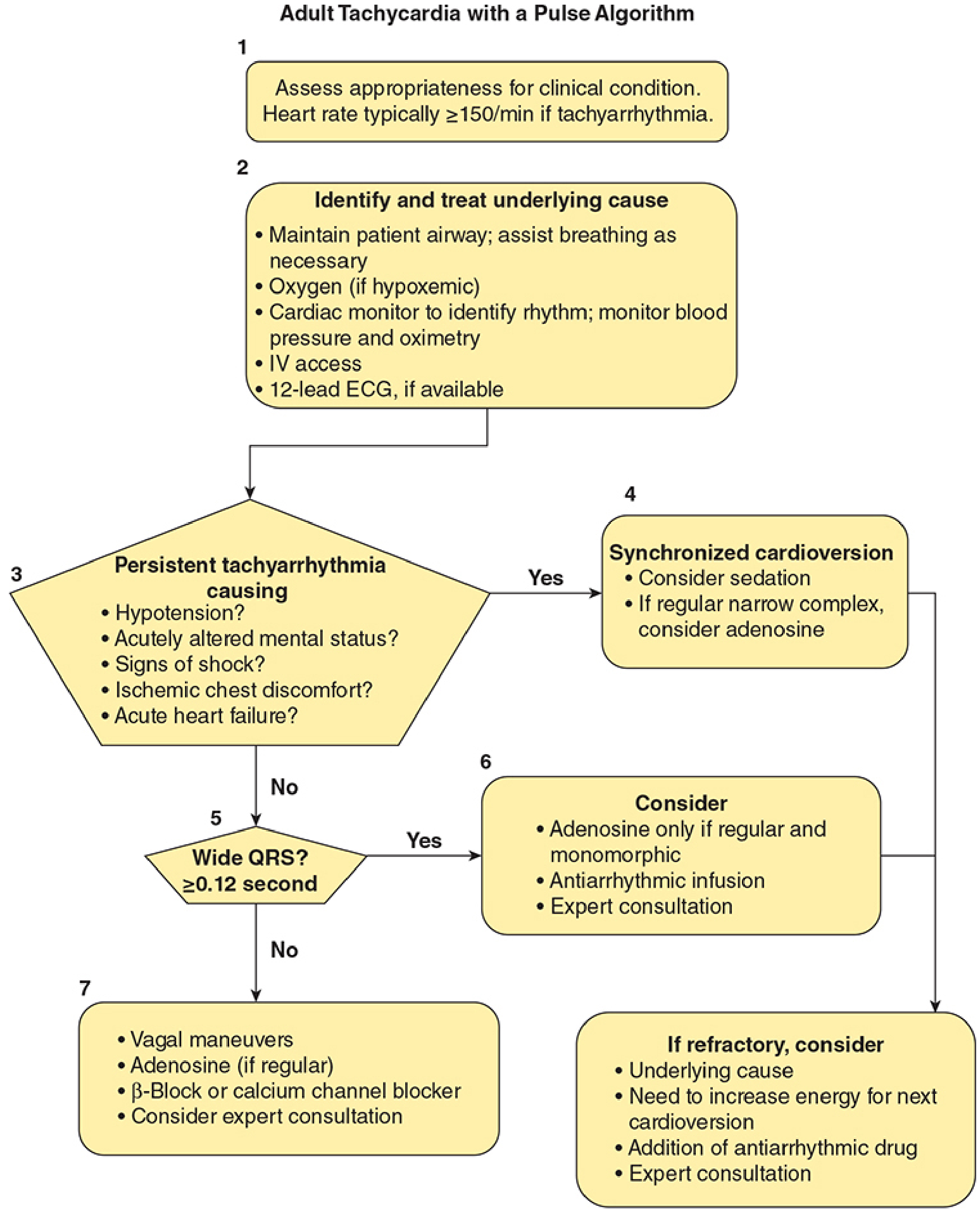
A. Narrow-Complex Tachycardia (QRS <0.12 sec) - Regular
These are supraventricular tachycardias (SVT, AVNRT, AVRT, AT, atrial flutter).
Step 1: Vagal maneuvers
- Valsalva maneuver (preferred; modified supine with leg elevation is most effective)
- Carotid sinus massage
- Effective in ~40-50% of patients
Step 2: Adenosine (if vagal maneuvers fail)
- First dose: 6 mg rapid IV push + NS flush
- Second dose: 12 mg if required
- Warn the patient of transient chest tightness and sense of "impending doom"
- Diagnostic and therapeutic: may transiently reveal underlying flutter waves or diagnose VT vs. SVT
Step 3: AV nodal blocking agents (if adenosine fails)
- IV verapamil or diltiazem (non-DHP calcium channel blockers)
- IV beta-blocker (metoprolol or esmolol)
- DC cardioversion if hemodynamically unstable or all pharmacologic measures fail
- Braunwald's Heart Disease, p. 874
B. Narrow-Complex Tachycardia - Irregular
Likely atrial fibrillation or multifocal atrial tachycardia (MAT)
Rate control (acute):
| Drug | Route | Note |
|---|---|---|
| Metoprolol or esmolol | IV | First-line if BP adequate |
| Diltiazem | IV | Good rate control, avoid if severe LV dysfunction |
| Digoxin | IV | Slower onset; prefer when BP is tenuous |
| Amiodarone | IV | When beta-blockers/CCBs are contraindicated or ineffective |
Rate targets: Resting HR <80 bpm; moderate exercise HR <100 bpm (symptomatic patients)
Rhythm control: Electrical or pharmacological cardioversion - no survival advantage over rate control in randomized trials of non-valvular AF, but may be preferred in certain populations (younger patients, structural heart disease, severe symptoms)
Anticoagulation: Always consider before cardioversion - if AF duration >48 hrs or unknown, must rule out LA thrombus (TEE) or anticoagulate for 3 weeks before elective cardioversion
- Fuster and Hurst's The Heart, 15th ed., p. 184-186
C. Wide-Complex Tachycardia (QRS ≥0.12 sec) - Regular
Assume ventricular tachycardia (VT) until proven otherwise.
If uncertain whether VT or SVT with aberrancy:
- Give adenosine IV during ECG monitoring - it may clarify the diagnosis by transiently interrupting AV conduction
Pharmacologic termination (stable VT):
| Drug | Dose | Notes |
|---|---|---|
| Procainamide | 20-50 mg/min IV until arrhythmia suppressed, hypotension, or QRS widens >50% (max 17 mg/kg); maintenance 1-4 mg/min | More effective than amiodarone in one trial; avoid in prolonged QT or CHF; avoid in end-stage renal disease (metabolite accumulation) |
| Amiodarone | 150 mg IV over 10 min, repeat if VT recurs; then 1 mg/min infusion x 6 hrs | Use when procainamide is contraindicated |
| Sotalol | 100 mg (1.5 mg/kg) IV over 5 min | Avoid if prolonged QT |
| Lidocaine | Less effective than amiodarone | Reserve as alternative |
Key rule: Use only ONE antiarrhythmic drug. Do NOT add a second without expert consultation. All three agents cause hypotension in up to 30% of patients.
Special cases:
- Idiopathic VT (no structural heart disease): Beta-blockers often terminate it
- Left ventricular fascicular reentrant tachycardia: IV verapamil or diltiazem (do NOT give calcium channel blockers for other VTs - risk of hemodynamic collapse)
- Braunwald's Heart Disease, p. 2714-2716
D. Wide-Complex Tachycardia - Irregular
Consider pre-excited AF (WPW) or polymorphic VT
- WPW + AF: Avoid AV nodal blocking agents (adenosine, verapamil, digoxin, beta-blockers) - they can accelerate conduction down the accessory pathway and precipitate VF. Use procainamide or cardioversion
- Polymorphic VT / Torsades de pointes: Correct electrolytes (K+, Mg++); IV magnesium sulfate 2g over 15 min; stop QT-prolonging drugs; overdrive pacing if recurrent
Reversible Causes to Correct in All VT
Before or during pharmacologic/electrical therapy, always seek and correct:
- Hypoxemia / hypercapnia
- Hypokalemia / hypomagnesemia
- Digitalis toxicity
- Acidosis
- Acute myocardial ischemia (check ECG + troponin)
- Increased sympathetic tone from pain, fever, or acute illness
Long-Term / Definitive Management
| Rhythm | Definitive Strategy |
|---|---|
| Recurrent AVNRT / AVRT | Catheter ablation (curative) |
| Recurrent AF | Pulmonary vein isolation; AV node ablation + pacemaker if refractory |
| Sustained VT with structural disease | ICD implantation ± catheter ablation |
| Idiopathic VT | Catheter ablation or long-term beta-blocker/CCB |
| Sinus tachycardia | Treat the underlying cause (pain, fever, hypovolemia, thyroid, anemia) |
Management of tachycardia
I just covered this in detail in my previous response. Here's a concise summary for quick reference:
Management of Tachycardia - Quick Reference
Step 1: Initial Steps (All Patients)
- O₂, monitoring, 12-lead ECG
- Identify and correct reversible causes (hypoxia, pain, hypovolemia, electrolytes)
Step 2: Stable or Unstable?
UNSTABLE (hypotension, AMS, chest pain, shock, HR >150)
→ Immediate Synchronized Cardioversion
| Rhythm | Energy |
|---|---|
| AF | 120-200 J biphasic |
| AFL / other SVT | 50-100 J |
| Monomorphic VT | 100 J |
| Polymorphic VT | Unsynchronized defibrillation (high energy) |
STABLE → Classify by QRS width
Narrow Complex (QRS <0.12s) - Regular SVT
- Vagal maneuvers (Valsalva, carotid massage) - effective ~40-50%
- Adenosine 6 mg IV rapid push → 12 mg if needed
- Verapamil / Diltiazem or Beta-blocker (metoprolol/esmolol) IV
Narrow Complex - Irregular (AF/MAT)
- Rate control: Metoprolol, diltiazem, digoxin (if BP tenuous), or amiodarone
- Rhythm control: Electrical or pharmacological cardioversion (anticoagulate if AF >48 hrs)
- Target HR: <80 bpm rest, <100 bpm exercise
Wide Complex (QRS ≥0.12s) - Regular → Assume VT
- Adenosine IV if uncertain VT vs SVT with aberrancy (diagnostic + may be therapeutic)
- Procainamide 20-50 mg/min IV (max 17 mg/kg) - preferred pharmacologic agent
- Amiodarone 150 mg IV over 10 min if procainamide contraindicated
- Sotalol 100 mg IV over 5 min
- Use only ONE antiarrhythmic at a time
Wide Complex - Irregular
- WPW + AF: Avoid AV nodal blockers → use Procainamide or cardioversion
- Torsades de pointes: Magnesium 2g IV + stop QT-prolonging drugs
Long-Term Definitive Treatment
| Rhythm | Strategy |
|---|---|
| AVNRT / AVRT | Catheter ablation (curative) |
| AF | Pulmonary vein isolation; AV node ablation + pacemaker |
| Sustained VT | ICD ± catheter ablation |
| Sinus tachycardia | Treat the underlying cause |
For full details including drug doses, energy levels, and special populations, see my previous response above.
This is a shared conversation. Sign in to Orris to start your own chat.