
Pathophysiology of gangrenous gall bladder
gangrenous cholecystitis pathophysiology
gangrenous cholecystitis
gangrenous cholecystitis gallbladder necrosis pathology gross specimen
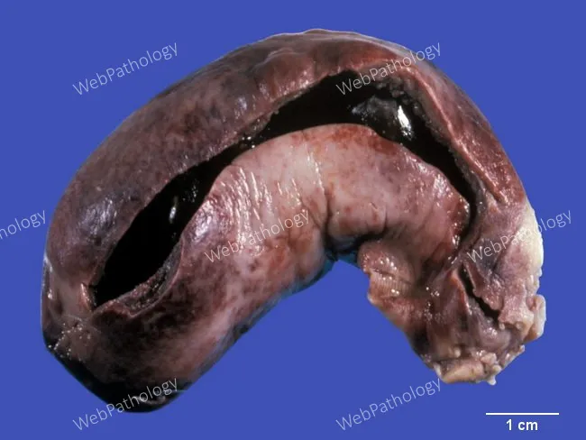
Gross pathology photograph of a gallbladder specimen showing gangrenous cholecystitis. The organ is enlarged and markedly distended, opened to reveal intraluminal gallstones. The serosal surface is dusky red-brown from congestion and hemorrhage, with transmural inflammation and necrosis evident in the gallbladder wall. The walls are friable and may show edema while the mucosa is disrupted by necrotic debris. External serosa appears thickened and mottled; the lumen contains pigmented bile and debris. Overall, features are consistent with acute calculous cholecystitis complicated by gangrene, with possible early perforation risk. This macroscopic appearance correlates with ischemic injury to the gallbladder wall and extensive inflammatory infiltration. The image illustrates a classic teaching example for gross surgical pathology and hepatobiliary disease. Clinically, gangrenous cholecystitis represents a severe, life-threatening progression from simple cholecystitis, often in patients with comorbidities such as diabetes, cardiovascular disease, and advanced age. The photo supports differential considerations including emphysematous cholecystitis or perforated cholecystitis and emphasizes urgent surgical management and histopathologic confirmation of transmural necrosis. Note the scale is conveyed by a contrasting background and a centimeter ruler present in the image, supporting gross measurements and education. This representation facilitates correlation with surgical findings and guides learning in medical school, pathology residency, and radiology-pathology conferences.
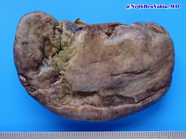
Gross pathology photograph illustrating gangrenous cholecystitis. The gallbladder is enlarged and distended, with a thickened wall and friable consistency. The serosal surface appears dull and dusky red‑brown, reflecting profound congestion and ischemia. Depressed necrotic foci are present on the mucosal and serosal surfaces, consistent with transmural necrosis typical of gallbladder gangrene. The organ shows irregular, hemorrhagic, devitalized areas interspersed with relatively preserved tissue. Exterior faces mild edema with focal surface ulcers at the fundus and body. The lumen contents are not visible in this specimen, as processing often removes bile. This macroscopic image was captured from an excised gallbladder to illustrate severe inflammatory biliary disease in surgical pathology. The blue background and centimeter scale provide reference for size and orientation. Imaging technique includes gross pathology photography, color‑calibrated macro imaging, and high‑resolution digital capture under standardized lighting. Diagnostic significance lies in confirming advanced gallbladder necrosis with gangrene, which elevates risk of perforation, peritonitis, and sepsis; it supports a clinical diagnosis of acute gangrenous cholecystitis. Educationally, this image aids recognition of gross features, correlates with clinical presentation, and enhances training in surgical pathology interpretation. This image is suitable for medical students, residents, and fellows studying gallbladder pathology, inflammatory hepatobiliary disease, and surgical specimen assessment.
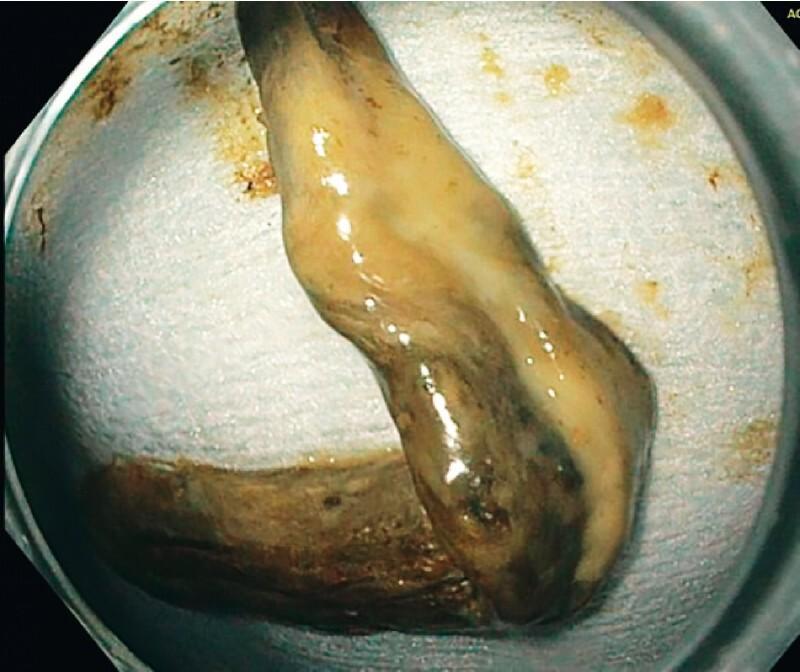
This clinical photograph shows a gross surgical specimen of a necrotic gallbladder wall removed during an endoscopic procedure. The specimen is elongated and measures approximately 9 x 1.5 cm. It exhibits significant pathological changes characteristic of gangrenous cholecystitis, including a non-uniform, mottled appearance with colors ranging from yellowish-tan to dark brown and black, indicating extensive tissue necrosis. The surface is moist and glistening, suggesting a sloughing or mucoid texture. This visual serves as an educational example of severe biliary pathology and the complications of stent obstruction following endoscopic ultrasound-guided gallbladder drainage (EUS-GBD). The specimen was retrieved via endoscopy after a patient presented with recurrent symptoms and a hydropic gallbladder, highlighting the clinical importance of identifying necrotic debris in cases of stent dysfunction.
Pathophysiology of Gangrenous Gallbladder
1. Initiating Event: Cystic Duct Obstruction
2. Chemical Irritation and Mucosal Injury (Early Phase)
- Mucosal phospholipases hydrolyze luminal lecithins into toxic lysolecithins
- The normal protective glycoprotein mucus layer is disrupted
- This exposes the mucosal epithelium to the direct detergent action of bile salts
- Prostaglandins released within the distended gallbladder wall amplify mucosal and mural inflammation
3. Mechanical Compression of Mural Blood Supply (Critical Step)
- Distension of the gallbladder raises intraluminal pressure
- Increased pressure compromises blood flow to the mucosa and wall
- The cystic artery is an end artery with no collateral circulation - this anatomical feature makes the gallbladder uniquely vulnerable
- Venous outflow is obstructed first → mucosal congestion and edema → then arterial compromise → ischemia → necrosis
4. Progression to Full-Thickness Necrosis
| Stage | What Happens |
|---|---|
| Early | Mucosal edema, congestion, erosion; sparse neutrophils |
| Intermediate | Inflammatory cell infiltration through the wall; hemorrhage |
| Advanced | Transmural ischemic infarction; bacterial invasion |
| Gangrenous | Full-thickness necrosis; green-black discoloration; friable wall |
5. Superimposed Bacterial Infection
- Bile/gallbladder wall cultures are positive in >40% of cases
- Common organisms: E. coli, Klebsiella, Enterobacter, Proteus (aerobic gram-negatives); Enterococcus, Streptococcus (gram-positives)
- Anaerobes (notably Clostridia welchii) are identified in ~15% of isolates; when gas-forming organisms invade the necrotic wall, this becomes emphysematous cholecystitis - an even more severe variant
6. Acalculous Pathway - Pure Ischemic Mechanism
- The pathophysiology is primarily transmural ischemia without stone obstruction
- Systemic hypotension/hypoperfusion + the end-artery anatomy of the cystic artery = rapid wall necrosis
- Contributing factors: wall edema compressing microcirculation, biliary stasis with sludge, inspissated bile obstructing the cystic duct
- This form carries higher morbidity and mortality than calculous gangrenous cholecystitis because the underlying critical illness compounds the local ischemic injury
7. Gross and Histological Morphology
- Gross: Enlarged, tense organ with bright red to green-black discoloration; subserosal hemorrhages; fibrinous/fibrinopurulent serosal exudate; small-to-large perforations; lumen filled with turbid bile, fibrin, pus, and hemorrhage
- Micro: Transmural necrosis; hemorrhagic infarction; extensive neutrophilic infiltration (if bacterial superinfection); loss of mucosal architecture; vascular thrombosis in mural vessels
8. Risk Factors Predisposing to Gangrene
- Diabetes mellitus (microangiopathy + impaired neutrophil function)
- Cardiovascular disease (pre-existing vascular compromise)
- Advanced age (reduced reserve)
- Male sex
- Critical illness: sepsis, hypotension, multi-organ failure
- Immunosuppression
- Major trauma, burns
- Postoperative state (splanchnic hypoperfusion)
Summary Pathophysiological Sequence
Stone/sludge occludes cystic duct
        ↓
Gallbladder distension → raised intraluminal pressure
        ↓
Chemical injury (lysolecithins, bile salts, prostaglandins)
        ↓
Compression of mural microvasculature (cystic artery = end artery)
        ↓
Venous stasis → mucosal ischemia → arterial compromise
        ↓
Transmural ischemic necrosis → gangrene
        ↓
Bacterial superinfection (E. coli, Klebsiella, Clostridia)
        ↓
Perforation → pericholecystic abscess / peritonitis / sepsis