X ray of nose

Searching Images

X-ray of nose nasal bones radiograph

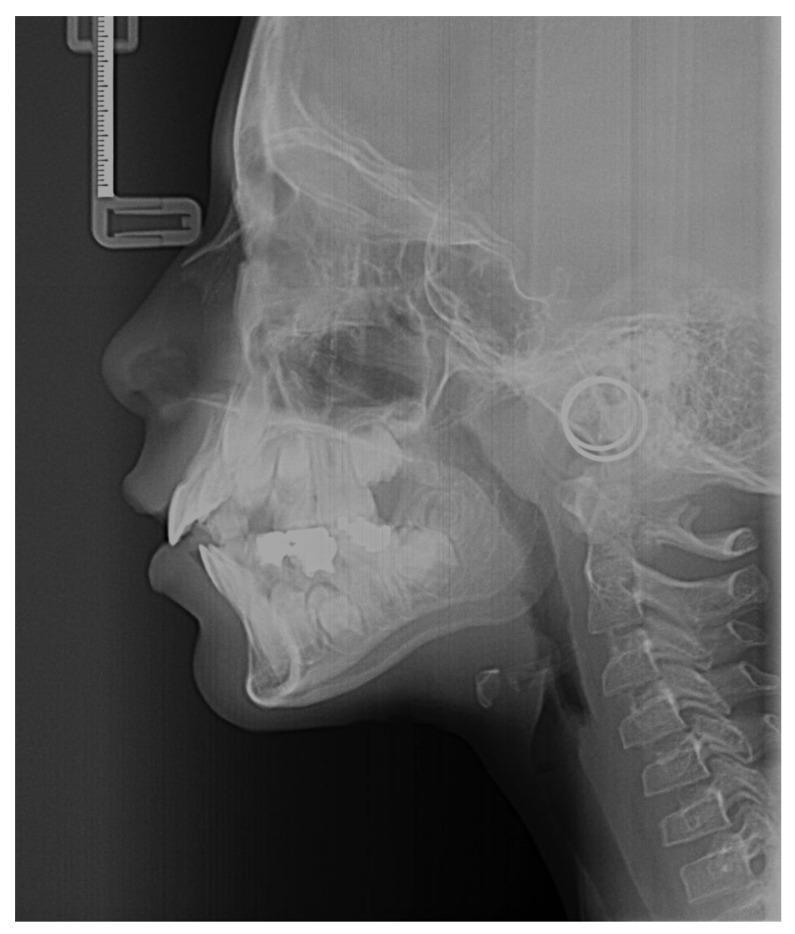
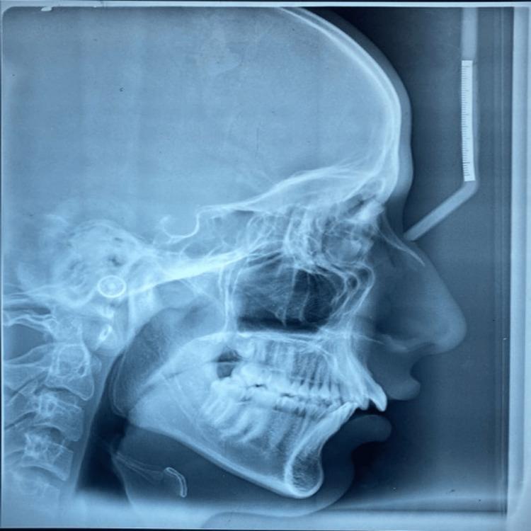
This diagnostic image is a lateral cephalometric radiograph, a specialized extraoral x-ray used primarily in orthodontics and maxillofacial surgery. The radiograph displays a lateral view of the human skull, including the facial bones, mandible, and maxilla. Visible anatomical landmarks include the sella turcica, nasal bone, and the anterior and posterior nasal spines. The dental arches are shown with evident dental restorations (radiopaque fillings). Importantly for skeletal maturity assessment, the cervical vertebrae (C2-C4) are clearly visible, showing the morphology of the vertebral bodies and inferior borders used in the Cervical Vertebral Maturation (CVM) method. The soft tissue profile is outlined, depicting the contours of the nose, lips, and chin relative to the underlying skeletal structures. Radiographic artifacts are present, including a nasion relator (ruler) near the frontal bone and ear rods (circular radiopaque markers) near the external auditory meatus to ensure standardized positioning.
This diagnostic image is a lateral cephalometric radiograph, a specialized extraoral x-ray used primarily in orthodontics and maxillofacial surgery. The radiograph displays a lateral view of the human skull, including the facial bones, mandible, and maxilla. Visible anatomical landmarks include the sella turcica, nasal bone, and the anterior and posterior nasal spines. The dental arches are shown with evident dental restorations (radiopaque fillings). Importantly for skeletal maturity assessment, the cervical vertebrae (C2-C4) are clearly visible, showing the morphology of the vertebral bodies and inferior borders used in the Cervical Vertebral Maturation (CVM) method. The soft tissue profile is outlined, depicting the contours of the nose, lips, and chin relative to the underlying skeletal structures. Radiographic artifacts are present, including a nasion relator (ruler) near the frontal bone and ear rods (circular radiopaque markers) near the external auditory meatus to ensure standardized positioning.

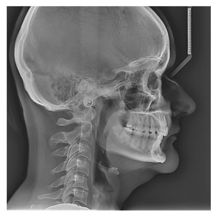
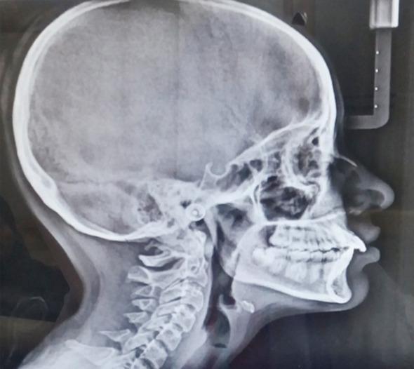
This diagnostic image is a lateral cephalogram (x-ray radiography) of the human skull and cervical spine. The imaging captures the sagittal profile, displaying the cranium (frontal, parietal, and occipital bones), facial bones, and the upper cervical vertebrae (C1-C5). Key anatomical landmarks visible include the sella turcica, nasal bone, maxilla, and mandible. The radiograph clearly demonstrates the relationship between the maxillary and mandibular dental arches, showing the occlusion of the anterior and posterior teeth. Soft tissue contours of the nose, lips, chin, and posterior airway space are also visible. In the upper right corner, a calibration ruler is present for linear measurements. This modality is primarily used in orthodontics and maxillofacial surgery to evaluate craniofacial growth, dentofacial proportions, and craniocervical posture, including the assessment of angles and distances such as the craniovertebral angle and hyoid bone position.
This diagnostic image is a lateral cephalogram (x-ray radiography) of the human skull and cervical spine. The imaging captures the sagittal profile, displaying the cranium (frontal, parietal, and occipital bones), facial bones, and the upper cervical vertebrae (C1-C5). Key anatomical landmarks visible include the sella turcica, nasal bone, maxilla, and mandible. The radiograph clearly demonstrates the relationship between the maxillary and mandibular dental arches, showing the occlusion of the anterior and posterior teeth. Soft tissue contours of the nose, lips, chin, and posterior airway space are also visible. In the upper right corner, a calibration ruler is present for linear measurements. This modality is primarily used in orthodontics and maxillofacial surgery to evaluate craniofacial growth, dentofacial proportions, and craniocervical posture, including the assessment of angles and distances such as the craniovertebral angle and hyoid bone position.

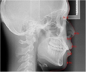
This diagnostic image is a lateral cephalometric radiograph, a standardized orthodontic x-ray used for skeletal and soft tissue profile analysis. The radiograph displays a lateral view of the human skull, including the cranium, cervical vertebrae, and facial bones, with a specific focus on the dentofacial structures in centric occlusion. Red annotations mark key soft tissue cephalometric landmarks along the facial profile: Ns (Soft tissue nasion) at the root of the nose; Pn (Pronasale) at the nasal tip; Sn (Subnasale) at the junction of the nose and upper lip; Ls (Labial superius) on the upper lip; Li (Labial inferius) on the lower lip; and Pos (Soft tissue pogonion) on the chin. The image demonstrates the relationship between the underlying hard tissue (maxilla, mandible, and teeth) and the overlying soft tissue drape. This visual is clinically significant for orthodontic diagnosis, treatment planning, and evaluating facial aesthetics and skeletal malocclusions.
This diagnostic image is a lateral cephalometric radiograph, a standardized orthodontic x-ray used for skeletal and soft tissue profile analysis. The radiograph displays a lateral view of the human skull, including the cranium, cervical vertebrae, and facial bones, with a specific focus on the dentofacial structures in centric occlusion. Red annotations mark key soft tissue cephalometric landmarks along the facial profile: Ns (Soft tissue nasion) at the root of the nose; Pn (Pronasale) at the nasal tip; Sn (Subnasale) at the junction of the nose and upper lip; Ls (Labial superius) on the upper lip; Li (Labial inferius) on the lower lip; and Pos (Soft tissue pogonion) on the chin. The image demonstrates the relationship between the underlying hard tissue (maxilla, mandible, and teeth) and the overlying soft tissue drape. This visual is clinically significant for orthodontic diagnosis, treatment planning, and evaluating facial aesthetics and skeletal malocclusions.

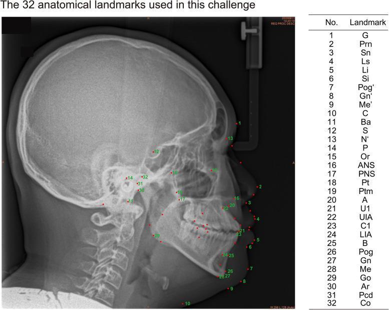
This diagnostic image is a lateral cephalometric radiograph, a specialized X-ray used in orthodontics and maxillofacial surgery to analyze skeletal, dental, and soft tissue relationships of the human head in profile. The radiograph displays clear views of the cranium, facial bones, mandible, upper cervical vertebrae, and the soft tissue outline of the nose, lips, and chin. Overlaid on the image are 32 numbered red dots representing standardized anatomical landmarks. These points are distributed across the cranial base (e.g., Sella, Nasion), midface (e.g., Anterior Nasal Spine, Subspinale), mandible (e.g., Menton, Gonion), dental structures (incisor positions), and facial soft tissues (e.g., Pronasale, Labrale). Adjacent to the radiograph is a reference table linking each number to its corresponding medical abbreviation, such as ANS, PNS, and Pog. This educational visual demonstrates the precise localization required for cephalometric tracing and clinical assessment of craniofacial morphology.
This diagnostic image is a lateral cephalometric radiograph, a specialized X-ray used in orthodontics and maxillofacial surgery to analyze skeletal, dental, and soft tissue relationships of the human head in profile. The radiograph displays clear views of the cranium, facial bones, mandible, upper cervical vertebrae, and the soft tissue outline of the nose, lips, and chin. Overlaid on the image are 32 numbered red dots representing standardized anatomical landmarks. These points are distributed across the cranial base (e.g., Sella, Nasion), midface (e.g., Anterior Nasal Spine, Subspinale), mandible (e.g., Menton, Gonion), dental structures (incisor positions), and facial soft tissues (e.g., Pronasale, Labrale). Adjacent to the radiograph is a reference table linking each number to its corresponding medical abbreviation, such as ANS, PNS, and Pog. This educational visual demonstrates the precise localization required for cephalometric tracing and clinical assessment of craniofacial morphology.

This diagnostic image is a lateral cephalometric radiograph (lateral cephalogram), a standardized extraoral X-ray used primarily in orthodontics and maxillofacial surgery. The image displays a profile view of the human skull, including the cranium, facial bones, and the cervical spine. Key anatomical structures visible include the maxilla, mandible, sella turcica, frontal sinus, and the nasal bone. The soft tissue profile of the nose, lips, and chin is discernible, allowing for the assessment of skeletal and dental relationships to the integumentary profile. Radiopaque features include the dentition within the alveolar processes and a vertical calibration ruler located anterior to the forehead, which is used for linear measurements and scale. A circular radiopaque ear rod (earpost) from the cephalostat is visible in the external auditory meatus region to ensure standardized head positioning. This modality is essential for cephalometric tracing to analyze growth patterns, dental malocclusion, and treatment planning for orthognathic surgery.
This diagnostic image is a lateral cephalometric radiograph (lateral cephalogram), a standardized extraoral X-ray used primarily in orthodontics and maxillofacial surgery. The image displays a profile view of the human skull, including the cranium, facial bones, and the cervical spine. Key anatomical structures visible include the maxilla, mandible, sella turcica, frontal sinus, and the nasal bone. The soft tissue profile of the nose, lips, and chin is discernible, allowing for the assessment of skeletal and dental relationships to the integumentary profile. Radiopaque features include the dentition within the alveolar processes and a vertical calibration ruler located anterior to the forehead, which is used for linear measurements and scale. A circular radiopaque ear rod (earpost) from the cephalostat is visible in the external auditory meatus region to ensure standardized head positioning. This modality is essential for cephalometric tracing to analyze growth patterns, dental malocclusion, and treatment planning for orthognathic surgery.

This diagnostic image is a lateral cephalogram, a standardized orthodontic X-ray providing a profile view of the human skull. The radiograph displays the cranium, facial bones, and the upper cervical spine (C1-C5). Key anatomical landmarks visible include the sella turcica, nasion, and the pterygomaxillary fissure. The dental and skeletal relationship demonstrates a Class II malocclusion, characterized by a retrognathic mandible relative to the maxilla. The maxillary incisors appear proclined, and there is a noticeable overjet. The soft tissue profile is clearly outlined, showing the nose, lips, and chin contour, which aids in assessing the patient's aesthetic harmony. The nasal bone is well-defined, and the pharyngeal airway space appears patent. This imaging modality is essential in orthodontics and maxillofacial surgery for cephalometric analysis, treatment planning, and monitoring craniofacial growth and development.

This diagnostic image is a lateral cephalogram, a standardized orthodontic X-ray providing a profile view of the human skull. The radiograph displays the cranium, facial bones, and the upper cervical spine (C1-C5). Key anatomical landmarks visible include the sella turcica, nasion, and the pterygomaxillary fissure. The dental and skeletal relationship demonstrates a Class II malocclusion, characterized by a retrognathic mandible relative to the maxilla. The maxillary incisors appear proclined, and there is a noticeable overjet. The soft tissue profile is clearly outlined, showing the nose, lips, and chin contour, which aids in assessing the patient's aesthetic harmony. The nasal bone is well-defined, and the pharyngeal airway space appears patent. This imaging modality is essential in orthodontics and maxillofacial surgery for cephalometric analysis, treatment planning, and monitoring craniofacial growth and development.

Here are X-ray images of the nose and nasal region:

Lateral Cephalometric Radiograph - Nasal Bone View
Lateral cephalogram showing nasal bone and facial profile
This lateral skull X-ray clearly shows the nasal bone in profile, along with the anterior and posterior nasal spines, soft tissue contour of the nose, maxilla, and mandible. The nasal bone appears as a distinct, slightly curved radiopaque structure at the bridge of the nose.

Lateral cephalogram with soft tissue landmarks
This annotated lateral cephalogram highlights key nasal soft tissue landmarks including:
  • Ns - Soft tissue nasion (root of the nose)
  • Pn - Pronasale (nasal tip)
  • Sn - Subnasale (base of the nose)

Cephalometric landmark mapping
This radiograph includes numbered anatomical landmark mapping used in orthodontic/surgical planning, with the nasal bone and ANS (Anterior Nasal Spine) clearly marked.

Key points about nasal X-rays:
  • The standard view for nasal bones is the lateral view (side profile), which best shows the nasal bones and any fractures
  • A PA (posteroanterior) or occipitomental view is also used to assess nasal deviation
  • The nasal bones appear as thin, paired, radiopaque structures at the bridge
  • Nasal X-rays are commonly ordered after trauma to detect fractures, or in orthodontic/surgical planning to assess the nasal spine and septum
This is a shared conversation. Sign in to Orris to start your own chat.