
In detail study of fever
"fever" AND "pathophysiology"
fever pathophysiology pyrogen hypothalamus temperature regulation diagram
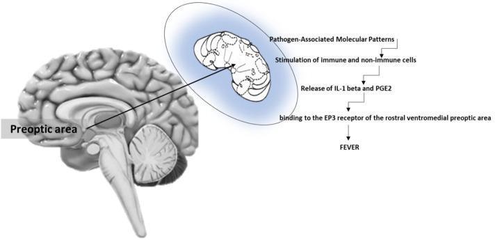
This medical illustration depicts the pathophysiology of fever induction at the brain level. On the left, a sagittal view of the human brain identifies the preoptic area (POA) within the hypothalamus, positioned anterior to the brainstem. An inset zoom highlights the rostral ventromedial preoptic area (rvmPOA) as the specific target for pyrogenic mediators. To the right, a sequential flowchart outlines the molecular mechanism: 1) Exposure to Pathogen-Associated Molecular Patterns (PAMPs); 2) Stimulation of immune and non-immune cells; 3) Release of pro-inflammatory cytokines, specifically Interleukin-1 beta (IL-1 beta), and Prostaglandin E2 (PGE2); 4) Binding of PGE2 to EP3 receptors in the rvmPOA. This signaling cascade leads to the clinical manifestation of fever. The diagram serves as an educational summary of the endocrine and neural pathways involved in thermoregulation and the systemic inflammatory response, highlighting the critical role of the hypothalamus and EP3 receptor signaling in pyrogenesis.
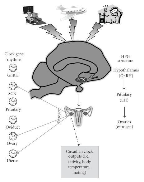
This pathophysiology diagram illustrates the integration of the hypothalamus-pituitary-gonadal (HPG) axis with circadian rhythm regulation in the female reproductive system. A sagittal section of the brain highlights the suprachiasmatic nucleus (SCN) and its relationship with GnRH neurons in the hypothalamus. The diagram outlines the hormonal cascade: Hypothalamus (GnRH) stimulates the Pituitary (LH), which acts on the Ovaries to produce Estrogen. External circadian disruptors—shift work, jet lag, and sleep deprivation—are shown influencing the SCN via lightning bolt symbols. Educational icons represent 'Clock gene rhythms' present in the GnRH neurons, SCN, Pituitary, Oviduct, Ovary, and Uterus, emphasizing peripheral molecular clocks. Solid arrows denote estrogen's feedback effects on reproductive tissues and the brain, while a dashed arrow indicates influence over circadian outputs like body temperature and activity levels. This visual serves as a summary of how environmental factors and internal clocks synchronize reproductive endocrinology.
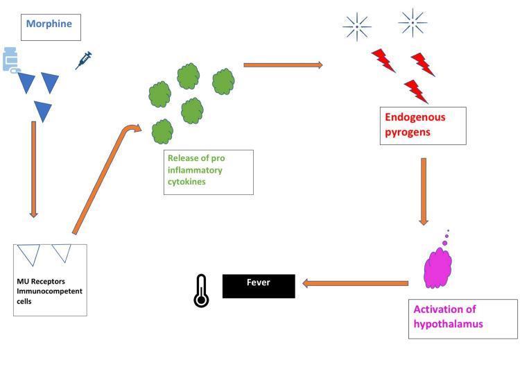
This pathophysiology diagram illustrates the biological mechanism of opioid-induced fever. The pathway is depicted as a series of steps connected by orange directional arrows. It begins with morphine (represented by blue triangles, a pill bottle, and a syringe icon) binding to Mu (μ) receptors on immunocompetent cells. This interaction triggers the release of pro-inflammatory cytokines, visualized as green spherical clusters. These cytokines promote the production of endogenous pyrogens, depicted by red lightning bolts and sunburst icons. The pyrogens act upon the central nervous system, specifically leading to the activation of the hypothalamus (shown as a purple anatomical cloud). The final physiological outcome is fever, symbolized by a black box labeled 'Fever' and a clinical thermometer icon. This schematic is designed for medical education to explain the rare adverse effect where opioids bypass typical thermoregulation to induce a febrile state via immune system mediation.
Fever - A Detailed Study
1. Definition and Normal Body Temperature
2. Fever vs. Hyperthermia - A Critical Distinction
| Feature | Fever | Hyperthermia |
|---|---|---|
| Mechanism | Active resetting of hypothalamic set point | Failure to dissipate heat; no set point change |
| Pyrogens involved | Yes | No |
| Responds to antipyretics | Yes | No |
| Example | Bacterial infection | Heat stroke, malignant hyperthermia |
3. Pathophysiology of Fever
3a. The Thermoregulatory Center
- Vasomotor changes (vasodilation / vasoconstriction)
- Shivering and metabolic heat production
- Behavioral thermoregulatory changes
3b. Pyrogens
- Bacterial endotoxins (lipopolysaccharide, LPS) - the classic example, from the outer membrane of gram-negative bacteria
- Viral products and toxins
- Other bacterial cell wall products
- Interleukin-1 (IL-1) - also called "leukocyte pyrogen" or "endogenous pyrogen" - the most potent
- Interleukin-6 (IL-6) - integral to acute-phase reactant production
- Tumor Necrosis Factor-alpha (TNF-α)
- Interferon-gamma (IFN-γ)
3c. The Fever Cascade (Step-by-Step)
- Pathogen entry - Bacteria, viruses, or their products enter tissues or bloodstream
- Phagocytosis - Blood leukocytes, tissue macrophages, and NK lymphocytes engulf and digest pathogen products
- Cytokine release - These cells release pyrogenic cytokines (IL-1, IL-6, TNF-α, IFN-γ) into circulation
- Hypothalamic signaling - Cytokines (especially IL-1β) reach the blood-brain barrier, specifically the organum vasculosum laminae terminalis (OVLT), a leaky capillary bed in the wall of the third ventricle above the optic chiasm
- PGE2 production - IL-1β triggers endothelial cells in the OVLT to produce prostaglandin E2 (PGE2)
- Set-point elevation - PGE2 binds EP3 receptors in the rostral ventromedial preoptic area (rvmPOA), inhibiting warmth-sensitive neurons, raising the thermoregulatory set point
- Heat conservation and production - The body, now "below" the new set point, activates:
- Peripheral vasoconstriction
- Shivering ("chills/rigors")
- Brown adipose tissue thermogenesis
- Behavioral heat-conserving changes (curling up, seeking warmth)
4. Characteristics of a Febrile Episode
4a. Chills (Rigor)
- The patient feels intensely cold (chills/rigors)
- Skin becomes cold (cutaneous vasoconstriction)
- Shivering occurs (to generate heat) This continues until body temperature reaches the new set point.
4b. Plateau Phase
4c. Crisis / "Flush" (Defervescence)
- Intense sweating
- Cutaneous vasodilation (hot, flushed skin)
- Rapid heat dissipation
5. Fever Patterns (Clinical Types)
| Pattern | Description | Associated Diseases |
|---|---|---|
| Continuous/Sustained | Temperature stays elevated >38°C with <1°C daily variation | Lobar pneumonia, typhoid fever |
| Remittent | Daily temperature variation >1°C but does not touch normal | Most bacterial infections |
| Intermittent | Fever spikes alternating with normal temperature periods | Malaria, septicemia, pyemia |
| Hectic/Septic | Wide swings (>2°C) between fever spikes and normal/subnormal | Septicemia, abscesses, TB |
| Pel-Ebstein | Recurrent periodic fever - fever for days/weeks then afebrile for days/weeks | Hodgkin's lymphoma (classic) |
| Relapsing | Periods of fever alternating with afebrile periods | Relapsing fever (Borrelia), malaria |
6. Clinical Manifestations of Fever
Systemic effects:
- Tachycardia - approximately 2 to 5 beats per minute per 1°F rise in temperature (the most consistent sign)
- Increased respiratory rate (to assist heat dissipation)
- Peripheral vasodilation contributing to relative tachycardia
- Decreased blood pressure (peripheral vasodilation + potential septic shock)
- Increased metabolic rate - each 1°C rise increases metabolic rate by ~10-13%
- Diaphoresis during defervescence
- Myalgias and arthralgias (mediated by cytokines)
- Headache, malaise, anorexia (cytokine-mediated)
- Confusion/delirium - especially in the elderly and with very high temperatures
Temperature-Pulse Dissociation (Faget's sign)
-
Typhoid fever
-
Leptospirosis
-
Rickettsiosis
-
Dengue
-
Legionellosis
-
Babesiosis
-
Goldman-Cecil Medicine, p. 2937
7. Causes of Fever
Infectious Causes (most common):
- Bacterial infections (pneumonia, UTI, meningitis, septicemia, endocarditis, abscesses)
- Viral infections (influenza, dengue, HIV, EBV, CMV)
- Fungal infections (histoplasmosis, coccidioidomycosis, candidiasis - especially in immunocompromised)
- Parasitic infections (malaria, leishmaniasis, toxoplasmosis)
- Mycobacterial infections (tuberculosis)
Non-Infectious Causes (important):
- Acute myocardial infarction
- Pulmonary embolism / infarction
- Intracranial hemorrhage / stroke
- Neuroleptic malignant syndrome
- Thyroid storm
- Acute adrenal insufficiency
- Transfusion reaction
-
Drug fever
-
Malignancy (especially lymphoma, leukemia, renal cell carcinoma, hepatoma)
-
Connective tissue diseases (SLE, RA, vasculitis)
-
Inflammatory conditions (pancreatitis, gout, DVT, pulmonary embolism)
-
Sarcoidosis
-
Tissue necrosis (post-MI, bowel infarction)
-
ROSEN's Emergency Medicine, Box 8.1
8. Major Causes of Hospital-Associated (Nosocomial) Fever
| Common | Less Common |
|---|---|
| INFECTIOUS: C. difficile enterocolitis, Pneumonia, Surgical wound, UTI, Vascular catheter | INFECTIOUS: Biliary tract disease, Endometritis, Intra-abdominal abscess, Mediastinitis, Sinusitis |
| NON-INFECTIOUS: Drug-induced fever, Hematoma, Immediate post-op state, Transfusion reaction, VTE | NON-INFECTIOUS: Adrenal insufficiency, Gout, MI, Organ infarction, Pancreatitis |
9. Fever of Unknown Origin (FUO)
- Temperature >38.3°C (101°F) on multiple occasions
- Duration >3 weeks
- No diagnosis after 1 week of intensive in-hospital investigation
- Classic FUO - community-acquired, duration >3 weeks
- Nosocomial FUO - hospital-acquired, >3 days without diagnosis
- Immune-deficient FUO - in neutropenic patients
- HIV-associated FUO
- Infections (~30-40%) - TB, endocarditis, abscesses, brucellosis, typhoid, HIV
- Inflammatory/Autoimmune (~20-30%) - Adult-onset Still's disease, SLE, vasculitis, RA, IBD
- Malignancy (~15-20%) - Lymphoma, leukemia, renal cell carcinoma, hepatoma
- Miscellaneous (~15%) - Drug fever, sarcoidosis, Crohn's disease, familial Mediterranean fever
- Undiagnosed (~10-15%)
10. Diagnosis and Evaluation of Fever
Clinical Assessment:
- Detailed history: onset, duration, pattern, associated symptoms, travel history, exposures (animals, insects, sick contacts), medications, prior vaccinations, immunosuppression
- Physical examination: vital signs (especially pulse-temperature relationship), skin rash, lymphadenopathy, mucous membranes, cardiac examination (murmurs), abdominal examination, neurological status
Key Investigations:
- Full blood count with differential (neutrophilia - bacterial; lymphocytosis - viral; eosinophilia - parasitic/drug)
- CRP, ESR (acute phase reactants)
- Blood cultures (x2 sets, before antibiotics)
- Urine analysis and culture
- Chest X-ray
- Lumbar puncture (suspected meningitis)
- Molecular testing (PCR) of body fluids for respiratory/enteric infections
- Liver function tests, renal function
- Peripheral blood smear (malaria, babesiosis)
- Serological tests (dengue NS1, leptospira, Weil-Felix)
- CT chest/abdomen/pelvis (lymphoma, abscesses, malignancies)
- Echocardiography (endocarditis)
- Bone marrow biopsy
- PET scan (increasingly used for occult malignancy/infection)
- Tissue biopsy (lymph node, liver)
11. Management of Fever
When to Treat:
- Temperature >39-40°C causing discomfort
- High-risk patients: elderly, cardiac disease, neurological conditions, febrile seizures in children
- Metabolic compromise or altered consciousness
Antipyretics - Mechanism:
| Drug | Mechanism | Notes |
|---|---|---|
| Paracetamol (Acetaminophen) | COX inhibition (CNS-selective) | First-line; safe in most patients; avoid in liver failure |
| Aspirin | Non-selective COX inhibition | Avoid in children (Reye's syndrome risk), GI side effects |
| Ibuprofen | Non-selective COX-1 and COX-2 inhibition | Effective anti-inflammatory; avoid in renal impairment |
| Naproxen | COX inhibition (longer acting) | Used for sustained fever control |
Physical Measures:
- Tepid sponging (avoid cold water - causes peripheral vasoconstriction)
- Hydration (critical in prolonged fever - prevents dehydration and ensures supportive care)
- Cooling blankets for extreme hyperthermia
Treating the Underlying Cause:
- Appropriate antibiotics for bacterial infection (guided by culture/sensitivity)
- Antivirals for specific viral infections (e.g., oseltamivir for influenza)
- Antifungals (fluconazole, voriconazole) for fungal infections
- Antiparasitics (artemisinin-based for malaria)
- Withdrawal of offending drug in drug fever
- Corticosteroids for inflammatory/autoimmune causes of FUO
12. Special Considerations
Fever in the Elderly:
- May not mount a significant fever even with severe infection
- Leukocytosis may be the primary clue
- Higher risk of atypical presentations (confusion without fever in sepsis)
Post-operative Fever ("The 5 W's"):
- Wind (Days 1-2): Atelectasis
- Water (Days 3-5): Urinary tract infection
- Wound (Days 4-7): Surgical site infection
- Walking (Days 5+): Deep vein thrombosis
- Wonder drugs (Any time): Drug fever
Drug Fever:
- Any drug can cause drug fever
- Typically: sustained temperature 38.5-40°C
- Patient paradoxically looks "well" despite high fever
- Eosinophilia and elevated aminotransferases may be clues
- Resolves within 72-96 hours of drug withdrawal
Fever with Rash:
- Petechial/purpuric: Meningococcemia (medical emergency), Rocky Mountain Spotted Fever, DIC
- Maculopapular: Viral exanthems, drug reactions, measles, syphilis
- Vesicular: Varicella, herpes zoster, hand-foot-mouth
- Erythema migrans: Lyme disease
13. Beneficial vs. Harmful Effects of Fever
| Beneficial Effects | Harmful Effects |
|---|---|
| Inhibits growth of some pathogens | Increased metabolic demand |
| Enhances immune cell function | Dehydration and electrolyte imbalance |
| Increases neutrophil activity at infection sites | Febrile convulsions (in children) |
| Augments T-cell activation | Cardiovascular stress (tachycardia) |
| Promotes acute phase protein synthesis | Confusion/delirium in elderly |
| Risk of brain damage at >41°C |
Summary
- Guyton and Hall Textbook of Medical Physiology (9780443111013) - Physiology of fever and thermoregulation
- Goldman-Cecil Medicine (9780323930345) - Pathobiology, clinical manifestations, and diagnosis
- ROSEN's Emergency Medicine (9780323757898) - Emergency approach, differential diagnosis
- Medical Physiology - Boron & Boulpaep (9780323319737) - Fever as regulated hyperthermia
- Bailey & Love's Surgery (9780367548117) - Post-operative fever