Reading File
Finding Sources
Finding Sources
Finding Sources
Finding Sources
Finding Sources
Reading File
Reading File
Finding Sources
Reading File
Finding Sources
Reading File
Reading File
Finding Sources
Reading File
Finding Sources
Loading Image
Loading Image
Loading Image
I now have thorough content from authoritative textbooks for all four topics. Here are your comprehensive study notes:
Anatomy Basics - Study Notes
1. Anatomical Planes, Position & Terms
(Gray's Anatomy for Students)
The Anatomical Position
The standard reference position: body erect, face looking forward, feet together with toes forward, hands by the sides with palms facing forward.
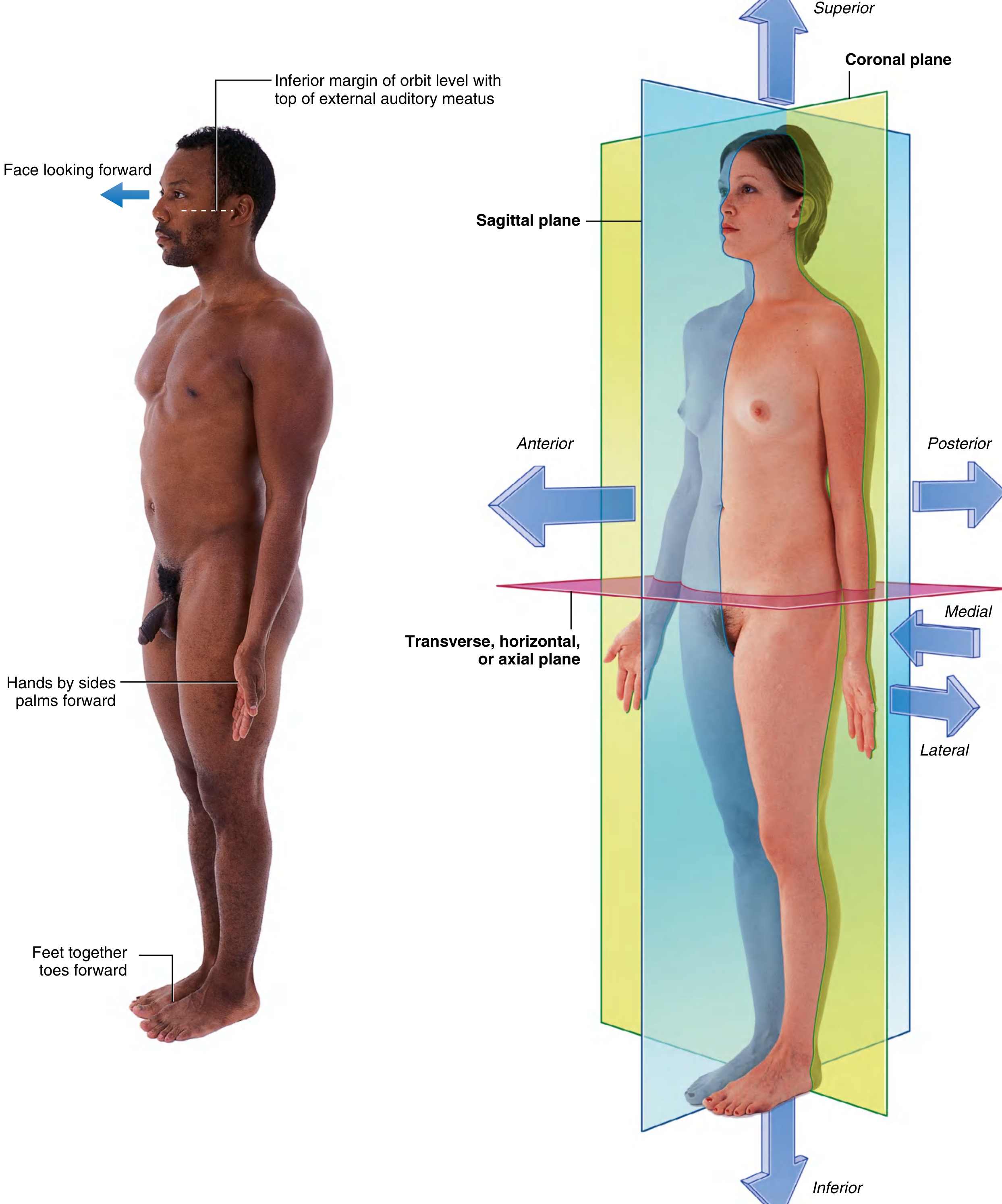
The Three Major Planes
| Plane | Orientation | Divides Body Into |
|---|---|---|
| Coronal (Frontal) | Vertical | Anterior and posterior parts |
| Sagittal | Vertical, perpendicular to coronal | Right and left parts |
| Median Sagittal | Sagittal through body midline | Equal right and left halves |
| Transverse (Horizontal / Axial) | Horizontal | Superior and inferior parts |
Directional Terms
Pair 1 - Anterior/Posterior
- Anterior (ventral) = toward the front (e.g., nose, sternum)
- Posterior (dorsal) = toward the back (e.g., vertebral column)
Pair 2 - Medial/Lateral
- Medial = closer to the median sagittal plane (e.g., nose is medial to the eyes)
- Lateral = farther from the median plane (e.g., thumb is lateral to the little finger)
Pair 3 - Superior/Inferior
- Superior = higher up (e.g., head is superior to shoulders)
- Inferior = lower down (e.g., knee is inferior to hip)
Additional Terms
- Proximal = closer to a structure's origin or point of attachment (e.g., the shoulder is proximal to the elbow)
- Distal = farther from the origin (e.g., the hand is distal to the elbow)
- Cranial = toward the head (synonym for superior)
- Caudal = toward the tail (synonym for inferior)
- Rostral = toward the nose, used especially in the head (e.g., the forebrain is rostral to the hindbrain)
- Superficial = external to the outer layer of deep fascia (skin, superficial fascia, mammary glands)
- Deep = enclosed by the outer layer of deep fascia (most skeletal muscles, viscera)
2. Bones and Cartilage
(Firestein & Kelley's Textbook of Rheumatology; Imaging Anatomy Atlas Vol. 3)
Bone
Bone is the main rigid structural tissue of the skeleton. Key features:
- Provides mechanical support, protection, and locomotion
- Serves as a mineral reservoir (especially calcium and phosphate)
- Houses bone marrow for hematopoiesis
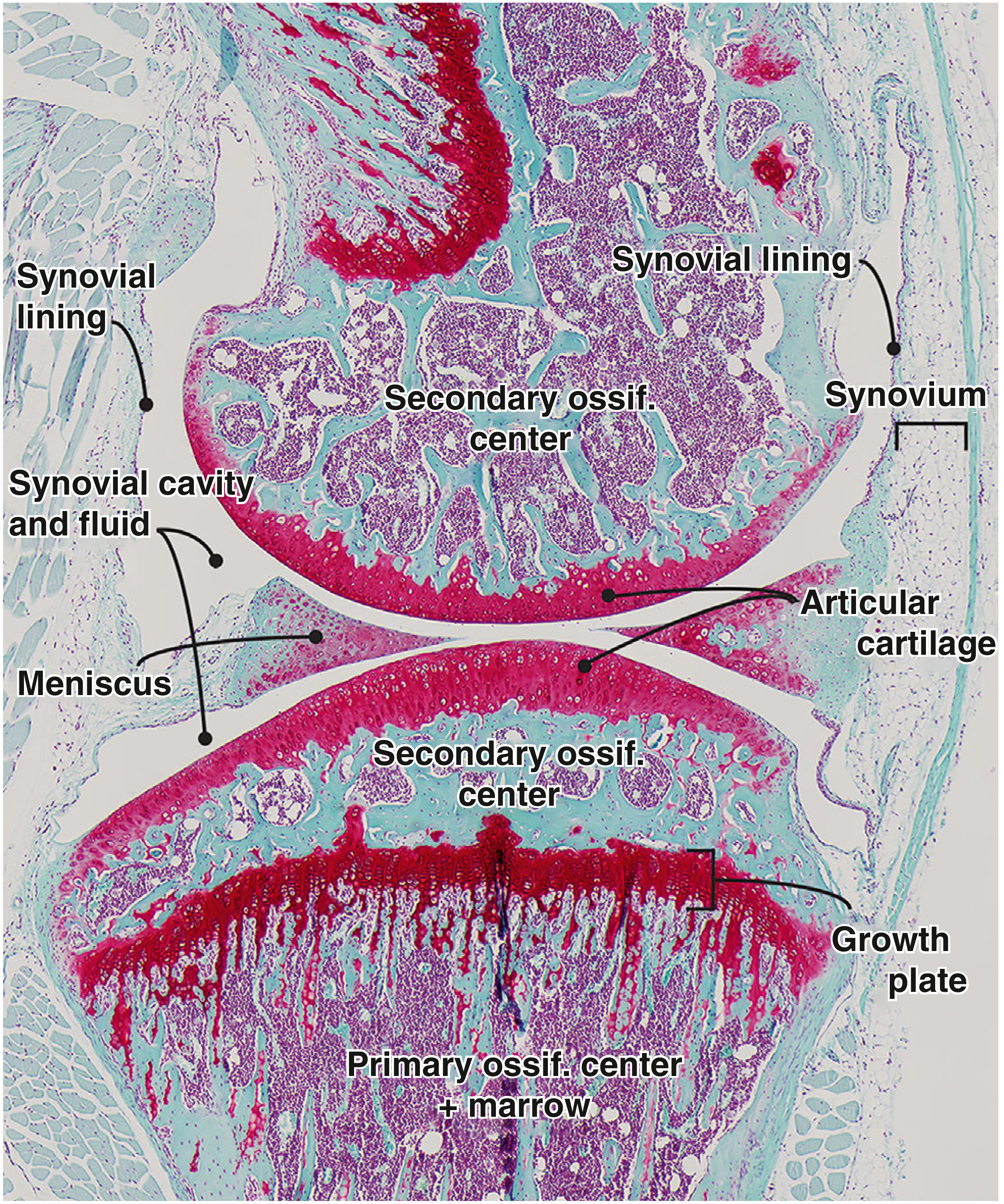
Ossification centers:
- Primary ossification center - forms in the diaphysis (shaft), contains marrow
- Secondary ossification center - forms in the epiphysis (ends of long bones)
- Growth plate (epiphyseal plate) - cartilaginous zone between ossification centers where longitudinal bone growth occurs in children
Cartilage
Cartilage is an avascular connective tissue with three main types:
| Type | Composition | Location | Features |
|---|---|---|---|
| Hyaline | Type II collagen + proteoglycans | Articular surfaces, costal cartilages, tracheal rings, growth plates | Most common; smooth, glassy appearance |
| Fibrocartilage | Type I + II collagen | Intervertebral discs, pubic symphysis, menisci | High tensile strength |
| Elastic | Elastic fibres + type II collagen | Ear pinna, epiglottis | Flexible and resilient |
3. Joints
(Firestein & Kelley's Textbook of Rheumatology)
Classification by Histology and Mobility
| Class | Also Called | Connective Tissue | Mobility | Examples |
|---|---|---|---|---|
| Fibrous | Synarthroses | Dense connective tissue | Minimal/none | Skull sutures, gomphoses (teeth), syndesmoses (ulna-radius) |
| Cartilaginous | Amphiarthroses | Hyaline cartilage or fibrocartilage | Slight | Pubic symphysis, intervertebral discs, 1st rib-sternum, growth plates |
| Synovial | Diarthroses | Synovial cavity + fluid | Freely moveable | Knee, hip, shoulder, elbow, wrist, ankle |
The Synovial Joint in Detail
Synovial joints are the most common functional joints in the skeleton and contain:
- Synovial cavity filled with synovial fluid (water, plasma filtrate, hyaluronic acid, lubricin, phospholipids)
- Articular cartilage covering the opposing bone ends
- Fibrous capsule enclosing the joint
- Synovial lining (synovium) producing the fluid
Synovial Joint Sub-types (by Shape)
| Shape | Axes of Motion | Example |
|---|---|---|
| Ball and socket | Multiaxial | Hip, shoulder |
| Hinge | Uniaxial | Elbow (humeroulnar) |
| Saddle | Biaxial | 1st carpometacarpal |
| Plane | Gliding | Patellofemoral |
| Condyloid/Ellipsoid | Biaxial | Wrist |
Movements possible at synovial joints: flexion, extension, abduction, adduction, rotation, circumduction.
4. Muscles - Basics
(Harper's Illustrated Biochemistry, 32nd Ed)
Three Types of Muscle
| Type | Appearance | Control | Force Direction |
|---|---|---|---|
| Skeletal | Striated | Voluntary (conscious) | Unidirectional (like a coil spring) |
| Cardiac | Striated | Involuntary | Unidirectional |
| Smooth | Non-striated | Involuntary | All directions (like a balloon deflating) |
The striated appearance of skeletal and cardiac muscle comes from the parallel alignment of contractile fibrils. Smooth muscle lacks this because its contractile fibrils are more randomly oriented.
Structure of Skeletal Muscle (Hierarchical)
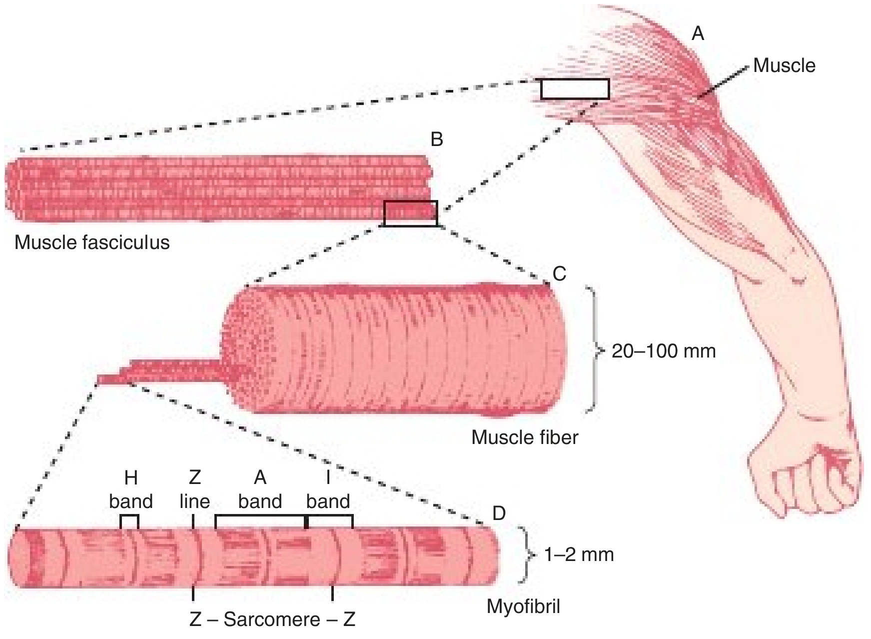
From largest to smallest:
- Muscle - the whole organ
- Fasciculus (fascicle) - bundle of muscle fibers
- Muscle fiber (cell) - a single multinucleated muscle cell; enclosed in the sarcolemma (electrically excitable plasma membrane); may extend the entire length of the muscle
- Myofibril - longitudinal bundles of overlapping thick and thin filaments within the fiber, embedded in sarcoplasm
- Sarcomere - the repeating functional unit of a myofibril, defined as the region between two Z lines (1500-2300 nm depending on contraction state)
The Sarcomere - Band Pattern
| Band/Line | Contents | Appearance |
|---|---|---|
| A band | Thick (myosin) + thin (actin) filaments | Dark |
| I band | Thin filaments only | Light |
| H band | Thick filaments only (center of A band) | Less dense zone within A band |
| Z line | Anchors thin filaments; marks sarcomere boundary | Dense, narrow line bisecting I band |
Sliding Filament Theory
When muscle contracts, the H zone and I bands shorten while the thick and thin filaments themselves remain the same length - the filaments slide past each other. Tension developed is proportional to the degree of filament overlap.
Sources:
- Gray's Anatomy for Students (anatomical planes & terms)
- Firestein & Kelley's Textbook of Rheumatology (joints)
- Harper's Illustrated Biochemistry, 32nd Ed (muscle basics)
- Imaging Anatomy Text and Atlas Vol. 3 (bones, joints, cartilage)
This is a shared conversation. Sign in to Orris to start your own chat.