
discuss internal anatomy of spinal cord
I'll start by reading the medical library skill, then search for spinal cord anatomy content.
Reading File
Now let me search the library for spinal cord internal anatomy sections and paragraphs in parallel.
Finding Sources
Finding Sources
Excellent sources found. Let me now read the key sections in parallel - Gray's Anatomy (comprehensive), Localization in Clinical Neurology (cross-sectional), Histology Atlas, and Neuroscience Exploring the Brain.
Reading File
Reading File
Reading File
Reading File
Excellent content. Now let me fetch the key anatomical images and read more about Rexed's laminae and the white matter tracts in parallel.
Loading Image
Loading Image
Reading File
Reading File
I now have comprehensive information from multiple authoritative textbooks. Let me also fetch the Gray's Anatomy cross-section diagram and get the descending tracts content.
Loading Image
Reading File
I now have all the information I need from authoritative textbooks, along with excellent diagrams. Let me compile the comprehensive answer.
Internal Anatomy of the Spinal Cord
Overview
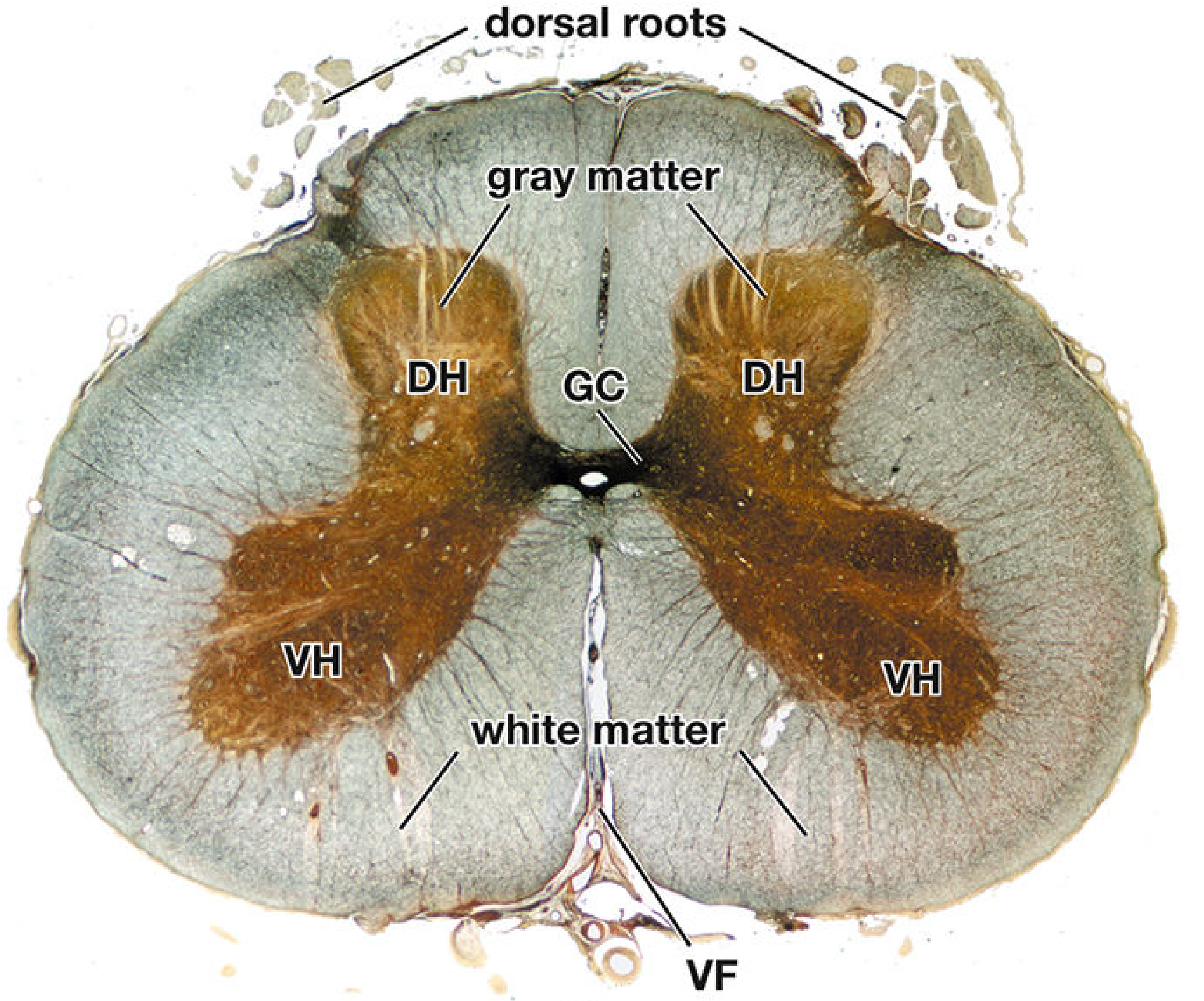
A cross-section of the spinal cord reveals two fundamental components: a centrally placed gray matter (butterfly- or H-shaped) surrounded by an outer white matter. The gray matter contains neuronal cell bodies, dendrites, and synapses; the white matter houses myelinated axonal tracts ascending to and descending from the brain.
1. Gray Matter
Shape and General Organization
The gray matter resembles a butterfly or modified "H" in cross section, with two lateral columns joined by a transverse gray commissure containing the central canal. Each lateral column has:
- A dorsal (posterior) horn - receives sensory input
- A ventral (anterior) horn - contains motor neurons
- An intermediate zone - between dorsal and ventral horns
Histological cross section of the spinal cord (Bielschowsky silver stain, lower lumbar level). DH = dorsal horn, VH = ventral horn, GC = gray commissure, VF = ventral fissure.
Dorsal (Posterior) Horn
Contains cell bodies of second-order sensory neurons that receive afferent input from primary sensory neurons entering via the dorsal roots. The dorsal horn is the primary site for processing pain, temperature, and mechanosensory information before relay to higher centers.
Ventral (Anterior) Horn
Contains the cell bodies of lower motor neurons (LMNs):
- Alpha (α) motor neurons - large basophilic cells that innervate extrafusal skeletal muscle fibers (the effectors of voluntary movement)
- Gamma (γ) motor neurons - innervate intrafusal fibers of neuromuscular spindles, regulating spindle sensitivity and the stretch reflex
A somatotopic organization exists: neurons for flexor muscles lie dorsal to those for extensor muscles; neurons for distal muscles (e.g., hand) lie lateral to those for trunk muscles.
Intermediolateral Column (Lateral Horn)
Present only at T1-L2 (thoracic and upper lumbar). Contains preganglionic cell bodies of the sympathetic nervous system. An equivalent sacral parasympathetic nucleus exists at S2-S4, providing preganglionic parasympathetic outflow to pelvic viscera.
Regional Enlargements
The spinal cord is not uniform in diameter:
- Cervical enlargement (C5-T1) - accommodates the vast number of neurons innervating the upper extremities
- Lumbar enlargement (L2-S3) - accommodates neurons innervating the lower extremities
2. Rexed's Laminae
Swedish anatomist Bror Rexed divided the gray matter cytoarchitecturally into 10 laminae (I-X):
| Lamina | Region | Name / Function |
|---|---|---|
| I | Dorsal horn tip | Nucleus posteromaginalis (posterior marginal nucleus) - pain/temperature relay |
| II | Dorsal horn | Substantia gelatinosa of Rolando - modulates pain input |
| III & IV | Dorsal horn | Nucleus proprius dorsalis - tactile and proprioceptive relay |
| V | Dorsal horn | Nucleus reticularis - receives nociceptive and visceral input |
| VI | Base of dorsal horn | Receives proprioceptive input from muscles and joints |
| VII | Intermediate zone | Nucleus dorsalis of Clarke (C8-L3) - origin of spinocerebellar tracts; also site of intermediolateral cell column at T1-L2 |
| VIII | Ventral horn (medial) | Interneurons; restricted to medial aspect at cervical and lumbar enlargements |
| IX | Ventral horn | Medial and lateral motor neuron columns (alpha and gamma LMNs) |
| X | Around central canal | Gray commissure; surrounds central canal |
The zone of Lissauer (posterolateral tract of Lissauer) separates the tip of the dorsal horn from the surface of the spinal cord - it carries fine pain and temperature fibers that ascend/descend 1-2 segments before synapsing.
3. White Matter
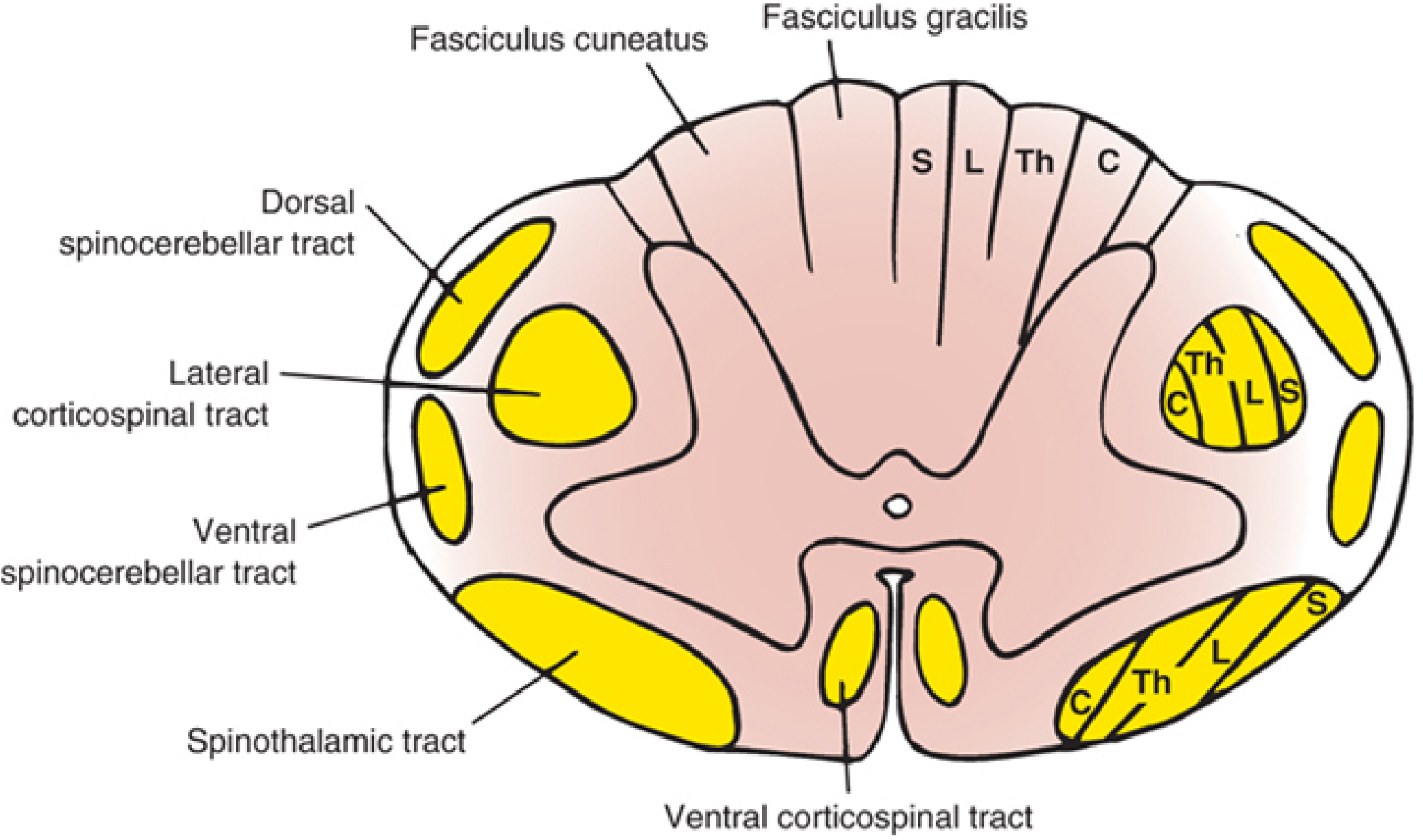
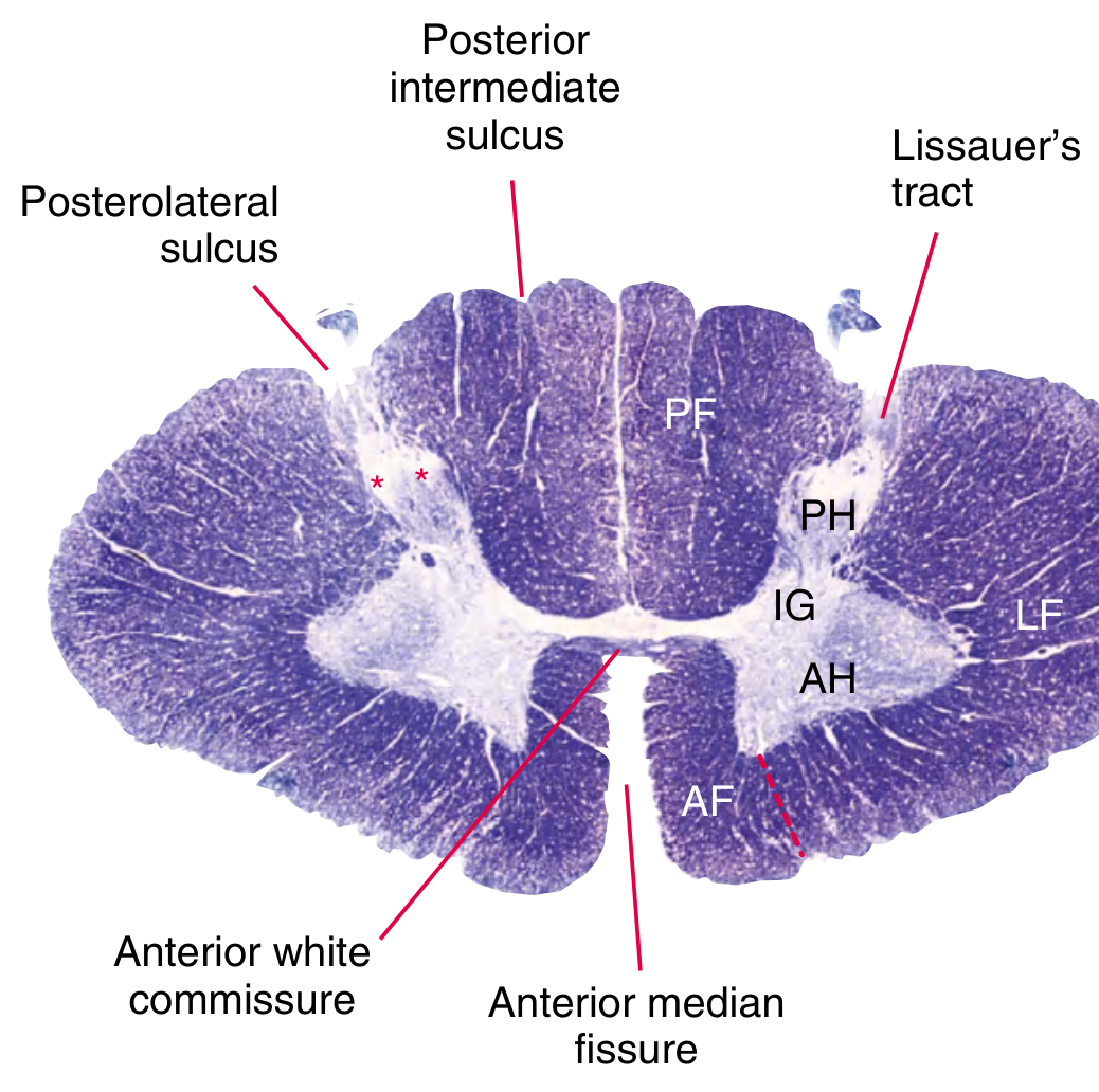
The white matter is organized into three funiculi (columns) on each side, separated by the gray matter horns and nerve root entry/exit zones:
| Funiculus | Location | Primary Contents |
|---|---|---|
| Dorsal (posterior) | Between dorsal median sulcus and dorsolateral sulcus | Ascending sensory tracts (fine touch, vibration, proprioception) |
| Lateral | Between dorsolateral and ventrolateral sulci | Mixed ascending and descending tracts |
| Ventral (anterior) | Between ventrolateral sulcus and ventral median fissure | Motor axons (descending), light touch (ascending) |
White commissures (dorsal and ventral) correspond to the gray commissure and allow crossing fibers to travel between sides.
Spinal cord cross-section diagram showing tract locations and somatotopic lamination (S=sacral, L=lumbar, Th=thoracic, C=cervical):
Spinal cord cross-section at C8 showing major landmarks (PF=posterior funiculus, PH=posterior horn, IG=intermediate gray, AH=anterior horn, AF=anterior funiculus, LF=lateral funiculus):
4. Major Ascending (Sensory) Tracts
Posterior Column - Medial Lemniscal Pathway
- Location: Dorsal funiculus (posterior columns)
- Subdivisions:
- Fasciculus gracilis (Goll's column) - medial; carries fine touch, vibration, conscious proprioception from lower limbs and lower trunk (sacral, lumbar, lower thoracic levels)
- Fasciculus cuneatus (Burdach's column) - lateral; carries same modalities from upper trunk, neck, and upper limbs (upper thoracic and cervical levels)
- Course: First-order axons ascend ipsilaterally to nucleus gracilis / nucleus cuneatus in the medulla; second-order fibers decussate as internal arcuate fibers and ascend as the medial lemniscus to the VPL nucleus of the thalamus; third-order fibers project to primary somatosensory cortex
- Modalities: Discriminative (fine) touch, vibration, two-point discrimination, conscious proprioception, stereognosis, position sense
Anterolateral (Spinothalamic) System
Located in the lateral and anterior funiculi. Contains three tracts:
-
Lateral spinothalamic tract (neospinothalamic) - carries sharp, localized pain and temperature. First-order neurons enter via dorsal root, synapse in laminae I and V; second-order neurons cross via the ventral white commissure (over 2-3 segments) and ascend contralaterally to the VPL nucleus of the thalamus. Somatotopy: cervical fibers are dorsomedial, sacral fibers are ventrolateral.
-
Ventral (anterior) spinothalamic tract - carries light (crude) touch; similarly crosses in the ventral commissure, ascends to VPL nucleus.
-
Spinoreticular tract (paleospinothalamic) - carries poorly localized, burning pain and the emotional/affective component of pain; projects to the brainstem reticular formation and intralaminar thalamic nuclei.
-
Spinomesencephalic tract - projects to the periaqueductal gray and superior colliculi; involved in central pain modulation.
Spinocerebellar Tracts (Unconscious Proprioception)
- Dorsal spinocerebellar tract (Clarke's column, C8-L3 origin) - ipsilateral; carries unconscious proprioception from lower limbs and trunk to the cerebellum
- Ventral spinocerebellar tract - carries similar information but crosses twice, ultimately reaching the cerebellum ipsilaterally
- Cuneocerebellar tract - equivalent of dorsal spinocerebellar for upper limbs (originates in accessory cuneate nucleus)
5. Major Descending (Motor) Tracts
Corticospinal Tracts
- Lateral corticospinal tract (~85-90% of fibers) - in the lateral funiculus; decussates at the pyramidal decussation in the medulla; provides direct voluntary motor control to contralateral limbs; synapse on laminae V, VII, IX
- Ventral (anterior) corticospinal tract (~10-15%) - in the ventral funiculus; uncrossed; most fibers eventually cross at the segmental level via the anterior white commissure; primarily controls axial and proximal muscles
Rubrospinal Tract
- Originates in the red nucleus of the midbrain; crosses immediately; travels in the lateral funiculus alongside the corticospinal tract; facilitates limb flexor muscles
Vestibulospinal Tracts
- Lateral vestibulospinal tract - facilitates alpha and gamma motor neurons of antigravity (extensor) muscles; important for postural tone
- Medial vestibulospinal tract - bilateral; mainly cervical levels; coordinates head and neck posture
Reticulospinal Tracts
- Medial (pontine) reticulospinal tract - facilitates extensor motor neurons
- Lateral (medullary) reticulospinal tract - inhibits motor neurons
- Both modulate voluntary movement and control autonomic functions
Tectospinal Tract
Originates in the superior colliculus; crosses to the contralateral side; descends in the anterior funiculus; mainly controls head-turning reflexes in response to visual stimuli
6. Central Canal
The central canal is lined by ependymal cells and is continuous with the fourth ventricle superiorly. It contains cerebrospinal fluid (CSF). It lies within lamina X of the gray commissure and is often obliterated in adults.
Summary Table: White Matter Columns and Their Tracts
| Column | Ascending Tracts | Descending Tracts |
|---|---|---|
| Posterior | Fasciculus gracilis, Fasciculus cuneatus | Descending autonomic, corticospinal (minor) |
| Lateral | Dorsal & ventral spinocerebellar, spinothalamic (lateral) | Lateral corticospinal, rubrospinal, lateral reticulospinal |
| Anterior | Spinothalamic (anterior/ventral) | Anterior corticospinal, vestibulospinal, tectospinal, medial reticulospinal |
Sources:
- Localization in Clinical Neurology, 8e, Cross-Sectional Anatomy and Ascending/Descending Tracts chapters
- Gray's Anatomy for Students - Internal Features and Neuroanatomy
- Histology: A Text and Atlas - Organization of the Spinal Cord
- Neuroscience: Exploring the Brain, 5e - Sensory Organization of the Spinal Cord
This is a shared conversation. Sign in to Orris to start your own chat.