Monocular and binocular cuse of visual depth perception
Reading File
Searching the Web
monocular binocular cues visual depth perception
Finding Sources
Finding Sources
Reading File
Reading File
Reading File
Loading Image
Here is a thorough breakdown of monocular and binocular cues for visual depth perception:
Visual Depth Perception: Monocular and Binocular Cues
Depth perception is the brain's ability to reconstruct a three-dimensional world from two-dimensional retinal images. The cues used are divided into two major categories based on whether one or both eyes are needed.
Monocular Cues
These cues work with a single eye and are also called pictorial cues because they can be captured in a flat photograph or painting.
| Cue | Description |
|---|---|
| Relative size | Objects of the same known size appear smaller when they are farther away. The brain interprets smaller = more distant. |
| Interposition (occlusion) | When one object partially blocks another, the blocked object is perceived as farther away. |
| Linear perspective | Parallel lines (e.g., railroad tracks, roads) appear to converge toward a vanishing point as distance increases. |
| Texture gradient | Surface textures appear finer and more densely packed with increasing distance. A cobblestone path gets "smoother" looking as it recedes. |
| Height in the visual plane | Objects closer to the horizon appear higher in the visual field and are judged as more distant. |
| Light and shadow | Shadows and shading reveal the 3D shape and relative position of objects. Light sources above cast predictable shadow patterns the brain uses to infer depth. |
| Aerial (atmospheric) perspective | Distant objects appear hazier, lighter, and slightly blue-shifted due to scattering of light by the atmosphere. Mountains in the distance look faded compared to foreground objects. |
| Motion parallax | When the observer moves, nearby objects shift faster across the visual field than distant objects. This is a powerful dynamic cue - particularly useful for animals with laterally placed eyes (e.g., rabbits) that have little binocular overlap. If the direction and velocity of movement are known, motion parallax can provide absolute depth information. |
| Accommodation | The lens of the eye changes shape (thickens) to focus on near objects. Proprioceptive signals from the ciliary muscles are fed back to the brain as a weak distance cue, effective mainly for distances under ~2 meters. |
Binocular Cues
These require both eyes and are the strongest cues for depth at close to moderate distances (especially within arm's reach).
1. Retinal (Binocular) Disparity
The two eyes are horizontally separated by roughly 6-7 cm. This means each eye receives a slightly different image of the same scene - the closer an object, the greater the difference (disparity) between the two retinal images. The brain compares these two images and uses the amount of disparity to calculate depth. This process is called stereopsis.
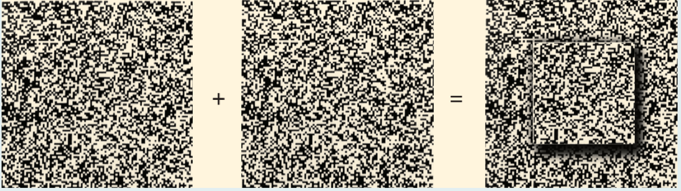
A key demonstration of this is Bela Julesz's random-dot stereogram (1960), shown below. When the left image is shown to the left eye and the right image to the right eye, a 3D square "pops out" - even though neither image alone contains any depth information whatsoever. This proved that stereopsis operates before object recognition, not after it.
Stereopsis is especially important for tasks performed at arm's length (threading a needle, grasping objects), and is thought to enable fine motor control that requires precise distance estimation.
2. Convergence
To fixate on a near object, both eyes rotate inward (converge). The degree of convergence - sensed through the proprioception of the extraocular muscles (medial recti) and efference copy of the motor commands - tells the brain how close the object is. The more convergent the eyes, the closer the target. This cue is effective primarily for distances up to about 6 meters.
Neural Substrate
The neurological basis for combining binocular inputs lies in primary visual cortex (V1):
- Neurons in layer 4C of V1 are strictly monocular - they respond only to input from one eye (either left or right), mirroring the segregated inputs from the lateral geniculate nucleus (LGN).
- Neurons in layers 2, 3, 5, and 6 above and below layer 4C are predominantly binocular - they receive converging input from both eyes and respond to light in either eye.
- These binocular neurons have two precisely aligned receptive fields (one in each eye), both pointing to the same location in the contralateral visual field.
- Binocular neurons tuned for disparity are the neural substrate for stereopsis - their response varies depending on the slight positional offset between the two retinal images, allowing the brain to compute depth from disparity.
Comparison: Monocular vs. Binocular
| Feature | Monocular | Binocular |
|---|---|---|
| Eyes required | One | Both |
| Effective range | All distances | Short to medium range (best < 10m) |
| Accuracy | Moderate | Higher (esp. retinal disparity) |
| Works in still images/photos | Yes | No |
| Works with one eye patched | Yes | No |
| Key cues | Size, perspective, shadow, motion parallax | Retinal disparity, convergence |
Retinal disparity is far more accurate than any monocular cue alone, which is why people with monocular vision (or amblyopia with suppression) have noticeably impaired depth judgment at close range but can still navigate effectively using monocular cues at distance.
- Neuroscience: Exploring the Brain, 5th Ed., pp. 960-962 (Binocularity, Stereopsis, Box 10.1)
This is a shared conversation. Sign in to Orris to start your own chat.