
Cryphorchidism reproductive system phisiology long question
Reading File
Now let me search the medical library in parallel for cryptorchidism content.
Finding Sources
Finding Sources
Finding Sources
Excellent - multiple rich sources found. Let me read the most comprehensive ones in parallel.
Reading File
Reading File
Reading File
Reading File
Good content. Now let me get more physiological detail - testicular descent phases, hormones, reproductive physiology aspects, and the S Das manual which typically has very exam-style content.
Reading File
Finding Sources
Reading File
Excellent - I now have rich content. Let me get the key physiology detail from Campbell Walsh and also fetch the embryology diagram.
Reading File
Loading Image
I now have comprehensive content from multiple authoritative textbooks. Let me compile the full long-answer response.
Cryptorchidism: Reproductive System Physiology - Long Answer
Definition
Cryptorchidism (Greek: kryptos = hidden, orchis = testis) is the failure of one or both testes to descend from the abdominal cavity into the scrotal sac. It is the most common congenital anomaly of the male genitalia and a significant cause of male infertility and testicular malignancy.
Embryology and Normal Testicular Descent
Origin of the Testis
- At ~32 days postovulation, gonads arise as thickenings on the medial aspect of the urogenital ridge (posterior abdominal wall)
- The SRY gene (sex-determining region, Y chromosome) triggers testicular differentiation - it activates SOX9, which drives Sertoli cell specification
- By 6-8 weeks, primordial germ cells (PGCs) migrate from the yolk sac and localize within forming testicular cords
- Leydig cells appear in the interstitium by weeks 8-10 and begin testosterone secretion under hCG stimulation
Two Phases of Testicular Descent
(Robbins, Cotran & Kumar Pathologic Basis of Disease)
Phase 1 - Transabdominal Phase (weeks 10-15):
- The testes migrate from the posterior abdominal wall to the lower abdomen/pelvic brim
- Controlled by Müllerian-inhibiting substance (MIS)/Anti-Müllerian Hormone (AMH) secreted by Sertoli cells
- The gubernaculum (a fibromuscular band) swells and anchors the testis to the inguinal region, while the cranial suspensory ligament regresses
- INSL3 (insulin-like factor 3, produced by Leydig cells) acting via its receptor RXFP2/LGRF8 is critical for gubernacular swelling and anchoring in this phase
Phase 2 - Inguinoscrotal Phase (weeks 25-35, third trimester):
- The testis passes through the inguinal canal into the scrotum
- This phase is androgen-dependent - testosterone (converted to DHT) acts via the androgen receptor
- Androgens are required to induce regression of the cranial suspensory ligament and drive gubernacular elongation/migration
- The genitofemoral nerve (GFN) provides essential innervation to the gubernaculum; calcitonin gene-related peptide (CGRP) released from GFN is the downstream mediator
- Complete descent into the scrotum is not achieved until the third trimester
Key hormonal regulators: INSL3 (Phase 1) + Androgens/DHT (Phase 2)
(Campbell Walsh Wein Urology)
Epidemiology
| Parameter | Data |
|---|---|
| Premature infants | ~30% affected at birth |
| Full-term infants | 3-5% at birth; ~1% by 1 year of age |
| Unilateral vs bilateral | Unilateral ~2x more common than bilateral |
| Bilateral accounts for | ~25% of cases |
| Spontaneous descent | Most inguinal UDT descend by end of Year 1 |
(The Developing Human; Robbins & Kumar)
Classification
1. Undescended Testis (True Cryptorchidism)
- Testis arrested along its normal path of descent
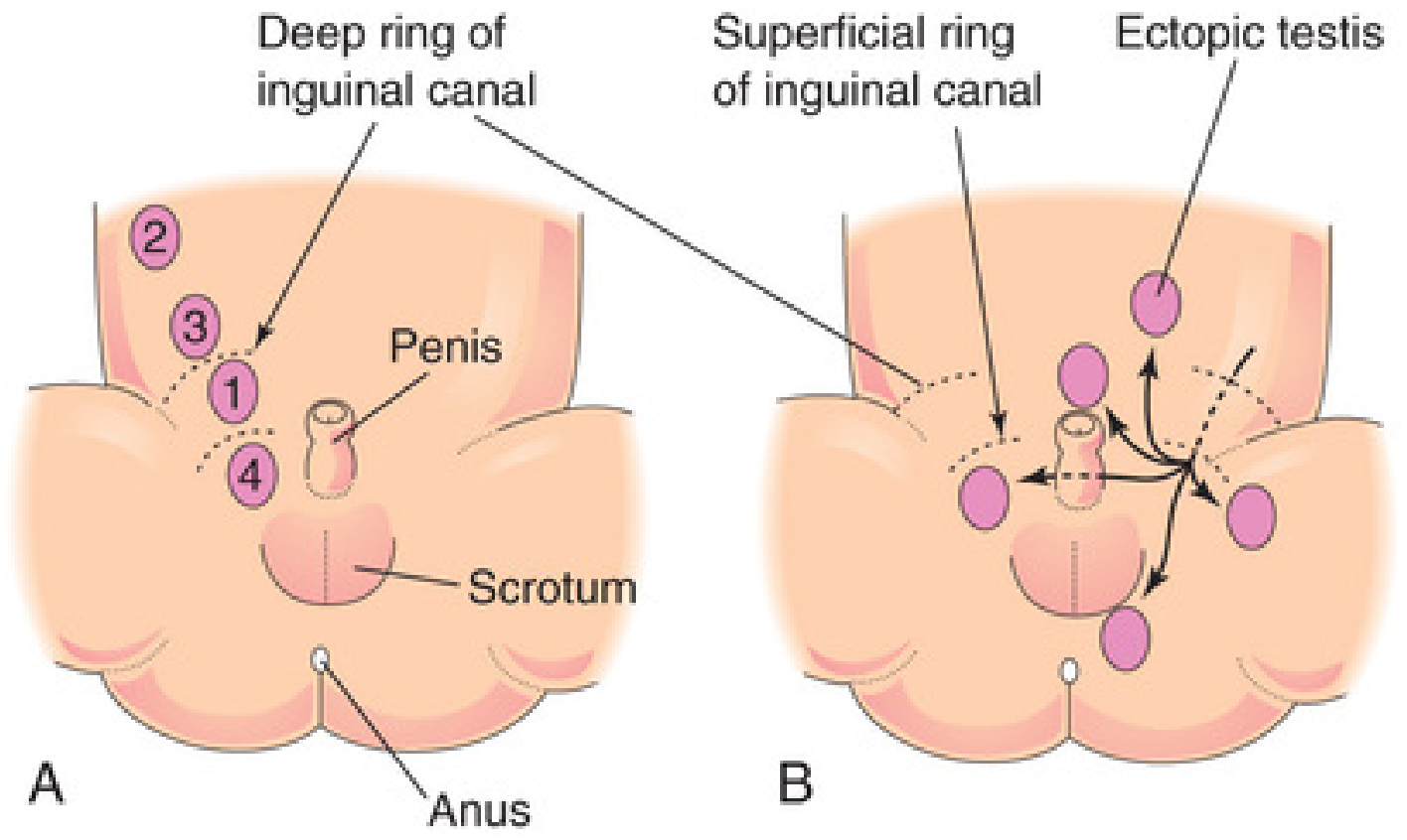
- Locations in order of frequency (see figure below):
- Inguinal canal (most common, ~70%)
- Just inside the deep inguinal ring
- Just outside the superficial inguinal ring
- High scrotal
2. Ectopic Testis
- Testis has deviated from the normal path of descent after exiting the inguinal canal
- Sites: superficial inguinal pouch (most common), perineum, root of penis (pubic type), femoral triangle
- Due to rupture/dominance of an accessory tail of the gubernaculum (Lockwood's five tails: scrotal, pubic, perineal, inguinal, femoral)
- An ectopic testis is usually well-developed (unlike an undescended testis, which is small and atrophied)
3. Retractile Testis
- A normally descended testis that retracts into the inguinal canal due to hyperactive cremasteric reflex
- Can be manually brought into the scrotum - does NOT require surgery
Pathophysiology and Consequences
Why the Scrotum Matters - Thermoregulation
Normal spermatogenesis requires a temperature 2-4°C below core body temperature (33-35°C). The scrotum achieves this via:
- Pampiniform plexus (countercurrent heat exchange)
- Cremaster muscle (temperature-regulated elevation/lowering)
- Dartos muscle and thin scrotal skin
In cryptorchidism, the retained testis is exposed to normal abdominal temperature (~37°C), which is toxic to the germinal epithelium (spermatogonia are heat-sensitive) while Leydig cells are relatively spared.
Histological Changes (Robbins, Cotran & Kumar)
| Timeline | Histological Change |
|---|---|
| At birth | Usually normal |
| By 2 years | Basement membrane thickening of seminiferous tubules |
| Progressive | Loss of spermatogonia; tubules contain only Sertoli cells |
| Late stage | Hyalinized tubular cords; increased interstitial stroma |
| Any stage | Germ cell neoplasia in situ (GCNIS) may appear |
| Throughout | Leydig cells are SPARED → testosterone production relatively preserved |
Notably, similar histologic changes may appear in the contralateral normally descended testis in unilateral cryptorchidism - suggesting an underlying intrinsic gonadal developmental defect (testicular dysgenesis syndrome) rather than purely positional damage.
Etiology and Risk Factors
Hormonal causes:
- Deficiency of fetal testosterone or DHT production
- Deficiency of INSL3 or RXFP2 mutations
- Deficiency of AMH/MIS
Genetic/syndromic associations:
- Prader-Willi syndrome
- Noonan syndrome
- Androgen insensitivity syndromes
- HOX gene mutations
Environmental factors:
- Endocrine disruptors (estrogen-like compounds in the intrauterine environment)
- Twin studies suggest both maternal inheritance and intrauterine environment play roles
Most cases: etiology unknown (cryptorchidism is rarely associated with a well-defined hormonal disorder in clinical practice)
Clinical Features
- Asymptomatic - usually discovered by parent or physician noting an empty scrotum
- Unilateral > bilateral
- The undescended testis is smaller and less developed than the contralateral testis
Complications:
| Complication | Details |
|---|---|
| Infertility/subfertility | Due to impaired spermatogenesis (Sertoli cell dysfunction, heat damage) |
| Testicular malignancy | 4-10 fold increased risk (most common: seminoma) |
| Testicular torsion | Undescended testis frequently undergoes torsion - mimics strangulated hernia |
| Inguinal hernia | 10-20% of cryptorchid patients (patent processus vaginalis) |
| Psychological | Empty scrotum in older children |
Reproductive Prognosis
(Campbell Walsh Wein Urology)
| Condition | Paternity Rate | Inhibin B | Sperm Density |
|---|---|---|---|
| Normal controls | ~96% | Normal | Normal |
| Unilateral cryptorchidism (treated) | ~96% | ~2/3 of normal | Reduced |
| Bilateral cryptorchidism (treated) | ~70% | ~1/3 of normal | Markedly reduced |
- Fertility impairment in cryptorchidism is primarily due to Sertoli cell/seminiferous epithelium dysfunction (reflected by low inhibin B), rather than Leydig cell dysfunction (testosterone levels less severely affected)
- Bilateral cryptorchidism carries a significantly worse prognosis for fertility than unilateral
Diagnosis
Physical examination - primary tool:
- Examine in warm room; supine and frog-leg position; palpate inguinal canal and scrotum
- Palpable vs non-palpable testis guides management
Investigations:
- Ultrasound - for inguinal testes
- MRI - for suspected intra-abdominal testis
- Laparoscopy - gold standard for non-palpable testis (diagnostic and therapeutic)
- Hormonal stimulation test (hCG stimulation): rise in testosterone confirms functional testicular tissue; no response suggests absent testes (anorchia)
- FSH, LH, inhibin B - markers of Sertoli cell and spermatogenesis status
Treatment
Medical (Hormonal)
- hCG injections (human chorionic gonadotropin) - stimulates Leydig cells to produce testosterone, promoting descent
- GnRH (buserelin) intranasal - less commonly used
- Success rate is modest (~20-30%); not recommended as primary treatment by modern guidelines
Surgical - Orchidopexy (Definitive Treatment)
- Timing: Current recommendation = 6 to 12 months of age (before histologic changes become established)
- Sabiston: orchidopexy at 6-12 months for palpable inguinal testis
- Robbins: "hormone injections followed by early orchiopexy at 6-12 months if medical treatment fails"
Procedure:
- Standard dartos pouch orchidopexy - for palpable inguinal testis; testis brought down and fixed in a sub-dartos pouch in the scrotum
- Two-stage Fowler-Stephens orchidopexy - for high intraabdominal testis with short vessels:
- Stage 1: Ligation of testicular vessels (to allow collateral circulation to develop over 6 months)
- Stage 2: Orchidopexy via collateral blood supply
- Laparoscopic orchidopexy - increasingly preferred single-stage option for intra-abdominal testis
Outcomes of Orchidopexy
- Reduces (but does not eliminate) risk of malignancy
- Reduces risk of torsion
- Improves fertility outcomes if performed early
- Deficient spermatogenesis still reported in 10-60% even after orchidopexy
- Cancer risk remains elevated even after repositioning (linked to intrinsic testicular dysgenesis)
Relationship to Testicular Cancer
- Cryptorchidism is the most important risk factor for testicular germ cell tumors
- Risk is highest with abdominal cryptorchidism (>intra-canalicular)
- Mechanism: Germ cell neoplasia in situ (GCNIS) - malignant transformation of arrested spermatogonia/gonocytes
- The contralateral descended testis is also at higher risk for malignancy - supporting the testicular dysgenesis theory
- Most common tumor arising: seminoma
- Orchidopexy does NOT eliminate the cancer risk but does allow regular physical examination and earlier detection
Summary Table
| Feature | Key Point |
|---|---|
| Definition | Failure of testis to descend into scrotum |
| Incidence | 3-5% at birth; 1% at 1 year |
| Phase 1 hormone | INSL3 + AMH (transabdominal) |
| Phase 2 hormone | Androgens/DHT (inguinoscrotal) |
| Most common site | Inguinal canal |
| Pathology | Spermatogonia lost; Leydig cells spared |
| Complications | Infertility, malignancy, torsion, hernia |
| Treatment timing | Orchidopexy at 6-12 months |
| Cancer risk | 4-10x increased risk |
| Fertility (bilateral) | ~70% paternity rate after treatment |
Sources: Robbins, Cotran & Kumar Pathologic Basis of Disease | Robbins & Kumar Basic Pathology | Campbell Walsh Wein Urology | The Developing Human: Clinically Oriented Embryology (Moore & Persaud) | Sabiston Textbook of Surgery | S Das Manual on Clinical Surgery
This is a shared conversation. Sign in to Orris to start your own chat.