
Diabetes melitus and it’s types
I'll start by reading the medical library skill, then search for comprehensive information on diabetes mellitus.
Reading File
Now I'll search the medical library in parallel for comprehensive diabetes mellitus content.
Finding Sources
Finding Sources
Excellent sources found. Now I'll read the most relevant sections in parallel - Guyton's physiology, Katzung's pharmacology, and Symptom to Diagnosis for comprehensive coverage.
Reading File
Reading File
Reading File
Now let me read more detail on complications, gestational DM, and MODY from the textbooks, and also search for any relevant images.
Reading File
Reading File
Finding Sources
I now have excellent, comprehensive information from multiple authoritative textbooks. Let me read the MODY section and complications section quickly.
Reading File
Finding Sources
I have comprehensive information from authoritative textbooks. Let me now compose a thorough, well-structured answer.
Diabetes Mellitus and Its Types
Definition
Diabetes mellitus (DM) is a syndrome of impaired carbohydrate, fat, and protein metabolism caused by either lack of insulin secretion or decreased sensitivity of tissues to insulin, resulting in chronic hyperglycemia. - Guyton and Hall Textbook of Medical Physiology
It is more precisely defined as an elevated blood glucose associated with absent or inadequate pancreatic insulin secretion, with or without concurrent impairment of insulin action. - Katzung's Basic and Clinical Pharmacology, 16th Edition
Classification
The ADA classifies diabetes mellitus into four main categories:
- Type 1 Diabetes Mellitus (T1DM)
- Type 2 Diabetes Mellitus (T2DM)
- Other Specific Types
- Gestational Diabetes Mellitus (GDM)
Type 1 Diabetes Mellitus
Mechanism: Selective beta cell destruction leading to severe or absolute insulin deficiency. The vast majority of cases (type 1a) are immune-mediated; a minority are idiopathic (type 1b). - Katzung's
Epidemiology:
- Represents 5-10% of all diabetics
- Most commonly presents in childhood (often called juvenile-onset DM or insulin-dependent DM)
- Highest incidence in northern Europeans and Sardinians
- Only 10-15% of patients have a positive family history
Autoantibodies present (key diagnostic markers):
- Islet Cell Antibodies (ICA)
- Insulin Autoantibodies (IAA)
- Anti-Glutamic Acid Decarboxylase (anti-GAD65)
- Anti-Tyrosine Phosphatase IA-2/ICA512
- Anti-Zinc Transporter 8 (ZnT8)
Genetics: Strong HLA association - particularly HLA-DR and HLA-DQ loci. - Quick Compendium of Clinical Pathology
Key features:
- Abrupt onset over days to weeks
- Prone to diabetic ketoacidosis (DKA)
- Requires insulin therapy from the outset
- Blood glucose can rise to 300-1200 mg/dL without treatment
LADA (Latent Autoimmune Diabetes in Adults): Up to 10-15% of patients labelled as "type 2" actually have a milder, slower autoimmune destruction. They can initially be managed with oral agents but eventually require insulin. - Katzung's
New treatment advance: Teplizumab-mzwv (anti-CD3 monoclonal antibody) has been approved for high-risk individuals with stage 2 T1DM (age 8+). A 14-day infusion course delays the onset of overt T1DM by approximately 25 months. - Katzung's, 16th Ed.
Type 2 Diabetes Mellitus
Mechanism: A heterogeneous condition characterized by tissue resistance to insulin combined with a relative deficiency in insulin secretion. Circulating insulin is sufficient to prevent ketoacidosis but inadequate to prevent hyperglycemia. Over time, progressive beta cell failure may occur requiring insulin. - Katzung's
Epidemiology:
- The most common form (~90% of all diabetes cases)
- Typically presents in adulthood, though increasingly seen in obese children/adolescents
- Strongly associated with obesity, physical inactivity, and central adiposity
Key features:
- Less prone to DKA than T1DM
- Initially managed with lifestyle changes, oral agents, and injectable non-insulin agents
- Insulin may be needed in later stages
Pathophysiology (simplified):
- Adiposity drives peripheral insulin resistance
- Compensatory hyperinsulinemia fails over time
- Beta cell exhaustion leads to absolute insulin deficiency in late disease
Management overview:
- Lifestyle modifications (diet, exercise, weight loss)
- Metformin - suppresses hepatic glucose production
- Sulfonylureas - increase insulin secretion
- Thiazolidinediones - increase insulin sensitivity
- GLP-1 receptor agonists - enhance insulin secretion + promote weight loss
- SGLT2 inhibitors (gliflozins) - increase urinary glucose excretion; also provide cardiovascular and renal protection
- Bariatric surgery for severe obesity with T2DM - Guyton & Hall
Type 3: Other Specific Types
This category encompasses multiple specific causes of hyperglycemia. - Quick Compendium of Clinical Pathology, Katzung's
| Subcategory | Examples |
|---|---|
| Exocrine pancreatic disease | Pancreatitis, pancreatectomy, cystic fibrosis |
| Endocrinopathies | Cushing's syndrome, acromegaly, pheochromocytoma, glucagonoma |
| Drug/chemical-induced | Corticosteroids, thiazide diuretics, antipsychotics |
| Monogenic defects (MODY) | See below |
| Genetic defects of insulin action | Insulin receptor mutations, acanthosis nigricans syndromes |
MODY - Maturity Onset Diabetes of the Young
MODY is a form of non-insulin-dependent (type 2) diabetes with autosomal dominant inheritance that usually arises in childhood or young adulthood. Affected individuals are rarely obese and have no autoantibodies. Accounts for 1-3% of all diabetes. - Quick Compendium of Clinical Pathology, Goldman-Cecil Medicine
MODY subtypes (by gene mutation):
| MODY Type | Gene | Protein |
|---|---|---|
| MODY 1 | HNF4A | Hepatocyte Nuclear Factor 4α |
| MODY 2 | GCK | Glucokinase |
| MODY 3 | HNF1A | Hepatocyte Nuclear Factor 1α |
| MODY 4 | IPF1 | Insulin Promoter Factor 1 |
| MODY 5 | HNF1B | Hepatocyte Nuclear Factor 1β |
MODY2 (glucokinase mutation) and MODY3 (HNF1A mutation) are the most common subtypes. MODY is important pharmacogenomically - MODY2 and MODY3 respond differently to sulfonylureas vs. insulin. - Emery's Elements of Medical Genetics and Genomics
Type 4: Gestational Diabetes Mellitus (GDM)
Definition: Any abnormality in glucose levels first noted during pregnancy. - Katzung's
Prevalence: Affects approximately 7% of all pregnancies in the United States.
Mechanism: The placenta and placental hormones (e.g., human placental lactogen) create progressive insulin resistance, most pronounced in the third trimester.
Screening:
- High-risk women: screen at the first prenatal visit
- Standard-risk women: screen at 24-28 weeks gestation
Risk factors: Obesity, prior GDM, family history of T2DM, advanced maternal age.
Significance: GDM increases the risk of macrosomia, birth complications, neonatal hypoglycemia, and future development of T2DM in both mother and child.
Pathophysiologic Effects of Hyperglycemia (Common to All Types)
| Effect | Mechanism |
|---|---|
| Glucosuria | Plasma glucose exceeds renal threshold (~200 mg/dL); glucose spills into urine |
| Osmotic diuresis & polyuria | Glucose in tubular fluid prevents water reabsorption |
| Polydipsia | Cellular dehydration due to hyperosmolar extracellular fluid |
| Weight loss | Impaired glucose utilization; shift to fat and protein catabolism |
| Polyphagia | Cellular starvation signals |
Diagnostic Criteria (ADA)
| Test | Pre-diabetes | Diabetes |
|---|---|---|
| Fasting Plasma Glucose | 100-125 mg/dL | ≥126 mg/dL |
| 2-hr OGTT (75g glucose) | 140-199 mg/dL | ≥200 mg/dL |
| HbA1c | 5.7-6.4% | ≥6.5% |
| Random glucose + symptoms | - | ≥200 mg/dL |
Sources: Quick Compendium of Clinical Pathology 5th Ed., Katzung's 16th Ed.
HbA1c reflects average plasma glucose over the preceding 8-12 weeks (corresponding to the ~120-day lifespan of red blood cells). - Katzung's
Chronic Complications
Chronic hyperglycemia damages blood vessels and nerves throughout the body:
- Microvascular: Diabetic retinopathy, nephropathy, neuropathy
- Macrovascular: Coronary artery disease, peripheral arterial disease, stroke
- Other: Diabetic foot, increased susceptibility to infections, gastroparesis
Summary Comparison
| Feature | Type 1 | Type 2 | GDM | MODY |
|---|---|---|---|---|
| Mechanism | Autoimmune beta cell destruction | Insulin resistance + relative deficiency | Placental hormone-induced resistance | Single-gene mutation |
| Age of onset | Childhood (any age) | Adulthood (now also youth) | During pregnancy | Childhood/young adult |
| Body habitus | Normal/thin | Often obese | Variable | Non-obese |
| Autoantibodies | Present | Absent | Absent | Absent |
| Ketosis risk | High | Low | Low | Low |
| Inheritance | Multifactorial (HLA linked) | Multifactorial | Multifactorial | Autosomal dominant |
| Treatment | Insulin (mandatory) | Lifestyle + oral agents ± insulin | Diet ± insulin | Gene-specific; often sulfonylureas |
Sources: Guyton and Hall Textbook of Medical Physiology; Katzung's Basic and Clinical Pharmacology 16th Ed.; Quick Compendium of Clinical Pathology 5th Ed.; Creasy & Resnik's Maternal-Fetal Medicine; Emery's Elements of Medical Genetics and Genomics; Goldman-Cecil Medicine.
Diabetes nephropathy, diabetes keto acidosis, neurological,complications of diabetes, and their management, management of DM
Searching all four topics in parallel now.
Finding Sources
Finding Sources
Finding Sources
Finding Sources
Now reading the best sections in parallel - Robbins for nephropathy, Goldman-Cecil for DKA, Adams & Victor for neuropathy, and searching for DM management.
Reading File
Reading File
Reading File
Finding Sources
Excellent data coming in. Now reading DKA management, nephropathy management, and DM overall management in parallel.
Reading File
Reading File
Finding Sources
Now reading the DKA treatment section and DM management from Katzung, plus the retinopathy section from Robbins.
Reading File
Reading File
Finding Sources
Now reading Goldman-Cecil for DM management overview, Lippincott for oral agents, and Harrison's for neuropathy treatment.
Reading File
Reading File
Reading File
Now reading Lippincott's oral agents section and Goldman-Cecil DM treatment section for management.
Reading File
Reading File
I now have comprehensive data from all needed sources. Let me also fetch the key pathology images and read DKA treatment details.
Reading File
Reading File
I now have comprehensive, authoritative content from multiple textbooks. Let me fetch key pathology images and finalize the answer.
Loading Image
Loading Image
Here is the comprehensive, textbook-sourced guide on all topics you asked about:
Diabetic Nephropathy, DKA, Neurological Complications, and Management of DM
PART 1: DIABETIC NEPHROPATHY
Overview
Diabetic nephropathy is the leading cause of end-stage kidney disease (ESKD) in many countries, surpassing glomerulonephritis in Japan since 1997 and now the leading cause of CKD in China as well. Approximately 30-40% of type 2 diabetic patients develop diabetic nephropathy. - Brenner and Rector's The Kidney
Pathological Lesions
The kidneys are prime targets of diabetes. Three categories of lesions develop: - Robbins & Kumar Basic Pathology
1. Glomerular Lesions (most important)
- Basement membrane thickening - Glomerular capillary BM thickens along its entire length, detectable by EM within a few years of onset, even before any change in renal function
- Diffuse mesangial sclerosis - Increase in mesangial matrix with cell proliferation and BM thickening; found in most patients with >10 years of disease; leads to nephrotic syndrome (proteinuria, hypoalbuminemia, edema)
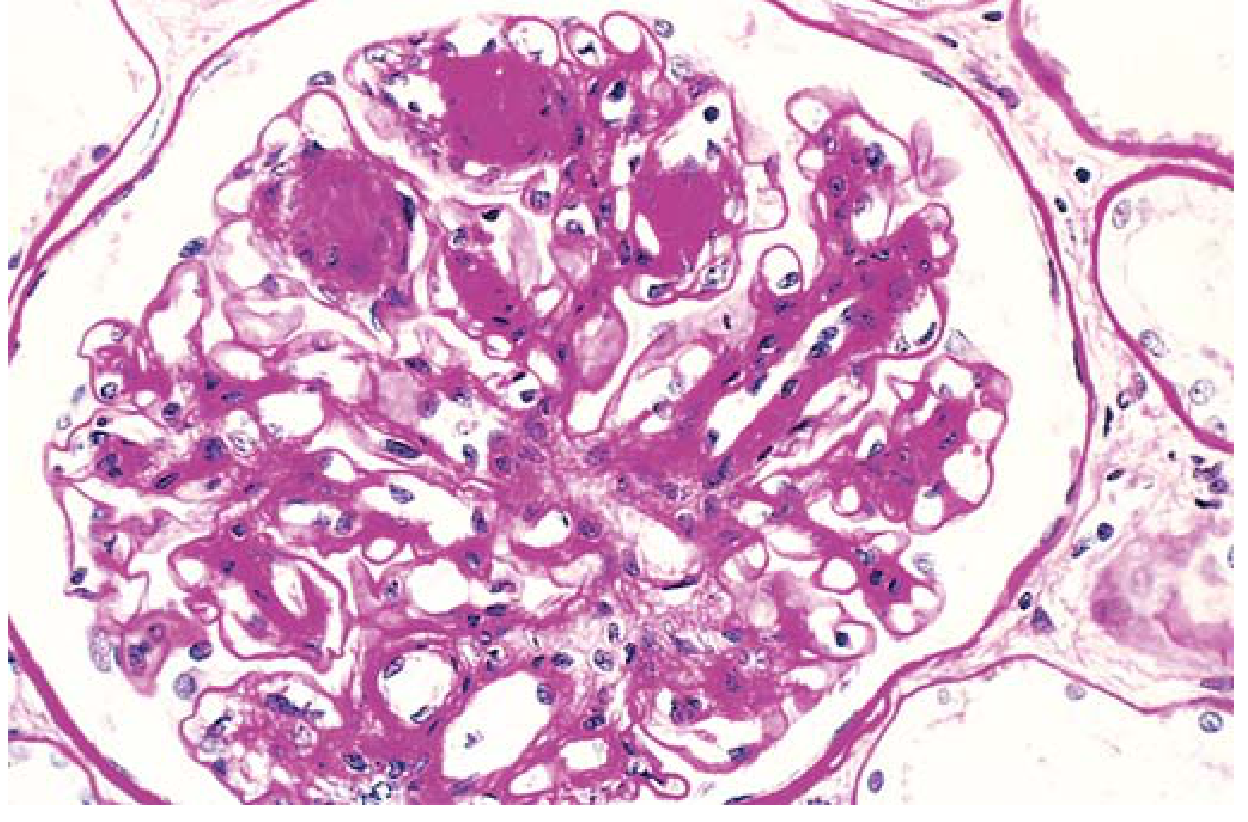
- Nodular glomerulosclerosis (Kimmelstiel-Wilson lesion) - Ball-like deposits of laminated matrix in the periphery of the glomerulus (PAS stain); occurs in ~15-30% of long-term diabetics; virtually pathognomonic of diabetes
2. Vascular Lesions
- Renal atherosclerosis and arteriolosclerosis
- Hyaline arteriolosclerosis affects both afferent AND efferent arterioles - the latter is virtually unique to diabetes
- Results in diffuse cortical scarring (nephrosclerosis) with finely granular cortical surface
3. Pyelonephritis
- Both acute and chronic forms are more severe in diabetics
- Necrotizing papillitis (papillary necrosis) is much more prevalent in diabetics
Pathogenesis
The Renin-Angiotensin-Aldosterone System (RAAS) plays a central role in progression of diabetic nephropathy. Genetic polymorphisms of the ACE gene (insertion/deletion), IL-6, IL-10, and ICAM-1 also contribute. - Brenner and Rector's The Kidney
Management of Diabetic Nephropathy
- RAAS blockers (ACE inhibitors / ARBs) are the standard of care - reduce proteinuria and slow rate of renal function deterioration
- SGLT2 inhibitors (gliflozins) have proven significant protection against kidney disease progression
- Tight glycemic control (HbA1c target <7%)
- Blood pressure control (target <140/80 mmHg; lower if proteinuric)
- Dietary protein restriction in advanced CKD
- Monitoring: Annual microalbuminuria, eGFR, electrolytes
- Caution with antidiabetic drugs in CKD: Metformin contraindicated if eGFR <30; many oral agents accumulate and risk hypoglycemia
PART 2: DIABETIC KETOACIDOSIS (DKA)
Definition - The "DKA Triad"
The three fundamental biochemical features are: - Goldman-Cecil Medicine
- Hyperglycemia (or known diabetes) - glucose variable, up to >1000 mg/dL
- Ketonuria ≥2+ OR serum ketones ≥3.0 mmol/L
- Acidosis - arterial/venous pH <7.3, serum bicarbonate <18 mmol/L
Pathophysiology
DKA results from combined insulin deficiency + excess counter-regulatory hormones (glucagon, catecholamines, cortisol, GH):
- Substrates (amino acids, lactate, pyruvate) delivered from muscle and adipose tissue to liver
- Liver converts them to glucose and ketone bodies (β-hydroxybutyrate, acetoacetate, acetone)
- Glucose and ketones enter circulation faster than they are utilized
- Hyperglycemia → osmotic diuresis → dehydration and electrolyte loss → hemoconcentration → worsening hyperglycemia (vicious cycle)
Common Precipitants
| Most Common | Other |
|---|---|
| Infection | Cerebrovascular accident |
| Inadequate insulin / non-adherence | Acute pancreatitis |
| New-onset diabetes | Cushing syndrome, thyrotoxicosis |
| Acute coronary syndrome | Drugs: corticosteroids, SGLT2 inhibitors, clozapine, cocaine, lithium |
Clinical Features
- Polyuria, polydipsia, weakness, lethargy, anorexia
- Nausea, vomiting, non-specific upper abdominal pain
- Kussmaul breathing - deep, rapid respirations (respiratory compensation for metabolic acidosis)
- Fruity/acetone breath
- Dry mucous membranes, reduced JVP, tachycardia, orthostatic hypotension
- Depressed consciousness, sometimes frank coma
Diagnosis
| Parameter | Mild DKA | Severe DKA |
|---|---|---|
| Blood glucose | Variable (may be near-normal) | Can exceed 1000 mg/dL |
| Arterial pH | 7.20-7.30 | <7.00 |
| Serum bicarbonate | <18 mmol/L | Markedly reduced |
| Anion gap | Elevated | High, proportional to HCO3 drop |
| Ketones | ≥2+ or ≥3.0 mmol/L | Markedly elevated |
Important lab notes:
- Serum Na+ is falsely low (osmotic shift of water from ICF)
- K+ may be normal, high, or low - but total body deficit is always present
- WBC is elevated even without infection (due to acidosis itself)
- Elevated amylase is usually non-pancreatic
- Nitroprusside-based ketone tests underestimate severity (detect acetoacetate, NOT β-hydroxybutyrate)
Treatment of DKA
The four pillars of DKA management:
1. Fluid Replacement
- Start with 0.9% NaCl (normal saline) - typically 1 L in first hour
- Correct dehydration over 24-48 hours
- Switch to hypotonic saline once osmolality normalizes
2. Insulin
- Regular insulin IV infusion (0.1 units/kg/hr)
- Do NOT give insulin bolus before adequate K+ replacement if K+ <3.5 mEq/L
- Switch to subcutaneous insulin + start oral feeding when glucose <200 mg/dL and patient is eating
- Continue IV insulin until anion gap closes
3. Potassium Replacement
- Replace aggressively - insulin drives K+ into cells, worsening hypokalemia
- Monitor K+ every 2 hours during treatment
- Target K+ 3.5-5.0 mEq/L
4. Identify and Treat Precipitant
- Blood cultures, chest X-ray, ECG
Bicarbonate: Generally NOT recommended unless pH <6.9
DKA vs. Hyperosmolar Hyperglycemic State (HHS) Comparison
| Feature | DKA | HHS |
|---|---|---|
| Typical patient | T1DM (younger) | T2DM (elderly) |
| Glucose | Elevated (variable) | Very high (often >600 mg/dL) |
| Ketones | Marked | Minimal/absent |
| Acidosis | Yes (pH <7.3) | Absent or mild |
| Consciousness | Less impaired | Severely impaired (up to coma) |
| Mortality | ~4% | Up to 20% |
Prevention
Education in "sick-day rules": frequent glucose monitoring, ketone testing, small frequent carbohydrate fluids, dose adjustment during illness. Diabetes educator review after every DKA admission. - Goldman-Cecil Medicine
PART 3: DIABETIC NEUROPATHY (Neurological Complications)
Epidemiology
- Estimated lifetime prevalence ~50% of all diabetics
- ~15% have symptoms/signs of polyneuropathy clinically; ~50% have abnormal nerve conduction studies
- The most important factor is duration of diabetes - <10% at diagnosis, rising to 50% after 25 years
- More common in patients >50 years; rare in childhood
- Rates are similar in T1DM and T2DM - Adams and Victor's Principles of Neurology, 12th Ed.
Pathophysiology
Two main mechanisms:
- Microangiopathy of vasa nervorum (small endoneurial blood vessels) → ischemia/infarction of nerve fascicles
- Metabolic - poorly understood but related to hyperglycemia, advanced glycation end products, oxidative stress, protein kinase C activation
- Recent evidence also supports an inflammatory process - Robbins & Kumar, Adams & Victor
Clinical Syndromes of Diabetic Neuropathy
Six distinct clinical syndromes have been delineated: - Adams and Victor's Principles of Neurology
1. Distal Symmetrical Sensory Polyneuropathy (DSPN) - MOST COMMON
- Affects feet and legs in chronic, slowly progressive manner
- "Stocking-glove" pattern - starts distally, ascends
- Symptoms: burning, tingling, numbness, pain (worse at night), loss of protective sensation
- Signs: reduced vibration, position sense; absent ankle reflexes
- Loss of protective sensation (LOPS) → foot ulceration risk
2. Acute Ophthalmoplegia (Cranial Mononeuropathy)
- Affects CN III most commonly (also CN VI)
- Diabetic CN III palsy: ptosis, eye deviated down and out, but pupil is spared (unlike compressive CN III palsy which spares pupils)
- Due to ischemia of the nerve fascicle
3. Acute Mononeuropathy of Limbs/Trunk
- Includes painful thoracolumbar radiculopathy
- Asymmetric, often self-limiting
4. Diabetic Amyotrophy (Proximal Motor Neuropathy)
- Acute/subacute painful, asymmetric, predominantly motor
- Affects upper lumbar roots and proximal leg muscles
- Presents with severe thigh pain, hip flexor weakness, difficulty rising from a chair
5. Symmetrical Proximal Motor Weakness
- More symmetrical, proximal wasting without pain
- Subacute/chronic course, variable sensory loss
6. Autonomic Neuropathy
| System | Manifestation |
|---|---|
| Cardiovascular | Resting tachycardia, orthostatic hypotension, painless MI |
| GI | Gastroparesis (nausea, vomiting, early satiety, bloating), nocturnal diarrhea, constipation |
| Genitourinary | Neurogenic bladder, erectile dysfunction, retrograde ejaculation |
| Sudomotor | Anhidrosis, gustatory sweating |
Diabetic Foot
The combination of sensory neuropathy + peripheral arterial disease leads to:
- Plantar ulceration (typically under metatarsal heads)
- Slow healing due to impaired blood flow + repetitive trauma + hyperglycemia
- Osteomyelitis if infection penetrates to bone
- Charcot foot - repeated undetected fractures destroy normal foot architecture
Management of Diabetic Neuropathy
Prevention (most important):
- Tight glycemic control is the primary prevention strategy
- Lifestyle modifications - exercise, diet
Pharmacological pain management (DSPN): - Harrison's Principles of Internal Medicine 22E
| Drug Class | Examples | Notes |
|---|---|---|
| Gabapentinoids (1st line) | Pregabalin, Gabapentin | FDA-approved for diabetic neuropathy pain |
| SNRIs (1st line) | Duloxetine, Venlafaxine, Desvenlafaxine | Duloxetine FDA-approved |
| TCAs | Amitriptyline, Nortriptyline | Effective but side effects limit use |
| Sodium channel blockers | Carbamazepine, Lidocaine patch | 2nd line |
| Capsaicin patch | 8% topical | Local effect |
| Opioids | Tapentadol | Last resort; addiction risk |
Autonomic neuropathy:
- Orthostatic hypotension: Salt/fluid intake, compression stockings, midodrine, droxidopa (FDA-approved)
- Gastroparesis: Prokinetics (metoclopramide, domperidone), dietary modification (small frequent low-fat meals)
- Beta blockers for resting tachycardia (use caution with hypoglycemia unawareness)
Foot care:
- Daily foot inspection
- Protective footwear (no bare feet)
- Aggressive debridement of ulcers + systemic antibiotics if infected
- Pressure off-loading (total contact casting)
- Revascularization if significant PAD
- Podiatry referral
Supplement considerations: Metformin reduces B12 absorption → monitor B12 especially if peripheral neuropathy present
PART 4: OTHER CHRONIC COMPLICATIONS
Retinopathy
- Leading cause of new blindness in adults in the USA
- Microangiopathy of retinal vessels → microaneurysms, hemorrhages, exudates → neovascularization (proliferative retinopathy) → vitreous hemorrhage and tractional retinal detachment
- Management: tight glycemic control, laser photocoagulation, anti-VEGF injections (bevacizumab, ranibizumab)
Macrovascular Disease (Cardiovascular)
- 2-4x higher mortality from cardiovascular disease vs non-diabetics; manifests at younger age
- Driven by: hyperglycemia, insulin resistance, dyslipidemia, elevated free fatty acids, endothelial dysfunction, prothrombotic state, advanced glycation end products
- Manifestations: coronary artery disease, stroke, peripheral arterial disease
- Management: statins, ACE inhibitors/ARBs, antiplatelet therapy, GLP-1 agonists and SGLT2 inhibitors (proven cardioprotective)
Pathobiology of Chronic Complications - Common Mechanism
The root cause is hyperglycemia-driven oxidative stress and vascular damage via: - Goldman-Cecil Medicine
- Advanced glycation end products (AGEs) - alter collagen/elastin → vascular stiffness + BM thickening; bind AGE receptors on macrophages/endothelium → inflammation + oxidative stress
- Protein kinase C activation (via diacylglycerol) → enhanced angiogenesis, vasoconstriction, vascular permeability (↑VEGF), cytokine activation, extracellular matrix expansion
- Polyol pathway flux (aldose reductase)
- Hexosamine pathway
PART 5: MANAGEMENT OF DIABETES MELLITUS
Treatment Goals
| Parameter | Target |
|---|---|
| HbA1c | ≤7.0% for most patients (individualized) |
| Fasting glucose | 80-130 mg/dL |
| Post-prandial glucose | <180 mg/dL |
| Blood pressure | <130/80 mmHg |
| LDL cholesterol | <70 mg/dL (high CV risk) |
1. Lifestyle Modifications (Foundation)
- Caloric restriction and weight loss (especially for T2DM)
- Regular aerobic exercise (150 min/week moderate intensity)
- Smoking cessation
- Alcohol moderation
2. Insulin Therapy (T1DM mandatory; T2DM progressive)
Standard vs. Intensive Therapy: - Lippincott Illustrated Reviews Pharmacology
- Standard: 2 injections/day
- Intensive: 3+ injections/day with frequent glucose monitoring
- ADA target: mean blood glucose ≤154 mg/dL (HbA1c ≤7%)
- Intensive therapy significantly reduces microvascular complications (retinopathy, nephropathy, neuropathy) but increases hypoglycemia risk
Types of Insulin:
| Type | Onset | Peak | Duration | Examples |
|---|---|---|---|---|
| Rapid-acting | 10-15 min | 1-2 hr | 3-5 hr | Lispro, Aspart, Glulisine |
| Short-acting (Regular) | 30 min | 2-4 hr | 5-8 hr | Regular insulin |
| Intermediate | 1-2 hr | 6-10 hr | 12-18 hr | NPH |
| Long-acting | 1-2 hr | None | 20-24 hr | Glargine, Detemir, Degludec |
3. Oral Antidiabetic Agents (T2DM)
| Drug Class | Examples | Mechanism | Key Side Effects |
|---|---|---|---|
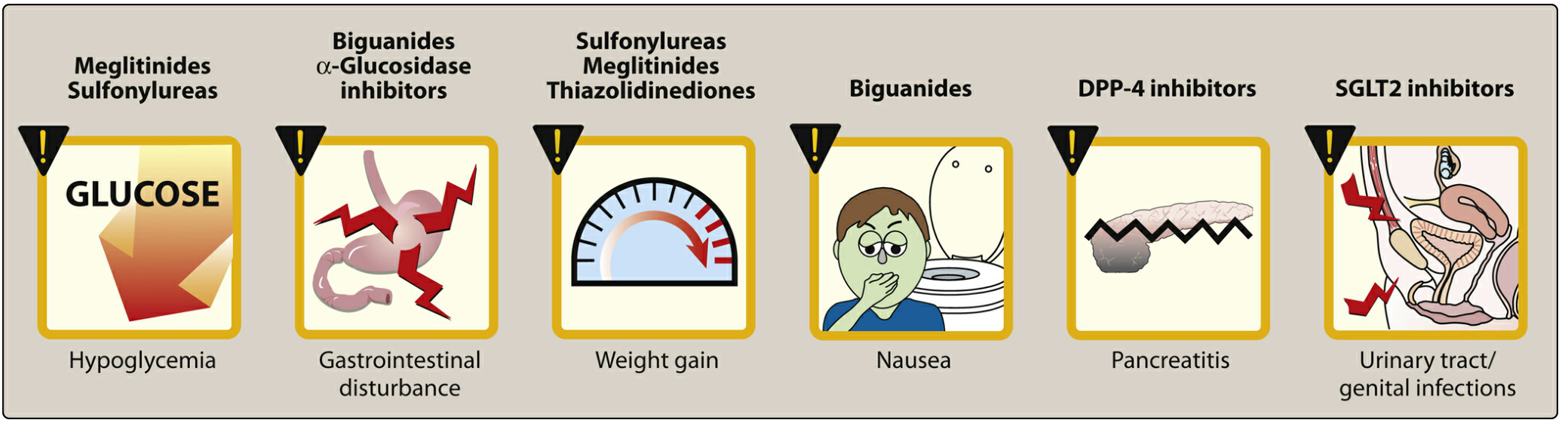
| Biguanides | Metformin (1st line) | ↓ Hepatic gluconeogenesis; ↑ insulin sensitivity | GI upset, lactic acidosis (rare), B12 deficiency. Contraindicated if eGFR <30 |
| Sulfonylureas | Glyburide, Glipizide, Glimepiride | Block β-cell K+-ATP channels → insulin secretion | Hypoglycemia, weight gain. Glipizide/glimepiride safer in renal impairment |
| Meglitinides | Repaglinide, Nateglinide | Similar to sulfonylureas, shorter-acting | Hypoglycemia (less than sulfonylureas), taken with meals |
| Thiazolidinediones (TZDs) | Pioglitazone, Rosiglitazone | PPAR-γ agonists → ↑ insulin sensitivity | Weight gain, fluid retention, heart failure, bone fractures |
| Alpha-glucosidase inhibitors | Acarbose, Miglitol | ↓ intestinal carbohydrate absorption | GI disturbance (flatulence, diarrhea) |
| DPP-4 inhibitors | Sitagliptin, Saxagliptin, Linagliptin | Block GLP-1/GIP degradation → ↑ insulin | Pancreatitis risk |
| GLP-1 receptor agonists | Semaglutide, Liraglutide, Dulaglutide, Exenatide | Mimic GLP-1 → ↑ insulin, ↓ glucagon, ↓ weight | GI side effects; cardioprotective in high CV risk |
| SGLT2 inhibitors | Empagliflozin, Dapagliflozin, Canagliflozin | Block renal glucose reabsorption → glucosuria | UTI/genital infections, DKA (rare euglycemic DKA), diuresis; cardio+renoprotective |
| Amylin analog | Pramlintide | ↓ Gastric emptying, ↓ glucagon, ↑ satiety | Nausea; reduce mealtime insulin by 50% |
4. Modern Combination Approach (T2DM)
Recommended by ADA/EASD guidelines:
- Start Metformin at diagnosis + lifestyle modification
- Add agents with proven cardiovascular benefit (GLP-1 RA or SGLT2 inhibitor) if ASCVD, CKD, or heart failure is present - regardless of HbA1c
- Intensify with 3-drug combinations as needed
- Add insulin if oral agents insufficient
5. Monitoring
- HbA1c every 3 months (intensively treated) or every 6 months (stable)
- Self-monitoring of blood glucose (SMBG) - 4x/day for intensive therapy
- Annual: microalbuminuria, eGFR, lipid panel, dilated eye exam, foot exam, blood pressure check
- Vaccinations: influenza (annual), pneumococcal, hepatitis B
Summary Diagram: Complications of DM
Hyperglycemia (Chronic)
         │
   ┌─────┴─────────────┐
   ▼                   ▼
Microvascular      Macrovascular
   │                   │
   ├─ Retinopathy    ├─ CAD / MI
   ├─ Nephropathy    ├─ Stroke
   └─ Neuropathy     └─ Peripheral Artery Disease
        │
        ├─ Peripheral sensory (DSPN)
        ├─ Autonomic (gastroparesis, orthostasis)
        ├─ Cranial (CN III palsy)
        └─ Diabetic foot → ulcer → amputation
Sources: Robbins & Kumar Basic Pathology; Goldman-Cecil Medicine 2-Volume Set; Adams and Victor's Principles of Neurology 12th Ed.; Harrison's Principles of Internal Medicine 22E (2025); Lippincott Illustrated Reviews: Pharmacology; Brenner and Rector's The Kidney; Katzung's Basic and Clinical Pharmacology 16th Ed.
This is a shared conversation. Sign in to Orris to start your own chat.