
Mei ek mbbs student hu agr mujhe yeh padhna hai ki baccha kaise bnta hai aur baccha sonography kaise dekhte hai ki uska body structure organ sb kaise hai like uski nostrils sahi bane hai head ka circumference sahi hai limbs sahi hai genital sahi hai ya nhi according to month wise toh yeh sb kaise pta karu
- Embryology - bachcha kaise banta hai (fertilization se fetal development tak)
- Obstetric Ultrasound - sonography mein month-wise fetal anatomy kaise assess karte hain
MBBS ke liye: Bachche ka Banana aur Fetal Sonography - Complete Guide
PART 1: BACHCHA KAISE BANTA HAI (Embryology)
Fertilization se Implantation tak (Week 1-2)
- Sperm oocyte se milta hai → secondary oocyte meiosis II complete karta hai → zygote banta hai
- Zygote mein 46 chromosomes hote hain (23 from each parent)
- Zygote cleavage karta hai → morula (16 cells) → blastocyst banta hai
- Blastocyst ke trophoblast cells endometrium mein ghuste hain
- Inner cell mass (embryoblast) se actual embryo banega
- Trophoblast → cytotrophoblast + syncytiotrophoblast → placenta ka basis
Embryonic Period (Week 3-8): Sab kuch banta hai
- 3 germ layers bante hain: Ectoderm, Mesoderm, Endoderm
- Notochord banta hai → neural plate induce karta hai
- Neural tube form hoti hai (brain + spinal cord ka precursor)
| Germ Layer | Kya Banega |
|---|---|
| Ectoderm | Skin, brain, spinal cord, eye lens, ear |
| Mesoderm | Heart, kidney, bone, muscle, blood vessels |
| Endoderm | Gut lining, liver, pancreas, lungs |
- Heart beating shuru (28th day)
- Limb buds appear hote hain
- Somites bante hain (vertebrae + muscles ka basis)
- Embryo C-shape mein fold hota hai
- Face develop hoti hai - nasal pits, optic vesicles, pharyngeal arches
- Limb buds mein digits differentiate hone lagte hain
- Urogenital system develop hona shuru
- Sab major organs form ho jate hain (isliye yeh period teratogenesis ke liye sabse sensitive hai)
- External genitalia abhi indifferent stage mein hain
- Crown-rump length ~30mm
- Embryo → Fetus ka transition week 9 mein
Fetal Period (Week 9-40): Growth aur Maturation
| Weeks | Key Development |
|---|---|
| 9-12 | Face recognizable, fingers alag, urine production shuru, external genitalia differentiate hone lagte |
| 13-16 | Rapid growth, bone ossification, eyes aur ears final position mein, 16 weeks par sex pata chal sakta hai |
| 17-20 | Movements mother feel karti hai (quickening), vernix caseosa aur lanugo appear, eyebrows |
| 21-24 | Lungs mature hone lagte (surfactant production), viability ki limit (~22-24 wks) |
| 25-28 | Eyes open, rapid brain development, subcutaneous fat deposit hona shuru |
| 29-36 | Pulmonary maturation, weight gain, lanugo disappear |
| 37-40 | Term pregnancy, all organs mature |
PART 2: FETAL SONOGRAPHY - MONTH-WISE
Sonography Kab Karte Hain - Standard Scans
| Trimester | Weeks | Purpose |
|---|---|---|
| 1st Trimester | 11-14 weeks | Nuchal Translucency (NT) scan, dating, anomaly screen |
| 2nd Trimester | 18-22 weeks | Anomaly scan (TIFFA/Level 2) - main structural scan |
| 3rd Trimester | 28-32 weeks | Growth scan, liquor, Doppler |
1st Trimester Scan (6-14 Weeks)
- Gestational sac intrauterine confirm karo (ectopic rule out)
- Fetal pole + cardiac activity - rate normal: 120-170 bpm
- CRL (Crown-Rump Length) - gestational age dating ke liye gold standard
- Nuchal Translucency (NT): Cervical skin fold ki thickness
- Normal: < 3.5 mm
- Increased NT → Down syndrome (Trisomy 21), cardiac defects, Turner syndrome ka risk
- Nasal bone: Present hona chahiye (absence → Down syndrome risk)
- Facial profile dekho
- Fetal heart rate, chorionicity (twins mein)
2nd Trimester - TIFFA Scan (18-22 Weeks) - MOST IMPORTANT
HEAD aur BRAIN
- BPD (Biparietal Diameter) - head ki width, dating ke liye
- HC (Head Circumference) - normal range charts se compare
- Intracranial anatomy:
- Lateral ventricles (ventriculomegaly rule out - normal atrium < 10mm)
- Cavum septum pellucidum (CSP) - absent → corpus callosum agenesis ka sign
- Cerebellum - "banana sign" absent hona chahiye (Chiari malformation mein abnormal)
- Cisterna magna - normal 2-10mm
- Posterior fossa
FACE aur NECK
- Lips - upper lip continuous hona chahiye (cleft lip screen)
- Nostrils/Nose - both nostrils present, nasal bone assess
- Profile - forehead, nose, lips, chin (micrognathia, flat face screen)
- Eyes - both orbits present, normal spacing (hypo/hypertelorism)
- Ears - position check
- Neck - cystic hygroma rule out
SPINE
- Longitudinal aur transverse views
- Vertebrae complete hone chahiye, skin cover ho
- Open spina bifida (NTD) rule out - "lemon sign" (head shape abnormal) aur "banana sign" cerebellar abnormality
HEART (Fetal Echocardiography)
- 4-Chamber view - most important, normally 2 atria + 2 ventricles equal size
- AVSD, hypoplastic left/right heart screen
- LVOT (Left Ventricular Outflow Tract) - aorta
- RVOT (Right Ventricular Outflow Tract) - pulmonary artery
- 3-vessel view (3VV) - aorta, pulmonary artery, SVC
- Normal heart: occupies ~1/3 of chest, axis ~45 degrees left
CHEST/LUNGS
- Lungs homogeneous echogenicity
- Diaphragm intact hona chahiye (CDH - Congenital Diaphragmatic Hernia rule out)
- CCAM/CPAM rule out
ABDOMEN
- AC (Abdominal Circumference) - growth parameter
- Stomach bubble visible hona chahiye (if not → esophageal atresia)
- Kidneys - both present, normal echogenicity, no hydronephrosis
- Bladder visible
- Cord insertion normal hona chahiye at umbilicus
- Liver, gallbladder assess
- Bowel - hyperechoic bowel flag karo
LIMBS
- All 4 limbs present hone chahiye
- Long bones:
- FL (Femur Length) - most important dating parameter
- Humerus, radius, ulna, tibia, fibula
- Short limbs → skeletal dysplasias (achondroplasia, thanatophoric dysplasia)
- Hands and feet - fingers aur toes count (polydactyly/oligodactyly)
- Club foot (talipes) rule out
- Rocker bottom feet (Trisomy 18 association)
GENITALIA
- 16 weeks se pata chal sakta hai, 20-22 weeks par clear
- Male: Scrotum + penis visible, testes descent later (3rd trimester mein)
- Female: Labia majora visible, "3 lines" sign
- Ambiguous genitalia → chromosomal/hormonal issue
- Note: India mein sex determination announce karna illegal hai (PCPNDT Act)
PLACENTA, LIQUOR, CORD
- Placental location - low-lying? (Placenta previa)
- AFI (Amniotic Fluid Index) - normal 5-25 cm
- Oligohydramnios (< 5) → renal agenesis, IUGR
- Polyhydramnios (> 25) → NTD, GI obstruction, diabetes
- Umbilical cord - 3 vessels (2 arteries + 1 vein), single umbilical artery → anomaly risk
Biometry Parameters - Dating aur Growth
| Parameter | Full Form | Use |
|---|---|---|
| CRL | Crown-Rump Length | 1st trimester dating (6-13 wks) |
| BPD | Biparietal Diameter | 2nd/3rd trimester dating |
| HC | Head Circumference | Head growth assess |
| AC | Abdominal Circumference | Growth, liver size |
| FL | Femur Length | Skeletal growth, dating |
| EFW | Estimated Fetal Weight | HC + AC + FL se calculate |
3rd Trimester Scan (28-40 Weeks)
- Growth monitoring - EFW charts pe plot karo, every 2-4 weeks if risk
- Liquor assessment (AFI ya Maximum Pool Depth)
- Placental maturity - Grannum grading (Grade 0 to III)
- Fetal presentation - cephalic/breech/transverse
- Doppler studies:
- Umbilical artery Doppler - S/D ratio, absent/reversed end-diastolic flow → fetal compromise
- Middle Cerebral Artery (MCA) Doppler - anaemia, brain-sparing
- Ductus Venosus - severe compromise mein
Kaunsi Books Padhni Chahiye (MBBS ke liye)
- Langman's Medical Embryology (T.W. Sadler) - MBBS standard
- The Developing Human (Moore & Persaud) - detailed
- DC Dutta's Textbook of Obstetrics - Chapter on Ultrasound in Obstetrics
- Ian Donald's Practical Obstetric Problems - classic reference
- ISUOG Guidelines - international standard practice guidelines
Summary - MBBS Exam ke liye Key Points
- Teratogenesis ka sensitive period = Week 3-8 (organogenesis)
- NT scan = 11-14 weeks, NT > 3.5mm abnormal
- TIFFA/Anomaly scan = 18-22 weeks, most comprehensive
- 4-chamber heart view = most common cardiac screening view
- Absent nasal bone + increased NT = Down syndrome risk
- Banana sign + Lemon sign = open spina bifida
- Single umbilical artery = anomaly association
- PCPNDT Act = India mein sex determination illegal
Like as a mbbs doctor agr mujhe genitals mei koi abnormalities toh nhi hai male ya female foetus mei differentiate kr saku pahale yeh toh aaye jisse phir agr male hai toh testicular any other disease related toh genital aur female hai toh labia majora labia minora cervix sahi develop ho rahe hai yeh sb konsi book se padhu bta sakte ho ?
Genital Development aur Abnormalities - MBBS ke liye Complete Guide
Recommended Books - Subject-wise
1. EMBRYOLOGY of Genitalia (PAHLE YEH PADHEIN)
"Indifferent Stage" - Dono sexes ek jaise dikhte hain (Week 5-7)
"The early genital system is indistinguishable between the two genders... both male and female fetuses have gonads with prominent cortical and medullary regions, dual sets of genital ducts, and external genitalia that appear similar."
- Wolffian (Mesonephric) duct → Male structures banega
- Mullerian (Paramesonephric) duct → Female structures banega
Key Decision - SRY Gene (Week 7 onward)
| Male (46,XY) | Female (46,XX) | |
|---|---|---|
| SRY gene | Present | Absent |
| Gonad | Testes develop (medulla predominates) | Ovary develops (cortex predominates) |
| Sertoli cells produce | AMH (Anti-Mullerian Hormone) | - |
| Leydig cells produce | Testosterone | - |
| AMH effect | Mullerian duct degenerates | - |
| Testosterone effect | Wolffian duct → vas deferens, epididymis, seminal vesicle | Wolffian duct degenerates |
| Without testosterone | - | Mullerian duct → uterus, cervix, upper vagina, fallopian tubes |
Yeh Diagram Library se hai - Female vs Male Internal Organs ka Development:
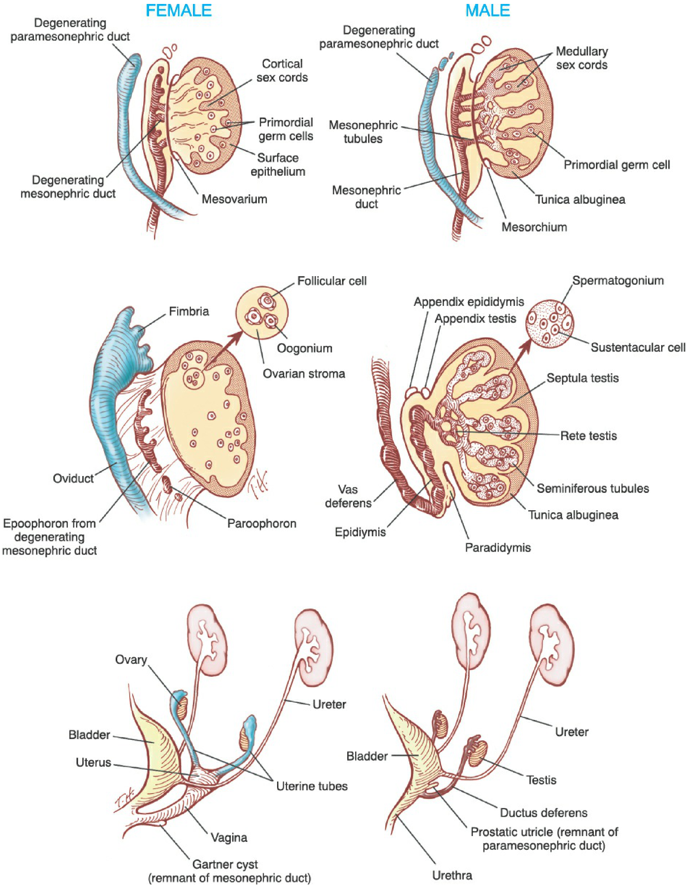
Female External Genitalia Development (Urogenital sinus se)
- Wolffian ducts regress → Week 9
- Mullerian ducts fuse → uterovaginal canal banta hai
- Lower vagina (20%) → sinovaginal bulbs se (urogenital sinus endoderm)
- Labia majora ← genital swellings (jo male mein scrotum banti)
- Labia minora ← urethral folds (jo male mein ventral penis banti)
- Clitoris ← genital tubercle (jo male mein penis banta)
Female Tract Development Step by Step:
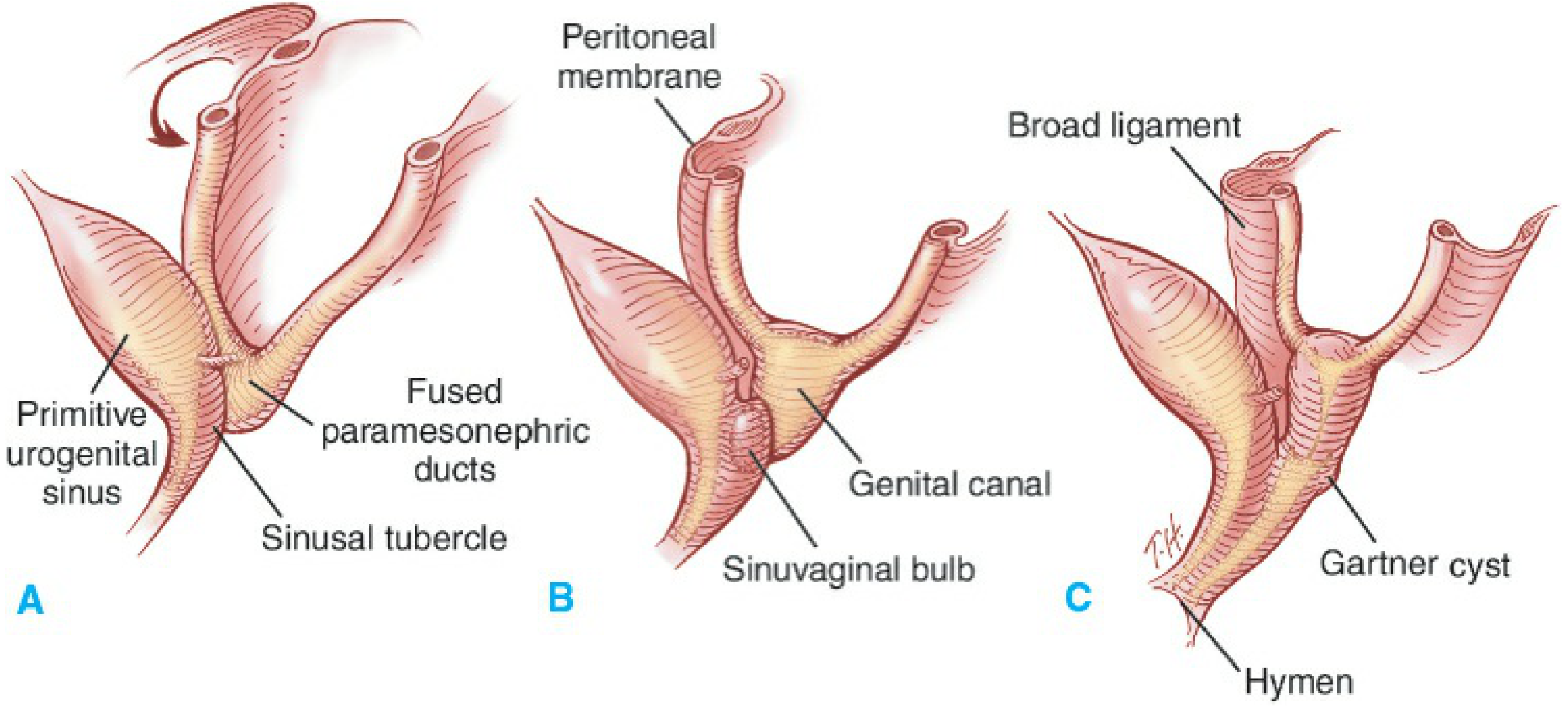
- Upper 80% → fused Mullerian ducts (paramesonephric)
- Lower 20% → sinovaginal bulbs (urogenital sinus)
- Hymen = remnant between these two parts
2. MALE GENITAL ABNORMALITIES
Common Male Genital Anomalies - Sonography + Clinic mein dekhte hain:
| Condition | Kya Hota Hai | Cause | Sonography |
|---|---|---|---|
| Hypospadias | Urethral meatus ventral surface par (glans/shaft/perineum) | Urethral folds ka incomplete fusion | Cord insertion se pata chalta hai |
| Epispadias | Urethral meatus dorsal surface par | Genital tubercle caudal displacement | Rare |
| Cryptorchidism | Undescended testes | Testosterone/gubernaculum failure | 3rd trimester mein scrotum empty |
| Micropenis | Stretched length < 2.5 SD | Testosterone insensitivity, hypogonadism | Seen on fetal scan |
| Bifid scrotum | Scrotum divided in middle | Genital swellings ka non-fusion | |
| Chordee | Ventral penile curvature | Normally resolves by 19th week | |
| Bladder exstrophy | Bladder exposed anteriorly | Cloacal membrane rupture | Absent bladder on scan |
"Failure of fusion of urethral folds leads to various grades of hypospadias. This defect never extends proximal to the bulbous urethra."
3. FEMALE GENITAL ABNORMALITIES
Mullerian Duct Anomalies (MDA) - Most Important Group
| Type | Defect | Result |
|---|---|---|
| Arcuate uterus | Minor fundal indentation | Usually normal fertility |
| Subseptate/Septate uterus | Incomplete/complete septum | Recurrent miscarriage |
| Bicornuate uterus | Incomplete fusion | Preterm labour risk |
| Didelphys | Complete non-fusion | Double uterus, double cervix |
| Unicornuate | One side only develops | One tube, high ectopic risk |
| Mayer-Rokitansky-Kuster-Hauser (MRKH) | Mullerian agenesis | No uterus, no upper vagina, normal ovaries |
Vaginal Anomalies
- Imperforate hymen - vaginal plate ka non-canalization → hematocolpos at puberty
- Transverse vaginal septum - sinovaginal bulbs + Mullerian junction par
- Vaginal agenesis - MRKH syndrome mein
4. DISORDERS OF SEX DEVELOPMENT (DSD) - Ambiguous Genitalia
| Old Name | New Name (DSD) | Chromosomes | Feature |
|---|---|---|---|
| Female pseudohermaphrodite | 46,XX DSD | XX | Clitoral enlargement, scrotalized labia (excess androgens) |
| Male pseudohermaphrodite | 46,XY DSD | XY | Micropenis, hypospadias, cryptorchidism (testosterone insensitivity) |
| True hermaphrodite | Ovotesticular DSD | Both/mosaic | Both ovary + testis tissue |
| XX sex reversal | 46,XX testicular DSD | XX | Phenotypic male, testicular tissue |
- Congenital Adrenal Hyperplasia (CAH) - 90% cases
- 21-hydroxylase deficiency (CYP21A2) - 90% of CAH
- Mechanism: Excess androgens → XX female virilize ho jaati hai
- Androgen Insensitivity Syndrome (AIS) - testosterone receptors kaam nahi karte
- Complete AIS mein: XY but phenotypically female (normal looking female with testes internally)
5. FETAL SONOGRAPHY - Genital Assessment
Normal Genitalia on Ultrasound:
| Sex | Sonographic Signs | What to Look |
|---|---|---|
| Male | "Turtle sign" - scrotum + penis visible | Both testes in scrotum (may be absent till 3rd trimester - normal), penis on midline |
| Female | "3 lines sign" - labia majora + minora | No protruding structures between legs |
Genital Abnormalities on Scan - Red Flags:
| Finding | Suspect |
|---|---|
| Ambiguous genitalia | DSD - chromosomal/hormonal |
| Enlarged clitoris (female fetus) | CAH (46,XX DSD) |
| No bladder + absent kidneys | Potter sequence (renal agenesis) |
| Absent scrotum or penis | DSD |
| Dilated vagina/hydrocolpos | Imperforate hymen, vaginal atresia |
| Cystic mass in pelvis (female) | Ovarian cyst, hydrocolpos |
| Cryptorchidism antenatally | Hypogonadism, DSD |
Subject-wise Reading Plan for MBBS
Step 1 - Foundation (2nd Year Anatomy)
- Langman's Medical Embryology - Chapter on Urogenital System
- The Developing Human (Moore) - Same chapter
- Focus: Normal development, duct systems, indifferent stage
Step 2 - Clinical Details (2nd Year Physiology)
- Ganong's Review of Medical Physiology - "Embryology of the Genitalia" chapter
- Focus: Hormonal control (AMH, testosterone, SRY)
Step 3 - Pathology of Genitalia (3rd Year)
- Robbins Pathology - Reproductive system chapter
- Focus: Tumours, DSD pathology
Step 4 - Clinical Application (Final Year)
- Obstetrics - DC Dutta ya Creasy & Resnik - Fetal anomalies chapter
- Gynecology - Berek & Novak's - Chapter 5 (Genital System Development) + Chapter on Reproductive Anomalies
- Urology (for male anomalies) - Smith & Tanagho - Chapter 2 (Development)
- Pediatrics - Nelson's - DSD/Intersex chapter
Quick Summary Table - Kahan Se Kya Padhein
| Topic | Best Book |
|---|---|
| Embryology basics, ducts, SRY | Langman's / Moore & Persaud |
| Female development detail, Mullerian anomalies | Berek & Novak's Gynecology |
| Male development, hypospadias, cryptorchidism | Smith & Tanagho's Urology / Campbell-Walsh |
| DSD / Ambiguous genitalia | Creasy & Resnik's MFM / Nelson's Pediatrics |
| Fetal USG - genital anomalies | Creasy & Resnik's MFM |
| CAH, hormonal basis | Ganong's Physiology |

Jaise mujhe mere teacher ne case study k liye photo de then unhone yeh sawaal pucha genital kaise hai eske thik hai and how you can differentiate between then if you find any abnormalities find first it is male or if yes then he is normal genital or not and if she is female then what abnormalities she has or she has any kind of abnormalities?
Case Analysis - RAVINA, GA 21 weeks 0 days (3D/4D Ultrasound)
Panel-wise Analysis:
Panel 1 (Top Left) - Fetal Face, Profile View (3D)
- Fetal face ka lateral/oblique 3D rendering
- Forehead - normal slope, no prominent bossing
- Nose - profile mein visible, normal nasal bridge
- Lips - upper aur lower lip dono visible, no cleft lip apparent
- Chin - normal size (micrognathia nahi hai)
- Eyes - closed eyelids visible
Panel 2 (Top Right) - Fetal Face, Frontal/3-Quarter View (3D)
- Face ka anterior/frontal 3D view
- Both eye orbits visible
- Nose - both nostrils visible, normal width
- Upper lip - intact, midline continuous - no cleft lip or palate visible
- Forehead - normal contour
- Ears partially visible
Panel 3 (Middle Left) - SPINE (3D Rendering - "Coronal/Dorsal" View)
- Yeh golden/amber colored 3D spine image hai
- Complete vertebral column visible from cervical to sacral region
- Vertebrae individually clearly outlined
- Posterior elements intact - skin cover dikh raha hai
- No gap/defect visible in spine
- No open spina bifida (NTD) apparent
- Spine appears intact and continuous
- No "lemon sign" ya "banana sign" noted (jo open NTD mein hote hain)
- Sacral region normal appearing
Panel 4 (Middle Right) - Fetal Face, Another Angle (3D)
- Face ka slightly different angle
- Profile aur 3-quarter view combined
- Nose, lips, chin normal dikh rahe hain
- Possibly hand/fingers visible near face (fetus typically puts hand near face)
Panel 5 (Bottom Left) - Face with Hand (3D)
- Fetal hand clearly visible near face/mouth
- Fingers - multiple fingers visible and separated (no obvious syndactyly/polydactyly at this view)
- Face partially visible
Panel 6 (Bottom Right) - Body/Lower Torso View (3D)
- Fetal body ka lower portion - abdomen/lower body region
- Yeh panel mein lower limbs aur body partially visible
- Buttocks/perineal region visible
Overall Structural Assessment (21 Weeks - Academic Teaching Analysis)
| Structure | Finding | Normal/Abnormal |
|---|---|---|
| Face | Intact, bilateral symmetry | ✅ Normal |
| Nose | Both nostrils visible, normal bridge | ✅ Normal |
| Lips | Upper lip intact, no cleft | ✅ Normal |
| Chin | Normal size | ✅ Normal (no micrognathia) |
| Spine | Complete, no defect visible | ✅ Normal |
| Hand/Fingers | Visible, no obvious abnormality | ✅ Normal |
| Overall 3D | Symmetric, well-developed | ✅ Appropriate for 21 weeks |
Genital Assessment at 21 Weeks - Academic Approach
- "Turtle sign" - penis aur scrotum visible between legs
- Scrotum mein testicular echogenicity (3rd trimester mein)
- "3 lines sign" - labia majora bilaterally
- No midline protrusion
Teacher ke Question ka Answer - Academic Framework
- 21 weeks par sex clearly visible hona chahiye
- Dedicated perineal/genital view - "hamburger sign" (female) ya "turtle sign" (male)
- India mein: Sonographer dekhta hai but legally reveal nahi karta (PCPNDT Act 1994)
- Penis - present, normal size (micropenis?)
- Scrotum - present, bifid nahi hai?
- Hypospadias - 2D mein urethral plate assess karna hota hai (3D se difficult)
- Cryptorchidism - 3rd trimester mein assess hota hai
- Labia majora - present bilaterally?
- Clitoral size - enlarged nahi hai? (CAH screen)
- Uterus/ovaries - 2nd trimester mein difficult to see
- Vaginal atresia - hydrocolpos se pata chalta hai
Kya Yeh Scan Normal Hai?
Aapke Teacher ke Liye Answer Summary
"This is a 3D ultrasound of a 21-week fetus (RAVINA, GA 21w0d). The visible structures - face (nose, lips, chin), spine, and hands - appear structurally normal on 3D surface rendering. No cleft lip, no open spina bifida, no obvious facial dysmorphism noted. For complete genital assessment and sex differentiation, dedicated 2D perineal views are required which are not present in this image set. In India, sex determination disclosure is prohibited under PCPNDT Act 1994."