
A 45-year-old man presented with gross, irregular, semi-purposeful movements of face and limbs. Underlying causes include, A. Huntington disease B. Hypothyroidism C. Infarction in subthalamic nucleus D. Salbutamol therapy E. Wilson disease
Movement Disorder Identified: Chorea
Analysis of Each Option
✅ A. Huntington Disease - CORRECT
❌ B. Hypothyroidism - INCORRECT
✅ C. Infarction in the Subthalamic Nucleus - CORRECT
- STN output falls → GPi inhibition of thalamus decreases → thalamic (and cortical) excitation increases → hyperkinetic movements result.
❌ D. Salbutamol Therapy - INCORRECT
✅ E. Wilson Disease - CORRECT
"Neurometabolic disorders: Wilson disease, Lesch-Nyhan syndrome, lysosomal storage disorders..."
Summary Table
| Option | Cause | Correct? | Reason |
|---|---|---|---|
| A. Huntington disease | Hereditary neurodegeneration | ✅ Yes | Classic hereditary chorea |
| B. Hypothyroidism | Metabolic | ❌ No | Hyperthyroidism causes chorea, not hypo |
| C. Subthalamic nucleus infarct | Vascular | ✅ Yes | Hemiballismus/hemichorea |
| D. Salbutamol | Drug-induced | ❌ No | Causes tremor, not chorea |
| E. Wilson disease | Neurometabolic | ✅ Yes | Copper deposition in basal ganglia → chorea |
Correct Answers: A, C, E
- Huntington disease - hereditary striatal degeneration
- Infarction of the subthalamic nucleus - hemiballismus/hemichorea via loss of GPi inhibition
- Wilson disease - copper deposition causing basal ganglia dysfunction
Movement disorders
What do you need on movement disorders? · What level of detail?
basal ganglia direct indirect pathway movement disorders diagram
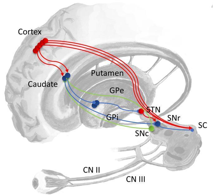
This medical diagram illustrates the basal ganglia circuitry and its contributions to eye movement control. The schematic highlights the functional pathways originating in the Cortex and projecting to the Striatum (Caudate and Putamen). The 'direct pathway' is depicted as an inhibitory connection from the striatum to the Substantia Nigra pars reticulata (SNr) or Globus Pallidus internus (GPi). The 'indirect pathway' involves sequential projections from the striatum to the Globus Pallidus externus (GPe), the Subthalamic Nucleus (STN), and then to the SNr or GPi. The SNr serves as a critical convergence point, sending inhibitory signals to the Superior Colliculus (SC) in the midbrain to regulate saccades. The Substantia Nigra pars compacta (SNc) is shown providing dopaminergic modulation to the striatum. Below the brainstem, the diagram integrates peripheral visual components, including the eye, Optic Nerve (CN II) projecting to the SC, and Oculomotor Nerve (CN III). A color-coded legend indicates functional dynamics: red for excitatory, blue for inhibitory, and green for modulatory connections.
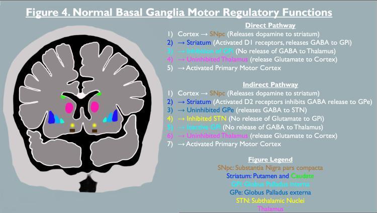
A pathophysiology diagram illustrating the motor regulatory functions of the basal ganglia. The image consists of a coronal brain section paired with text-based flowcharts detailing the 'Direct' and 'Indirect' pathways. In the anatomical illustration, key structures are color-coded: the Substantia Nigra pars compacta (SNpc) in brown/yellow, the Striatum (putamen and caudate) in pink and green, the Globus Pallidus interna (GPi) in cyan, the Globus Pallidus externa (GPe) in blue, and the Subthalamic Nuclei (STN) in gold. The 'Direct Pathway' list describes a sequence of dopaminergic potentiation and GABAergic inhibition involving the D1 receptors, leading to thalamic uninhibition and motor cortex activation. The 'Indirect Pathway' list outlines the more complex sequence involving D2 receptors, the GPe, and the STN, which normally modulates motor activity. The diagram highlights the role of neurotransmitters including dopamine, GABA, and glutamate in regulating muscle tone and movement. This educational material is relevant for understanding movement disorders like Parkinson’s disease and neuroleptic malignant syndrome.
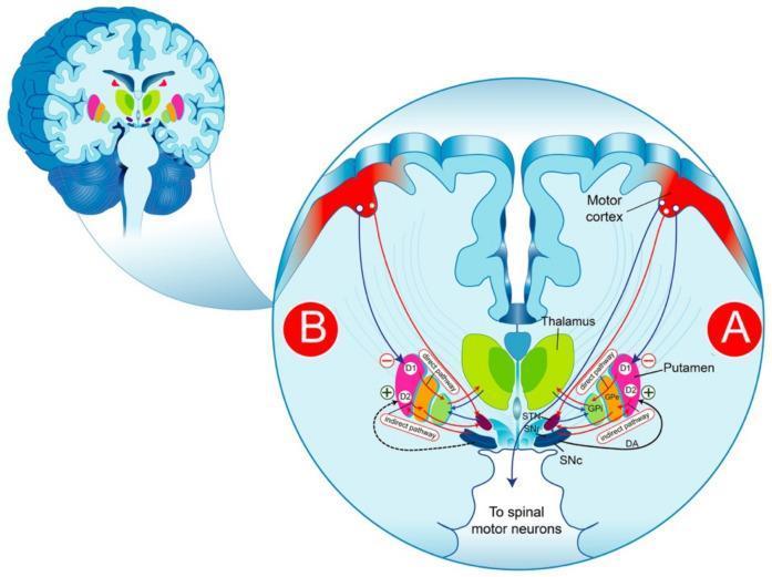
This medical illustration presents a comparative pathophysiology diagram of the basal ganglia circuitry in a physiological state (A) and Parkinson’s Disease (B). The diagram utilizes a coronal brain section view to detail the dopaminergic (DA) regulation of movement. Key anatomical structures labeled include the Motor Cortex, Thalamus, Putamen (striatum), Substantia Nigra pars compacta (SNc), Substantia Nigra reticulata (SNr), Subthalamic Nucleus (STN), Globus Pallidus externa (GPe), and Globus Pallidus interna (GPi). The 'direct pathway' is depicted via D1-receptor-mediated inhibitory projections from the putamen directly to the GPi/SNr, facilitating movement. The 'indirect pathway' is shown via D2-receptor-mediated projections through the GPe and STN, ultimately inhibiting movement. Comparison A highlights healthy dopamine release from the SNc, while Comparison B illustrates the neurodegenerative depletion of dopamine (indicated by dashed lines), resulting in overactivity of the indirect pathway and increased inhibition of the thalamocortical loop. This educational visual explains the mechanistic basis for bradykinesia and motor symptoms in Parkinson’s Disease.
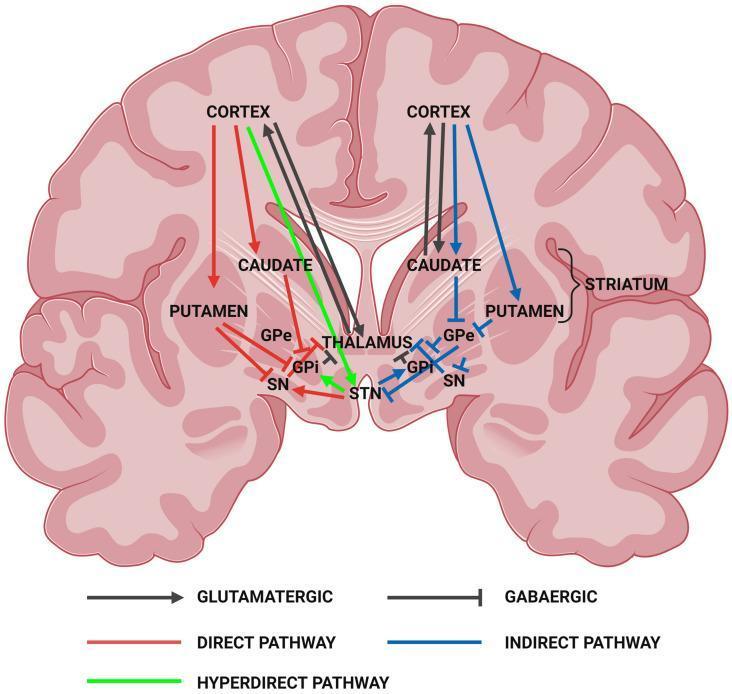
This medical illustration presents a pathophysiology diagram of the human brain in coronal section, focusing on the motor circuitry of the basal ganglia. The diagram maps the structural and functional relationships between the cerebral cortex, striatum (caudate nucleus and putamen), globus pallidus externa (GPe), globus pallidus interna (GPi), subthalamic nucleus (STN), substantia nigra (SN), and thalamus. Three primary signaling pathways are color-coded: the Direct Pathway (red), which facilitates movement; the Indirect Pathway (blue), which inhibits movement; and the Hyperdirect Pathway (green). The visual also utilizes distinct arrowheads to indicate neurotransmitter types: pointed arrows represent excitatory glutamatergic projections (e.g., from the cortex and STN), while T-shaped bars represent inhibitory GABAergic projections (e.g., from the striatum and globus pallidus). This anatomical model is essential for understanding neurodegenerative movement disorders such as Huntington’s Disease and Parkinsonism, specifically how striatal degeneration alters cortical excitation and inhibition loops.
Parkinson disease Huntington disease movement disorder comparison clinical features
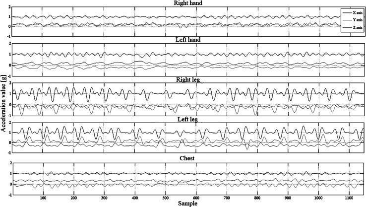
This Comparison Chart displays triaxial acceleration signals recorded for clinical assessment, likely in the context of movement disorders such as Parkinson’s disease. The figure presents five stacked time-series plots corresponding to different anatomical regions: Right hand, Left hand, Right leg, Left leg, and Chest. Each plot tracks acceleration values (measured in g) across X, Y, and Z axes against a sample timeline (0 to 1100 samples). Key visual features include: - Hand Signals: Characterized by relatively low-amplitude, stable oscillations, indicating smoother movement patterns. - Leg Signals: Demonstrate significantly higher amplitude fluctuations and increased frequency, particularly in the X-axis, representing more dynamic or irregular gait and limb acceleration. - Chest Signals: Show the most stable, low-amplitude patterns across all three axes, reflecting central body mass stability during activity. This data is used in neurology and rehabilitation medicine to objectively quantify motor symptoms (e.g., tremor, bradykinesia, or gait disturbances) using wearable sensors. It supports automated symptom recognition and condition monitoring through algorithmic analysis of movement symmetry and intensity.
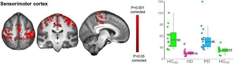
This composite educational graphic presents a comparison of functional connectivity in the sensorimotor cortex between patients with Huntington’s disease (HD) and Parkinson’s disease (PD). The left panel displays three orthogonal MRI views (axial, coronal, and sagittal) in radiological orientation, highlighting regions of interest in the sensorimotor cortex. Red-colored clusters indicate areas with statistically significant differences in functional connectivity (P < 0.05 to P < 0.001, corrected). The right panel features corresponding box plots showing parameter estimates (functional connectivity values) across four cohorts: healthy controls for HD (HCHD), HD patients, PD patients, and healthy controls for PD (HCPD). The data illustrates divergent pathological trends: HD carriers exhibit significantly lower functional connectivity (median = 14) compared to their controls (median = 50), whereas PD patients demonstrate higher functional connectivity (median = 45) compared to their matched controls (median = 23). Individual data points superimposed on the box plots indicate the variability within each group. This visual serves to teach the contrasting pathophysiology of sensorimotor network alterations in these two movement disorders.
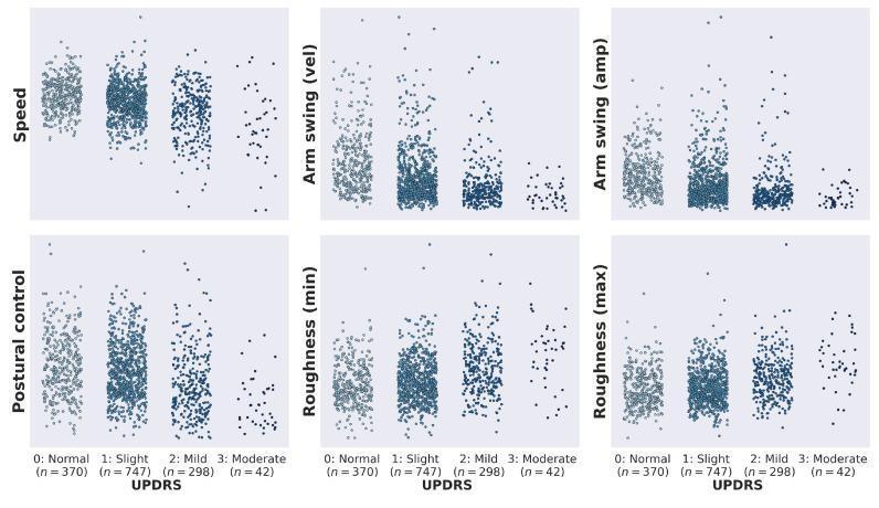
A comparison chart containing six swarm plots illustrating the distribution of automated gait features across four MDS-UPDRS (Unified Parkinson's Disease Rating Scale) gait item 3.10 categories: 0 (Normal), 1 (Slight), 2 (Mild), and 3 (Moderate). The six analyzed gait features are Speed, Arm swing (velocity), Arm swing (amplitude), Postural control, Roughness (min), and Roughness (max). Each subplot shows individual data points (n=1457 total across categories) representing specific gait assessments. For Speed and Arm swing features, the data clusters show a clear downward trend as UPDRS severity increases, indicating reduced movement amplitude and velocity in more advanced Parkinsonian gait. Conversely, Roughness (min) and Roughness (max) plots demonstrate an upward trend and increased variance in higher UPDRS categories, indicating more erratic or irregular walking patterns. The visualization demonstrates the clinical relevance of these digital biomarkers in characterizing gait impairment levels, with each feature showing a statistically significant (p<0.001) difference in means between the clinical rating groups.
Movement Disorders - Complete USMLE Overview
1. WHAT ARE MOVEMENT DISORDERS?
Key distinction: Corticospinal lesions → spasticity (velocity-dependent); Basal ganglia lesions → rigidity, dyskinesia, bradykinesia.
2. BASAL GANGLIA CIRCUITS - THE ESSENTIAL FRAMEWORK
Direct Pathway (facilitates movement - "GO")
- Dopamine at D1 receptors: EXCITES striatum → MORE inhibition of GPi → MORE thalamic output → MORE movement
Indirect Pathway (inhibits movement - "STOP")
- Dopamine at D2 receptors: INHIBITS striatum → LESS inhibition of GPe → GPe inhibits STN → LESS GPi output → LESS thalamic inhibition → MORE movement

3. CLASSIFICATION: HYPOKINETIC vs. HYPERKINETIC
| Category | Speed | Key Feature | Prototype |
|---|---|---|---|
| Hypokinetic | SLOW | Too little movement | Parkinson's disease |
| Hyperkinetic | FAST | Too much movement | Chorea, tics, tremor |
Speed Spectrum (slow → fast):
4. INDIVIDUAL MOVEMENT TYPES
A. BRADYKINESIA / HYPOKINESIA / AKINESIA
- Definition: Slowed, decreased, or absent movements (not due to UMN or LMN lesion)
- Mechanism: Increased inhibitory basal ganglia output to thalamus (GPi overactivity)
- Cause: Loss of dopaminergic input (Parkinson's) or striatal dysfunction
- USMLE tip: Bradykinesia + rigidity + resting tremor + postural instability = Parkinson's tetrad
B. RIGIDITY
- Definition: Uniform resistance throughout passive range of motion ("lead pipe" or "plastic" rigidity)
- Cogwheel rigidity: Lead pipe + superimposed tremor - pathognomonic of Parkinson's
- vs. Spasticity: Spasticity is velocity-dependent (UMN), with clasp-knife release
C. DYSTONIA
- Definition: Co-contraction of agonist AND antagonist muscles → sustained abnormal posture
- Types:
- Focal: Torticollis (neck), blepharospasm (eyes), writer's cramp (hand), spasmodic dysphonia (larynx)
- Generalized: DYT-1 mutation (TOR1A gene deletion), Ashkenazi Jewish population
- Dopa-responsive dystonia (DRD): Child with dystonia → responds dramatically to low-dose levodopa
- Treatment: Anticholinergics (trihexyphenidyl), botulinum toxin for focal, deep brain stimulation (GPi) for generalized
- Also seen in: Wilson's disease, Huntington's, Parkinson's, drug-induced (antipsychotics, metoclopramide)
D. ATHETOSIS
- Definition: Slow, writhing, sinuous movements of the fingers, hands, toes - intermediate between dystonia (slower) and chorea (faster)
- Seen in: Cerebral palsy, basal ganglia infarcts, kernicterus, Wilson's disease
- Choreoathetosis = mixed chorea + athetosis (common in Wilson's disease)
E. CHOREA
- Definition: Irregular, brief, semi-purposeful, flowing movements that randomly migrate from one body part to another - like a "dance"
- Mechanism: Preferential loss of indirect pathway striatal neurons → STN under-driven → GPi under-active → Thalamus over-active
Major Causes (mnemonic: WITCH + Drugs):
| Cause | Notes |
|---|---|
| W - Wilson's disease | Copper deposition; choreoathetosis + psychiatric features + Kayser-Fleischer rings |
| I - Immune (SLE, Sydenham's) | Sydenham's = post-streptococcal; SLE can be FIRST presentation |
| T - Thyrotoxicosis (hyperthyroidism) | Metabolic chorea; NOT hypothyroidism |
| C - Cerebrovascular (STN/striatal infarct) | Hemichorea/hemiballismus |
| H - Huntington's disease | Autosomal dominant; CAG repeat, HTT gene |
| Drugs | Levodopa, OCP, phenytoin, antipsychotics (tardive), cocaine |
| Pregnancy | Chorea gravidarum (often re-activates underlying SLE/Sydenham's) |
| Neuroacanthocytosis | Acanthocytes on blood film + chorea |
F. BALLISMUS / HEMIBALLISMUS
- Definition: High-amplitude, wild, flinging, rotatory movements of proximal limbs - like a "throw"
- Classic cause: Lacunar infarct of the subthalamic nucleus → STN underactive → GPi underactive → Thalamus overactive → hyperkinesis
- Side: Movements are contralateral to the lesion
- Other causes: Basal ganglia hemorrhage, tumor, non-ketotic hyperglycemia
- Treatment: Haloperidol (dopamine antagonist); usually self-limiting over weeks
G. TICS
- Definition: Sudden, brief, repetitive, stereotyped movements/sounds preceded by urge → followed by relief (suppressible)
- Types: Motor tics (face, neck > limbs) or vocal tics (grunting, barking, coprolalia)
- Tourette's syndrome: Both motor + vocal tics persisting > 1 year; 4:1 male; onset late childhood; associated with ADHD and OCD
- Treatment: CBIT (behavioral), clonidine/guanfacine, anti-dopaminergics in severe cases
H. MYOCLONUS
- Definition: Sudden, rapid, brief muscle jerk - the fastest movement disorder
- Asterixis (negative myoclonus): Brief lapses in sustained posture - "liver flap"; EMG shows brief silent periods in extensors
- Causes: Cortical (epileptic), metabolic/toxic encephalopathy, anoxic brain injury, CJD (prion), Alzheimer's (late), paraneoplastic (SCLC, ovarian)
I. TREMOR
- Definition: Rhythmic oscillating movement (agonist AND antagonist both activated - distinguishes it from myoclonus)
| Type | When Present | Causes | Hz |
|---|---|---|---|
| Resting tremor | At rest, disappears with action | Parkinson's (pill-rolling), Holmes tremor | 5-7 Hz |
| Postural tremor | Holding position against gravity | Essential tremor, physiological/toxic, anxiety, drugs | 5-8 Hz |
| Intention tremor | During voluntary movement, worsens near target | Cerebellar disease | Variable |
- Essential tremor: Most common movement disorder (5% prevalence); bilateral postural tremor of hands/arms; can affect head and voice; improves with alcohol; treated with propranolol or primidone
- Pill-rolling tremor of Parkinson's: 5-7 Hz, asymmetric resting tremor, worse when distracted
5. MAJOR DISORDERS - USMLE HIGH-YIELD SUMMARY
Parkinson's Disease
| Feature | Detail |
|---|---|
| Pathology | Loss of dopaminergic neurons in substantia nigra pars compacta |
| Pathognomonic lesion | Lewy bodies (alpha-synuclein aggregates) |
| Classic tetrad | Resting tremor, Bradykinesia, Rigidity (cogwheel), Postural instability |
| Additional features | Micrographia, masked facies, shuffling gait, hypophonia, pill-rolling tremor |
| Mechanism | Dopamine loss → direct pathway underactive + indirect pathway overactive → GPi overactive → thalamus inhibited → bradykinesia |
| Treatment | Levodopa/carbidopa, dopamine agonists (pramipexole), MAO-B inhibitors (selegiline), deep brain stimulation (STN or GPi) |
Huntington's Disease
| Feature | Detail |
|---|---|
| Genetics | Autosomal dominant; CAG trinucleotide repeat expansion in HTT gene (Ch 4); anticipation |
| Pathology | Degeneration of striatal neurons (caudate + putamen); caudate atrophy on MRI |
| Sequence | Early: Indirect pathway neurons die first → chorea; Late: Both pathways degenerate → rigid/parkinsonian state |
| Triad | Chorea + Dementia + Psychiatric disturbance |
| Age of onset | 30-50 years; juvenile form (<20 yrs) presents with rigidity |
| Death | ~15 years after onset; from respiratory infection, falls |
| Treatment | Tetrabenazine/deutetrabenazine (VMAT2 inhibitor) for chorea; no disease-modifying therapy |
Wilson's Disease
| Feature | Detail |
|---|---|
| Genetics | Autosomal recessive; ATP7B gene mutation → impaired biliary copper excretion |
| Organs | Liver (cirrhosis, acute liver failure) + Brain (basal ganglia, especially lenticular nucleus) + Eyes |
| Neurological | Dysarthria, dystonia, choreoathetosis, rigidity, tremor ("wing-beating" tremor), psychiatric disturbance, risus sardonicus |
| Eye sign | Kayser-Fleischer rings (brown corneal copper deposits) - seen in all with neurological involvement |
| Diagnosis | Low serum ceruloplasmin, high urine copper, slit-lamp exam, liver biopsy |
| Treatment | Penicillamine (chelation), trientine, zinc (blocks absorption); liver transplant cures the disease |
| Age | Liver disease: often <10 years; neurological: typically teens/early 20s |
6. DRUG-INDUCED MOVEMENT DISORDERS
| Drug Class | Movement Disorder | Mechanism |
|---|---|---|
| Antipsychotics (D2 blockers) | Acute dystonia (hours-days) | D2 blockade in striatum |
| Antipsychotics | Akathisia (restlessness) | D2 blockade |
| Antipsychotics | Parkinsonism (weeks) | D2 blockade reduces direct pathway |
| Antipsychotics (long-term) | Tardive dyskinesia (months-years) | Dopamine receptor upregulation; oro-lingual-buccal chorea |
| Metoclopramide | Same spectrum as antipsychotics | D2 antagonist |
| Levodopa | Dyskinesias/chorea | Excessive dopaminergic stimulation |
| OCP, phenytoin, cocaine | Chorea | Various |
7. QUICK COMPARISON TABLE
| Disorder | Speed | Suppressible? | Rhythmic? | Key Feature |
|---|---|---|---|---|
| Tremor (resting) | 5-7 Hz | No | Yes | Parkinson's, pill-rolling |
| Tremor (postural) | 5-8 Hz | No | Yes | Essential tremor |
| Tremor (intention) | Variable | No | Yes | Cerebellar |
| Chorea | Fast | No | No | Flowing, dance-like |
| Athetosis | Slow-medium | No | No | Writhing, fingers/toes |
| Ballismus | Fast | No | No | Flinging, proximal |
| Dystonia | Slow | Partially | No | Sustained posture |
| Tics | Fast | Yes (partially) | No | Preceded by urge |
| Myoclonus | Fastest | No | Sometimes | Brief jerk |
| Bradykinesia | Slow | N/A | N/A | Poverty of movement |