
Dengue in detail for CME, also provide probable cross question and answer . Also provide common patient question and answer in opd and er . And recent guidelines
dengue fever management guideline
latest dengue clinical management guidelines 2025 2026 WHO
https://communitymedicine4asses.wordpress.com/2025/07/11/who…
dengue fever petechial rash tourniquet test
WHO dengue classification warning signs severe dengue diagram
dengue skin rash exanthem
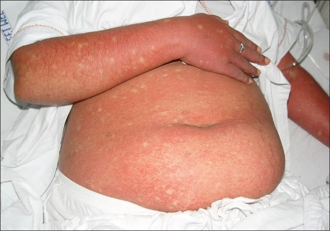
This clinical photograph displays a patient's abdomen and arm presenting a generalized, confluent erythematous rash, a characteristic cutaneous manifestation of Dengue Fever. The exanthem is marked by widespread, intense redness resulting from capillary dilatation and coalesced maculopapular lesions. A diagnostic hallmark visible here is the presence of 'islands of sparing'—small, rounded, or oval-shaped patches of normal-colored skin scattered throughout the erythematous background, often described as 'white islands in a sea of red'. These spared areas are randomly distributed across the affected trunk and limbs. The image serves as a key educational reference for recognizing the secondary rash phase of Dengue viral infection, typically occurring 3 to 6 days after the onset of fever. The visual presentation is essential for clinical diagnosis and differentiation from other viral exanthems, highlighting the specific immunologic response patterns seen in Dengue Fever (DF) and Dengue Hemorrhagic Fever (DHF).
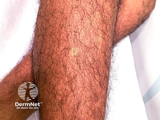
Clinical photography of a forearm demonstrates an acute, diffuse erythematous exanthem with pale islands of hypopigmentation or normal skin creating a white islands in a sea of red pattern. The imaging modality is surface, noninvasive dermatologic photography using standard lighting and color calibration; no stains or contrast agents applied. Anatomic context is the integumentary system, skin of the forearm (anterior-lateral aspect) in a febrile patient. Lesion morphology includes confluent erythema composed of macules and small papules merging to form broad red patches, with discrete islands of preserved or lighter skin within the inflamed background. The pattern may blanch with pressure and lacks overt vesiculation or necrosis in this view. Clinically this appearance is characteristic of viral exanthems seen in febrile illnesses such as dengue fever, chikungunya, or Zika; extension to trunk and limbs is common. Differential considerations include measles, rubella, drug eruption, pityriasis rosea, and pityriasis lichenoides. Diagnostic significance lies in recognizing a febrile exanthem pattern, prompting laboratory evaluation for dengue (NS1/IgM/IgG), complete blood count, and coagulation tests; monitor for warning signs. Potential educational uses include dermatology teaching, infectious disease triage, and documentation of rash evolution over time. This description supports rapid clinical triage and correlates with febrile dengue syndromes.
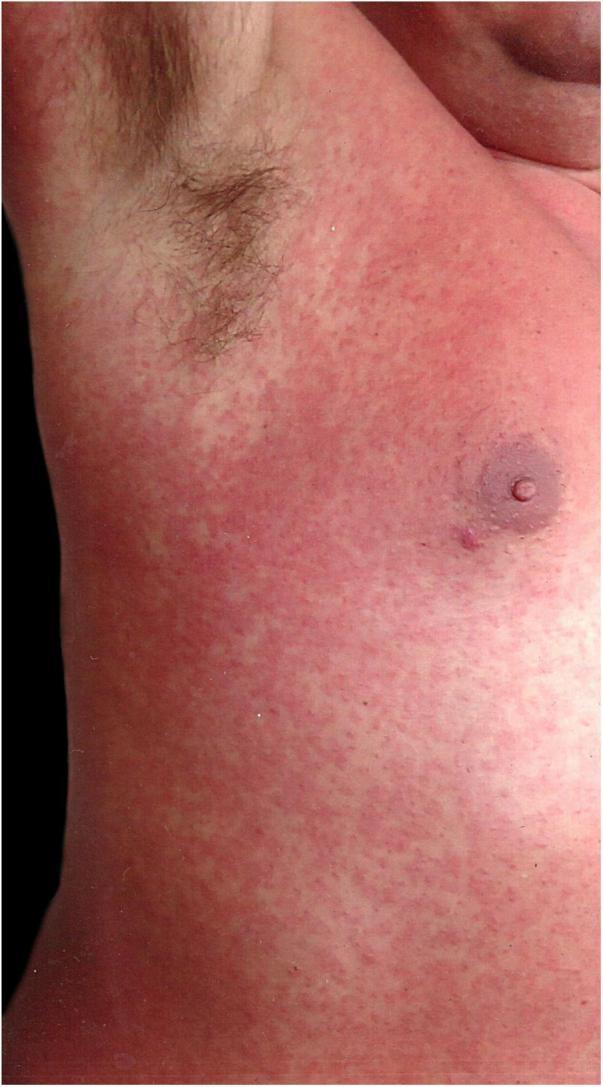
A clinical photograph showing a close-up of a patient's trunk and right axillary region on the third day of a rubelliform rash evolution. The image depicts a dense, generalized exanthem characterized by numerous small, erythematous maculopapular lesions. The morphology shows discrete reddish papules that are closely spaced, forming a nearly confluent, morbilliform-like pattern over the chest and abdomen. A distinctive clinical feature in this view is the relative sparing of the right axillary region, where the skin appears significantly less affected compared to the surrounding intensely red areas. The nipple and areola are visible and appear unaffected. The presentation is characteristic of an infectious viral exanthem, which in this clinical context was confirmed as a secondary dengue virus infection. The visual focus is on the morphology and distribution of the rash, providing an educational example of how systemic viral infections can manifest cutaneous signs while sparing certain anatomical regions.
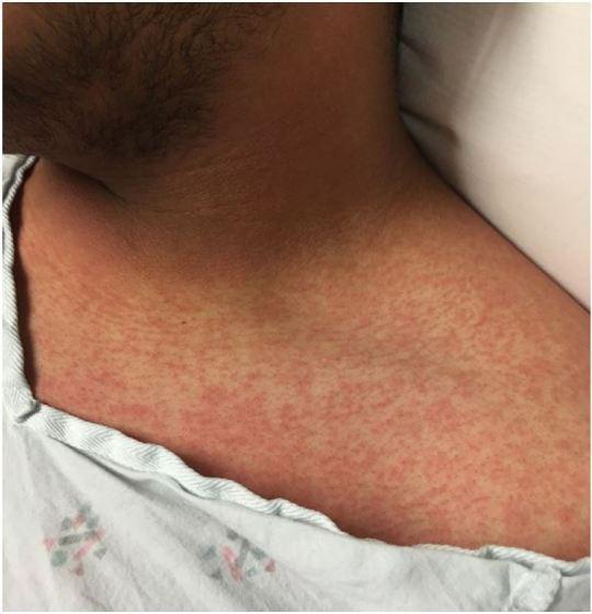
This clinical photograph displays a diffuse, erythematous maculopapular rash on the upper chest, neck, and shoulder of a patient. The dermatologic manifestation consists of numerous small, red macules (flat spots) and papules (slightly raised bumps) that are densely distributed across the skin surface. The pattern is characteristic of the exanthem seen in Zika virus infection, often described as a mild, dengue-like febrile illness. The image captures the anterior thoracic region near the clavicle and the lateral neck, illustrating the typical morphology and widespread distribution of the rash associated with flavivirus infections. This visual is a key educational resource for recognizing the clinical signs of emerging infectious diseases in a dermatology or emergency medicine context.
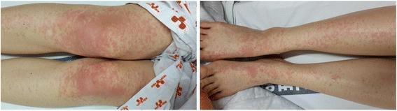
Clinical photograph of a patient's lower extremities demonstrating a widespread erythematous rash, characteristic of a viral exanthem. The left panel shows the thighs and knees, while the right panel shows the lower legs and dorsal feet. The morphology is primarily maculopapular, consisting of small, red, blanchable spots and slightly raised bumps. A distinct spatial variation is visible: the proximal lesions around the knees are highly confluent, forming large, poorly demarcated erythematous patches. In contrast, the distal lesions on the lower legs and feet are more discrete, individual maculopapules with intervening areas of normal-appearing skin. This presentation is consistent with the secondary stage of a Dengue virus infection (DENV-2), appearing several days after initial symptoms. The image serves as a clinical reference for identifying infectious disease-related skin manifestations and assessing the distribution and confluence of exanthematous eruptions.
Aedes aegypti mosquito
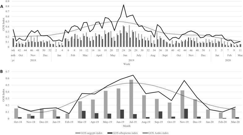
A dual-panel graph illustrating the temporal trends of Gravid Ovillanta System (GOS) indices for Aedes mosquitoes, a critical entomological indicator in dengue vector surveillance. Panel A presents weekly data for an intervention group, while Panel B displays monthly data for a control group, spanning October 2018 to March 2020. Both panels feature a solid black line representing the total GOS Aedes index, complemented by a black dotted trend decomposition curve. Bar charts categorize the data into GOS aegypti (light grey) and GOS albopictus (dark grey) indices. In the intervention group (A), the mosquito population shows significant fluctuation with a prominent peak in abundance around June 2019 (weeks 24–26), followed by a gradual downward trend. In the control group (B), Ae. aegypti consistently demonstrates higher relative abundance compared to Ae. albopictus. This comparative data is used to evaluate the efficacy of trap deployment and community-based interventions in reducing mosquito density, which is a primary risk factor for the transmission of arboviral diseases like dengue.
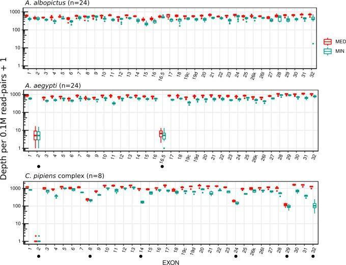
A comparison chart consisting of three logarithmic dot plots illustrating the targeted sequencing coverage of Voltage-Gated Sodium Channel (VGSC) exons in three mosquito species: Aedes albopictus (n=24), Aedes aegypti (n=24), and Culex pipiens complex (n=8). The y-axis represents the sequencing depth per 0.1M read-pairs plus 1 on a logarithmic scale (1 to 1000), while the x-axis lists specific exons from 1 to 32. For each exon, two data points are provided: Median depth (MED, red) and Minimum depth (MIN, teal). The chart demonstrates that A. albopictus exhibits high and consistent coverage across all exons. In contrast, A. aegypti shows a significant drop in coverage for exons 2 and 16.5, and the C. pipiens complex displays variability with notable coverage dips in exons 2, 8, 14, 24, 29, and 32 (indicated by black dot markers). This visual data is critical for evaluating the efficiency of a single probe set designed for insecticide resistance research across different medically relevant mosquito vectors.
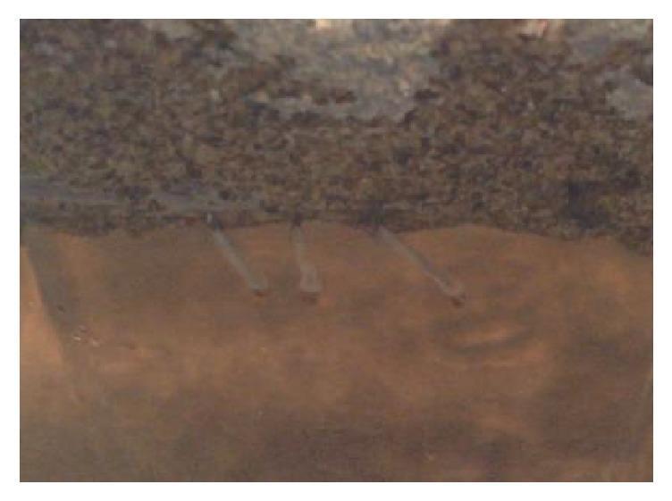
This clinical photograph/specimen image displays late third-stage Aedes aegypti larvae within an experimental aquatic environment. The image is divided into two distinct horizontal layers: a superior dark, particulate-rich layer composed of powdered Azolla pinnata, and an inferior lighter, translucent water layer. Three larvae are visible, exhibiting an elongated, slender morphology with a whitish, translucent appearance. The specimens are positioned at the interface between the particulate material and the clear water, a behavior relevant to studying the larvicidal effects and toxic accumulation of botanical biocontrol agents. This visual demonstrates the behavioral responses and morphological status of mosquito larvae during a 24-hour bioassay test, serving as a reference for public health entomology and vector control research focusing on the reduction of human disease vectors like Aedes aegypti, which transmits dengue, zika, and chikungunya viruses.
Dengue Fever - CME Review
1. Overview & Virology
2. Clinical Course - Three Phases (WHO framework)
3. WHO 2009 Classification (still the operational framework)
| Category | Criteria |
|---|---|
| Dengue without warning signs | Fever + 2 of: nausea/vomiting, rash, aches/pains, leukopenia, positive tourniquet test |
| Dengue with warning signs | Abdominal pain/tenderness, persistent vomiting, clinical fluid accumulation (effusion/ascites), mucosal bleeding, lethargy/restlessness, liver enlargement >2 cm, rising hematocrit with rapid platelet drop |
| Severe dengue | Severe plasma leakage → shock (DSS) or fluid accumulation with respiratory distress; severe bleeding; severe organ impairment (AST/ALT ≥1000, altered consciousness/encephalitis, cardiac impairment/myocarditis) |
- Red Book 2021 (AAP); Andrews' Diseases of the Skin; Harrison's Principles of Internal Medicine 22E
4. Diagnosis
- NS1 antigen: Positive from day 1, useful in the first 5 days of illness.
- IgM ELISA: Detectable from day 4-5; may be falsely negative early.
- RT-PCR / viral RNA: Most sensitive in the first 5 days (viremic phase).
- IgG: Rises in secondary infection, useful epidemiologically.
- Supportive labs: leukopenia, progressive thrombocytopenia, rising hematocrit (hemoconcentration), deranged LFTs, coagulopathy in severe disease.
- Note: prior dengue vaccination can confound serologic interpretation - Red Book 2021.
5. Management
- Non-severe dengue (outpatient): Oral hydration, paracetamol for fever/pain. Strictly avoid NSAIDs and aspirin (bleeding risk, and aspirin carries Reye syndrome risk in children). Daily review for warning signs, especially around defervescence; monitor hematocrit/platelets.
- Dengue with warning signs: Hospitalize. IV crystalloid rehydration titrated to urine output, vitals, and hematocrit trend; avoid fluid overload once leakage resolves (usually by 48 hours post-critical phase).
- Severe dengue/DSS: ICU-level care, aggressive but carefully titrated IV fluid resuscitation, blood/platelet products only for significant bleeding (not for isolated thrombocytopenia), organ support as needed. Steroids are not recommended - evidence remains inconclusive/low quality - Rosen's Emergency Medicine, Washington Manual.
6. Recent Guidelines (2025 update)
- Protocolized oral rehydration in non-severe dengue reduces hospital admissions with minimal harm.
- Lactate measurement alongside standard monitoring is now recommended to guide IV fluid therapy in suspected/confirmed severe arboviral disease (strong recommendation) - likely reduces mortality (not useful in liver failure/yellow fever).
- Passive leg raise (PLR) test suggested in shocked patients to judge fluid responsiveness before further boluses.
- Systemic corticosteroids - recommended against use in severe arboviral disease (very low certainty evidence).
- Paracetamol remains the antipyretic/analgesic of choice (avoid in yellow fever due to hepatic risk); metamizole/dipyrone suggested as an alternative where available.
- Reaffirms avoidance of NSAIDs/aspirin throughout.
- Qdenga (TAK-003) is the WHO-endorsed dengue vaccine, recommended for children aged 6-16 years in high-transmission settings (2 doses, 3 months apart).
- WHO Paediatric Drug Optimization (PADO) meeting (Oct 2025) is prioritizing pediatric-specific dengue therapeutics, reflecting the continued absence of a licensed antiviral.