
Explain in detail all aspects of primary survey and secondary survey in trauma management
ATLS trauma primary survey ABCDE assessment algorithm
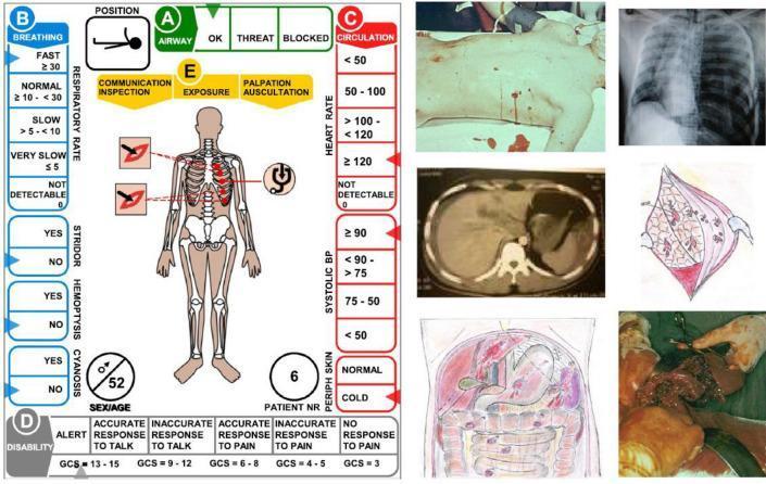
This composite educational infographic functions as a 'casualty card' for medical trauma simulation and mass casualty incident (MCI) training. The left side features a structured assessment template based on the ABCDE (Airway, Breathing, Circulation, Disability, Exposure) primary survey. It includes physiological parameters such as respiratory rate, presence of stridor/hemoptysis, heart rate (marked ≥120), systolic blood pressure (marked ≥90), and a Glasgow Coma Scale (GCS) section. An anatomical diagram indicates penetrating trauma to the left hemithorax. The right side contains clinical supporting evidence: a clinical photograph of a torso showing multiple puncture wounds with active bleeding; a chest X-ray demonstrating a massive left-sided hemothorax or pleural effusion with mediastinal shift; a CT scan of the abdomen; a surgical anatomical illustration of internal organ damage; and an intraoperative photograph showing severe abdominal visceral injury. The material is designed for advanced trauma life support (ATLS) training, illustrating the integration of clinical signs, diagnostic imaging, and surgical findings for urgent triage and decision-making.
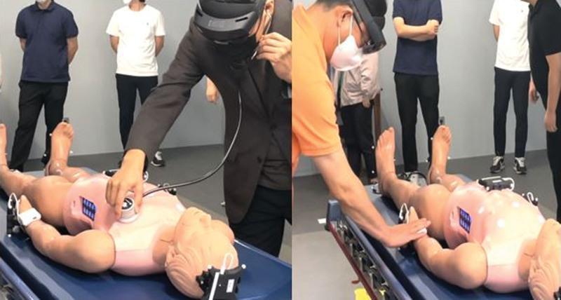
A composite clinical photograph illustrating a medical training simulation using Mixed Reality (MR) technology for trauma assessment. The left panel shows a practitioner wearing a head-mounted display (HMD) performing chest auscultation on a high-fidelity supine mannequin using an integrated MR stethoscope. The right panel demonstrates the palpation of the mannequin's radial pulse using an MR pulse band. The mannequin is equipped with electronic sensors and markers for spatial tracking, including white pads on the limbs and a chest-mounted device. The simulation is designed for trauma nursing and medical education, allowing for the overlay of virtual 'medical twin' patient data onto physical hardware. This procedural setup enables trainees to practice the primary survey (ABCDE approach) in a controlled, immersive environment. Other participants are shown in the background observing the clinical scenario, indicating a classroom or simulation lab setting for procedural competency training.
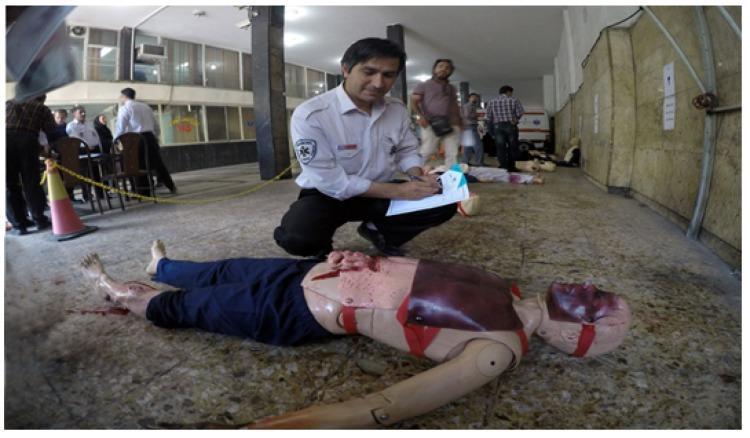
Clinical photograph of a high-fidelity medical simulation focused on emergency medical technician (EMT) training and mass casualty triage. In the foreground, a medical student or responder in uniform is kneeling next to a trauma mannequin, recording findings on a clipboard. The mannequin exhibits advanced moulage representing critical polytrauma, including evisceration of abdominal organs, a compound fracture of the lower leg with exposed bone and hemorrhage, and significant maxillofacial trauma with simulated hematomas and abrasions. The training scenario takes place in an indoor facility, illustrating a triage algorithm or primary assessment protocol (ABCDE). This visual serves as an educational tool for emergency medicine, pre-hospital trauma life support (PHTLS), and disaster response training, highlighting the identification of life-threatening injuries and the documentation of patient status in a simulated clinical environment.
This clinical photograph displays a primary skin lesion identified as melanoma (MEL). The visual demonstrates the ABCDE criteria used in dermatological assessment. The lesion is characterized by marked asymmetry and an irregular, poorly defined, and somewhat notched border. It exhibits significant color variegation, with shades of dark brown and black interspersed with areas of lighter tan and central reddish-pink hues. The surrounding skin is light-toned with normal texture and fine hair. To the right of the clinical image is a user interface panel from a diagnostic algorithm, displaying a 100% classification confidence for melanoma compared to other differential diagnoses such as melanocytic nevi, actinic keratoses, basal cell carcinoma, benign keratosis-like lesions, dermatofibroma, and vascular lesions. This content is intended for medical education regarding the clinical recognition of malignant skin conditions and the application of convolutional neural networks in dermatological diagnostics.
Primary Survey and Secondary Survey in Trauma Management
Overview and Guiding Principles
- Treat the greatest threat to life first
- The lack of a definitive diagnosis should never delay indicated treatment
- A detailed history is not essential to begin evaluation
PRE-ARRIVAL PREPARATION
PRIMARY SURVEY
- Airway obstruction
- Tension pneumothorax
- Open pneumothorax
- Flail chest
- Massive internal/external hemorrhage
- Cardiac tamponade
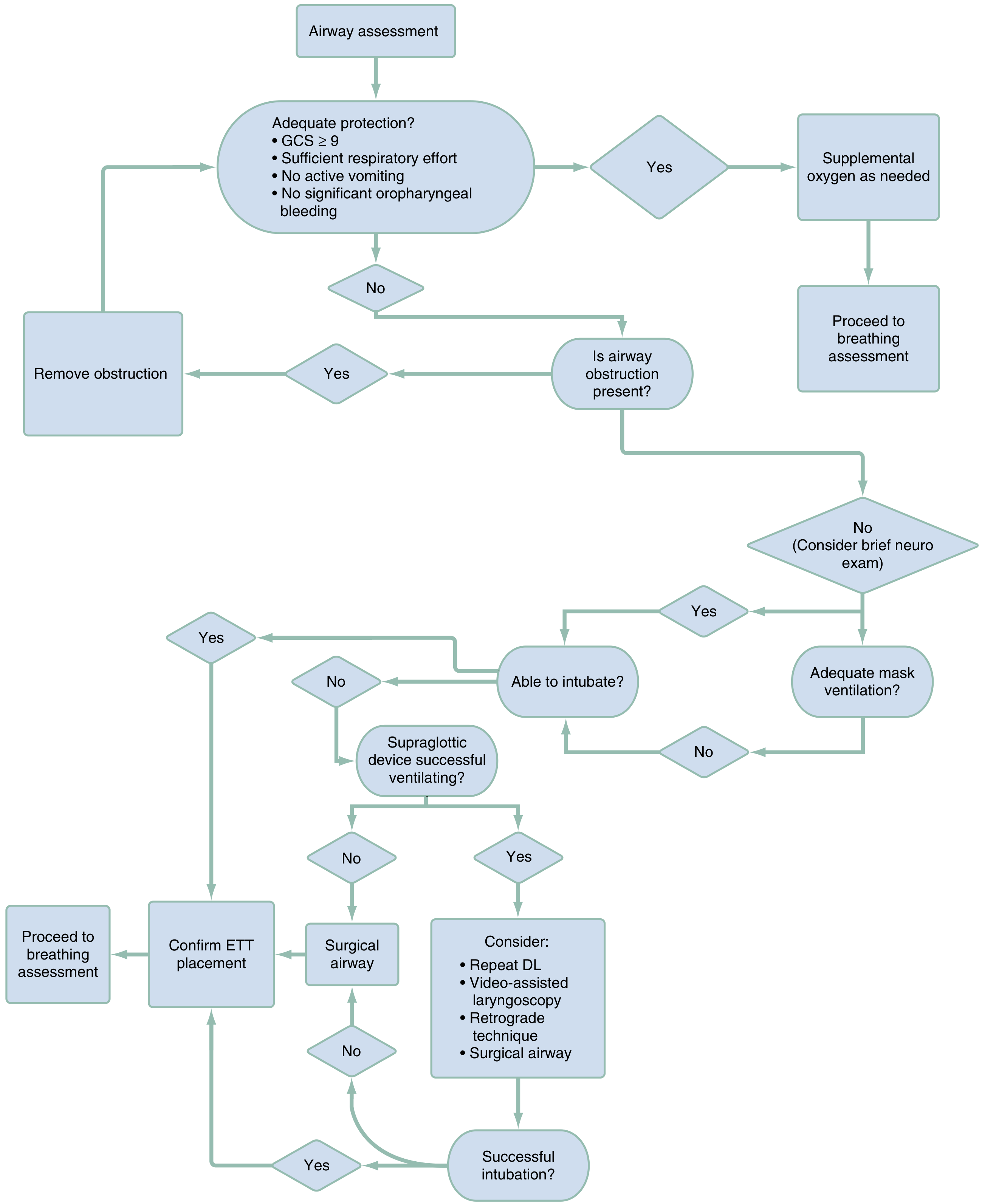
A - AIRWAY (with Cervical Spine Control)
- Inspect for foreign bodies, blood, vomit, loose teeth, maxillofacial fractures
- Check for stridor, gurgling, hoarseness (signs of obstruction)
- Ask the patient a question - a clear verbal response indicates a patent airway
- GCS ≥ 9
- Sufficient respiratory effort
- No active vomiting
- No significant oropharyngeal bleeding
- Jaw thrust / chin lift (with in-line cervical spine stabilization) - never hyperextend the neck
- Suction of oropharynx
- Oral or nasopharyngeal airway - OPA in unconscious patients without gag reflex; avoid NPA in basilar skull fracture
- Endotracheal intubation - indicated for GCS 3-8, inability to protect airway, or anticipated airway compromise. Use rapid sequence intubation (RSI). Video laryngoscopy minimizes cervical manipulation. A two-person technique is preferred (one maintains in-line stabilization, the other manages the airway)
- Surgical airway (cricothyroidotomy) - if anatomy precludes intubation, or in severe maxillofacial injury with significant bleeding/obstruction
B - BREATHING
- Ventilate with 100% oxygen; monitor oxygen saturation (SpO2)
- Inspect the thorax and neck for:
- Deviated trachea (tension pneumothorax, massive hemothorax)
- Open chest wounds / sucking chest wounds
- Abnormal chest wall motion (flail chest - paradoxical movement)
- Crepitus at neck or chest (subcutaneous emphysema)
- Respiratory rate, depth, and effort
- Auscultate bilaterally for breath sounds - absent/asymmetric suggests pneumothorax or hemothorax or right mainstem intubation
- Palpate for rib tenderness, crepitus, tracheal position
| Condition | Clinical Finding | Immediate Action |
|---|---|---|
| Tension pneumothorax | Absent breath sounds + hypotension + tracheal deviation | Needle thoracostomy (2nd ICS MCL or 4th/5th ICS anterior axillary line), then chest tube |
| Open pneumothorax | Sucking chest wound | Three-sided occlusive dressing, then chest tube |
| Massive hemothorax | Absent breath sounds + dullness to percussion + hemodynamic instability | Large-bore chest tube (36F); if >1500 mL initial output or >200 mL/hr, thoracotomy may be needed |
| Flail chest | Paradoxical chest wall movement | Oxygen, positive pressure ventilation, analgesia |
| Hemopneumothorax | Clinical/ultrasound findings | Tube thoracostomy |
C - CIRCULATION (with Hemorrhage Control)
- Level of consciousness - altered consciousness may indicate hypoperfusion
- Skin color and capillary refill - pallor, mottling, prolonged refill (>2 sec)
- Pulse character and rate - radial pulse (SBP ~80), femoral (SBP ~70), carotid only (SBP ~60); weak/thready = shock
- Blood pressure - note pulse pressure (SBP - DBP); narrowed pulse pressure is an early shock sign in young patients
- Assess for external hemorrhage
- Should be performed early in the primary survey
- Assesses pericardial space (tamponade), peritoneal cavity (hemoperitoneum), and pleural spaces (hemothorax, pneumothorax)
- More accurate than plain radiography for thoracic assessment
- Direct pressure to external bleeding sites
- Compression bandages, hemostatic dressings (e.g., QuikClot Combat Gauze - kaolin-impregnated)
- Tourniquet for exsanguinating extremity injuries - battlefield data showed tourniquet use reduced isolated limb exsanguination deaths from 9% (Vietnam) to 2% (Iraq)
- Two large-bore peripheral IV catheters (16G or larger)
- If peripheral access unavailable: central venous access or intraosseous (IO) access
- Warm crystalloid resuscitation - use permissive hypotension (target SBP ~90 mmHg) in penetrating trauma until hemorrhage is surgically controlled
- Transfusion in 1:1:1 ratio (plasma : platelets : packed RBCs) for massive hemorrhage
- Tranexamic acid (TXA): 1 g IV bolus followed by 1 g infusion over 8 hours - best within 1 hour of injury, benefit up to 3 hours
| Class | Blood Loss | Heart Rate | BP | Pulse Pressure | RR | Mental Status |
|---|---|---|---|---|---|---|
| I | <750 mL (<15%) | <100 | Normal | Normal | 14-20 | Normal |
| II | 750-1500 mL (15-30%) | 100-120 | Normal | Decreased | 20-30 | Anxious |
| III | 1500-2000 mL (30-40%) | 120-140 | Decreased | Decreased | 30-40 | Confused |
| IV | >2000 mL (>40%) | >140 | Very low | Very low | >35 | Lethargic/unconscious |
- Pericardial tamponade (Beck's triad: hypotension + muffled heart sounds + JVD) - pericardiocentesis or emergency thoracotomy
- Late-trimester pregnancy - left lateral decubitus position to relieve aortocaval compression
- Elderly patients on beta-blockers may not mount tachycardia despite significant blood loss
D - DISABILITY (Neurological Status)
- Glasgow Coma Scale (GCS) - rapid, reproducible score (3-15):
- Eye opening (1-4)
- Verbal response (1-5)
- Motor response (1-6)
- GCS ≤8 = severe TBI, intubation indicated
- Pupil size and reactivity - unilateral fixed/dilated pupil suggests herniation or ipsilateral CN III compression
- Limb movement and strength bilaterally - focal deficits suggest spinal cord or cerebral injury
- AVPU scale (Alert / Voice / Pain / Unresponsive) as a rapid alternative
- Capillary blood glucose in any patient with altered mental status (rule out hypoglycemia)
E - EXPOSURE / Environmental Control
- Completely disrobe the patient - cut off all clothing
- Remove prehospital splints and cervical collars briefly for examination, then reapply
- Inspect every square centimeter - anterior and posterior surfaces
- Log-roll the patient (maintaining in-line spinal stabilization) to inspect and palpate thoracic spine, flank, back, and buttocks
- Inspect for burns, toxic exposures, rashes, injection marks
- Immediately prevent hypothermia - use warm blankets, warm IV fluids, warmed resuscitation room. Hypothermia contributes to the "lethal triad" of coagulopathy, acidosis, and hypothermia
Adjuncts to the Primary Survey
- Pulse oximetry and continuous cardiac monitoring
- eFAST ultrasound (as above)
- Urinary catheter (unless urethral injury suspected: blood at meatus, scrotal/perineal hematoma, high-riding prostate)
- Nasogastric tube (unless basilar skull fracture suspected)
- Chest X-ray and pelvis X-ray - portable
- ABG / point-of-care labs - lactate, base deficit, hemoglobin, coagulation studies, type and screen
SECONDARY SURVEY
- Obtain pertinent history about the patient and mechanism of injury
- Identify and manage ALL significant injuries through a systematic head-to-toe examination
History - AMPLE
| Letter | Component |
|---|---|
| A | Allergies |
| M | Medications (especially anticoagulants, beta-blockers, steroids) |
| P | Past medical/surgical history |
| L | Last meal (aspiration risk) |
| E | Events/Environment related to injury (mechanism, circumstances, loss of consciousness) |
Head-to-Toe Physical Examination
HEAD
- Scalp: inspect and palpate for lacerations, hematomas, palpable skull defects
- Control scalp wound bleeding with direct pressure, sutures, or surgical clips
- Signs of basilar skull fracture:
- Periorbital ecchymosis ("raccoon eyes") - anterior fossa fracture
- Retroauricular ecchymosis (Battle's sign) - posterior fossa fracture
- Hemotympanum - middle cranial fossa fracture
- CSF rhinorrhea or otorrhea
- Pupils: size, shape, reactivity, visual fields
FACE
- Contusions, lacerations, conjunctival hemorrhage
- Midface stability - grasp midface and apply gentle pressure; instability suggests Le Fort fracture
- Malocclusion - mandible or maxillary fracture
- Avulsed teeth (aspiration risk)
- Nasal exam: epistaxis, septal hematoma (must be drained urgently to prevent avascular necrosis of septum)
NECK (maintain cervical immobilization throughout)
- Penetrating injuries, lacerations
- Tracheal deviation - tension pneumothorax (away from injury), massive hemothorax (variable)
- Jugular venous distension (JVD) - with hypotension suggests cardiac tamponade or tension pneumothorax
- Subcutaneous emphysema - tracheal/laryngeal fracture, pneumothorax
- Expanding hematoma - vascular injury (urgent surgical concern)
- Midline cervical tenderness - cervical fracture until proven otherwise
- Carotid bruit or thrill - vascular injury
CHEST
- Respiratory effort and excursion
- Contusions ("seatbelt sign"), lacerations
- Focal rib tenderness and crepitus
- Auscultation: air entry, heart sounds
- Cardiac contusion - suspected with sternal fracture or significant anterior chest trauma; check ECG
ABDOMEN
- Inspect for contusions, lacerations, distension, evisceration
- Palpate for tenderness, guarding, rigidity
- Seatbelt sign across abdomen - associated with bowel and mesenteric injuries, Chance fractures
- Penetrating injuries to chest, back, flank, or abdomen - all require surgical evaluation
- Note: early abdominal exam may be unreliable; serial exams, FAST, and CT are complementary
PELVIS
- Gentle single compression of iliac wings to assess for pelvic instability
- Do not repeatedly rock the pelvis - dislodges clot and worsens hemorrhage
- Inspect perineum for laceration or hematoma
- Inspect urethral meatus for blood - if present, urethral injury suspected, do NOT insert Foley without urology guidance
- Scrotal/vulval hematoma - suggests urethral or major vascular injury
- Pelvic wrap or binder for suspected open-book pelvic fracture with hemodynamic instability
RECTUM / GENITALIA
- Rectal examination: sphincter tone (neurological injury), gross blood (rectal perforation), high-riding or absent prostate (urethral disruption)
- Vaginal examination in female patients when indicated
EXTREMITIES
- Assess all four limbs for:
- Deformity (fractures, dislocations)
- Swelling, ecchymosis
- Open wounds
- Peripheral pulses - absent or asymmetric pulse = vascular injury, requires urgent management
- Capillary refill, sensation, motor function (neurovascular status)
- Immobilize open and closed fractures and dislocations
- Compartment syndrome - pain out of proportion, pain with passive stretch, tense compartment
NEUROLOGICAL (complete)
- Formal GCS score
- Cranial nerve examination
- Motor and sensory examination of all limbs
- Assess for spinal cord injury level
- Assess deep tendon reflexes
Concurrent Resuscitation During Secondary Survey
- Enhanced oxygenation as needed
- Adequate IV access confirmed
- Volume resuscitation ongoing
- Pain control
- On completion of the secondary survey, diagnostic laboratory and extended radiographic evaluation begins (CT scanning, formal radiographs, angiography as indicated)
TERTIARY SURVEY
- Obtunded or uncooperative patients who could not fully participate in the initial survey
- Patients undergoing emergent surgery (highest risk for missed injuries)
- Musculoskeletal injuries are most commonly missed (especially fractures of hands, feet, and spine)
SPECIAL CONSIDERATIONS
Elderly Patients
Pediatric Patients
Pregnant Patients
- Left lateral decubitus position from 20 weeks onwards to relieve aortocaval compression
- Fetal monitoring as indicated
- Perimortem cesarean section if cardiac arrest in third trimester patient does not respond to 4 minutes of CPR
Penetrating vs. Blunt Trauma
SUMMARY TABLE
| Phase | Timing | Goal | Key Actions |
|---|---|---|---|
| Primary Survey | Immediate (minutes) | Identify & treat life threats | ABCDE + simultaneous resuscitation |
| Adjuncts | During/after primary | Monitoring + screening | eFAST, ECG, X-ray, labs |
| Secondary Survey | After primary complete | Find all injuries | AMPLE history + head-to-toe exam |
| Tertiary Survey | 24h later | Missed injury detection | Repeat exam when patient cooperative |
- Tintinalli's Emergency Medicine: A Comprehensive Study Guide, Chapter 254
- Rosen's Emergency Medicine: Concepts and Clinical Practice, Chapter 32
- Rockwood and Green's Fractures in Adults, 10th ed. 2025, Chapter on Polytrauma
- Miller's Review of Orthopaedics, 9th ed.