
Endocrine system, pathology in detail, lamane lang, from textbook mbbs 3rd yr
-
Pituitary gland
- Pituitary adenomas
- Hyperpituitarism: acromegaly, gigantism, Cushing disease
- Hypopituitarism
- Diabetes insipidus
-
Thyroid gland
- Goitre and thyroiditis
- Graves disease
- Hypothyroidism
- Thyroid tumors: papillary, follicular, medullary, and anaplastic carcinoma
-
Parathyroid glands
- Primary, secondary, and tertiary hyperparathyroidism
- Hypoparathyroidism
- Parathyroid adenoma and hyperplasia
-
Adrenal glands
- Cushing syndrome
- Primary hyperaldosteronism (Conn syndrome)
- Adrenal insufficiency (Addison disease)
- Pheochromocytoma
- Neuroblastoma and adrenal cortical tumors
-
Endocrine pancreas
- Diabetes mellitus type 1 and type 2
- Diabetic complications
- Pancreatic neuroendocrine tumors: insulinoma, gastrinoma, etc.
-
Multiple endocrine neoplasia
- MEN 1
- MEN 2A and MEN 2B
1
pituitary adenoma AND pathology
pituitary gland disorders pathology Merck Manual Professional pituitary adenoma hypopituitarism diabetes insipidus
pituitary adenoma histology uniform cells reticulin stain pathology
https://www.merckmanuals.com/professional/endocrine-and-meta…
Pituitary gland pathology - MBBS 3rd year
| Part | Main hormones | Main role |
|---|---|---|
| Anterior pituitary (adenohypophysis) | GH, PRL, ACTH, TSH, FSH, LH | Growth, lactation, adrenal, thyroid, reproductive functions |
| Posterior pituitary (neurohypophysis) | ADH and oxytocin | Water balance and uterine/milk-ejection functions |
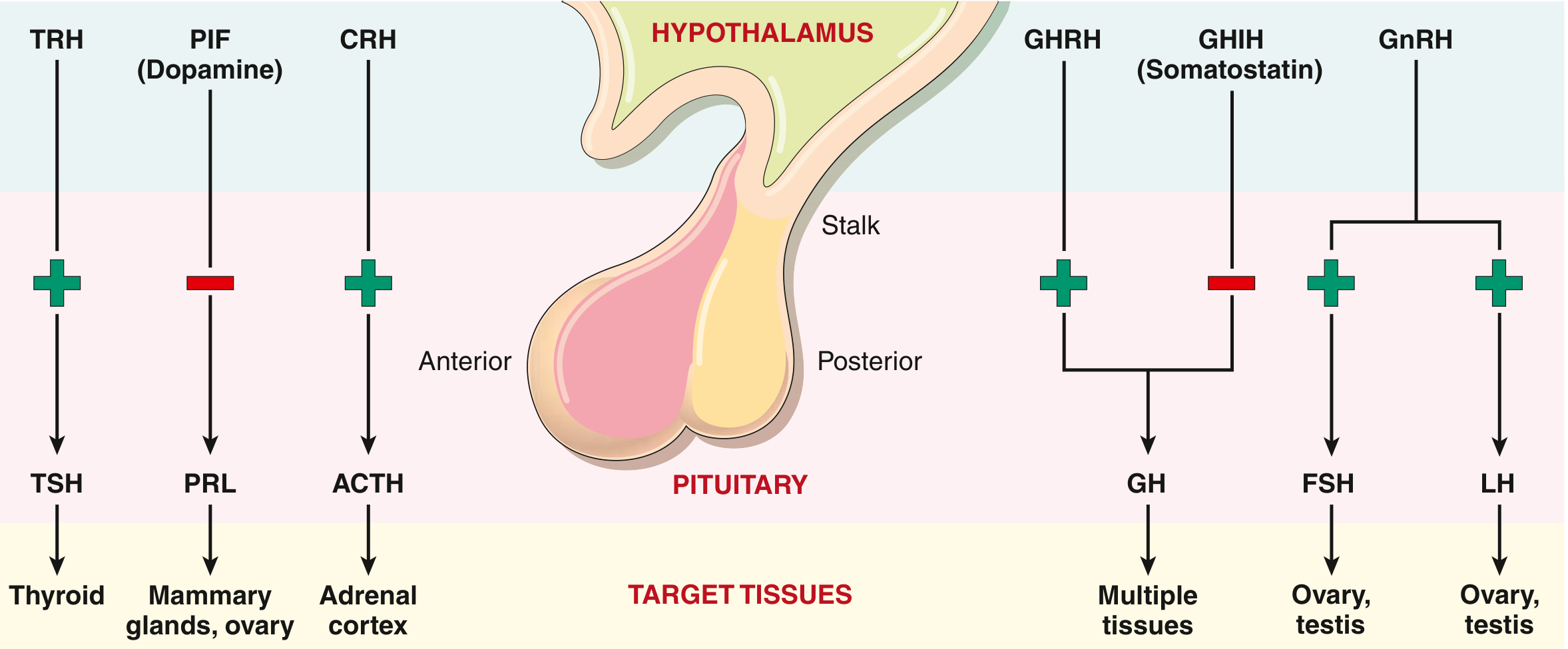
Robbins & Kumar Basic Pathology, pp. 723-724.
1. Clinical effects of pituitary disease
A. Hormone excess
- Excess PRL -> galactorrhea, amenorrhea
- Excess GH -> gigantism or acromegaly
- Excess ACTH -> Cushing disease
B. Hormone deficiency
C. Mass effect
- Headache
- Bitemporal hemianopia: loss of outer half of visual field of both eyes due to optic chiasma compression
- Nausea/vomiting from raised intracranial pressure
- Compression of normal pituitary -> hypopituitarism
Robbins & Kumar Basic Pathology, p. 724.
2. Pituitary adenoma / Pituitary neuroendocrine tumor (PitNET)
Definition
Classification
According to size
- Microadenoma: less than 1 cm
- Macroadenoma: more than 1 cm
According to function
- Functioning adenoma: secretes a hormone and produces a clinical syndrome
- Nonfunctioning adenoma: does not produce clinically obvious hormone excess; usually presents late as a large mass
- Silent adenoma: produces hormone demonstrable by immunohistochemistry but does not cause clinical hormonal excess
Pathogenesis
GNAS mutation -> increased cAMP signaling -> increased cell proliferation + increased hormone synthesisMorphology of pituitary adenoma
Gross appearance
- Usually soft, well-circumscribed tumor
- May expand the sella turcica
- Large tumors can compress the optic chiasma and normal pituitary tissue
- Large tumors may invade nearby structures
Microscopy
-
Monomorphic cells
Tumor cells look very similar to each other, unlike the normal anterior pituitary which contains a mixed population of acidophils, basophils, and chromophobes. -
Loss of reticulin network
Normal pituitary has a reticulin framework around cell nests. In adenoma, this framework is disrupted or absent.
One-line morphology answer: Pituitary adenoma shows a monotonous population of uniform pituitary cells with loss of normal reticulin architecture.
3. Functioning pituitary adenomas
A. Prolactinoma - lactotroph adenoma
Pathogenesis of symptoms
High prolactin -> low GnRH -> low FSH/LH -> hypogonadismClinical features in females
- Amenorrhea
- Oligomenorrhea
- Infertility
- Galactorrhea: milk secretion not related to normal breastfeeding
- Reduced libido
Clinical features in males
- Reduced libido
- Erectile dysfunction
- Infertility
- Decreased facial/body hair
- Gynecomastia may occur
- Often presents late with headache and visual problems because men may not notice hormonal symptoms early
Important point: stalk effect
Pituitary stalk compression -> less dopamine -> increased prolactinB. Somatotroph adenoma - GH-producing adenoma
GH excess -> increased IGF-1 -> abnormal tissue and bone growth1. Gigantism
- Marked increase in height
- Long limbs
- Generalized excessive growth
2. Acromegaly
- Enlarged hands and feet
- Ring and shoe size increase
- Coarse facial features
- Enlarged nose, lips, tongue
- Prognathism: protruding lower jaw
- Widely spaced teeth
- Thick oily skin and sweating
- Headache
- Hypertension
- Cardiomegaly and heart failure
- Arthritis
- Glucose intolerance or diabetes mellitus
- Increased risk of colonic neoplasia
Diagnosis
- Raised serum IGF-1
- GH does not suppress after an oral glucose load
- MRI of pituitary for tumor
| Gigantism | Acromegaly |
|---|---|
| Before epiphyseal closure | After epiphyseal closure |
| Increased height is prominent | Hands, feet, jaw, face enlarge |
| Child/adolescent | Adult |
C. Corticotroph adenoma - ACTH-producing adenoma
ACTH excess -> bilateral adrenal cortical hyperplasia -> cortisol excessCushing disease versus Cushing syndrome
- Cushing syndrome: clinical state due to excess cortisol from any cause
- Cushing disease: Cushing syndrome specifically due to a pituitary ACTH-secreting adenoma
Clinical features
- Central obesity
- Moon face
- Buffalo hump
- Thin skin and easy bruising
- Purple abdominal striae
- Proximal muscle weakness
- Hypertension
- Diabetes/glucose intolerance
- Osteoporosis
- Infections
- Depression or mood changes
- Menstrual irregularity
- Hyperpigmentation may occur with high ACTH
D. Thyrotroph adenoma - TSH-producing adenoma
- Hyperthyroidism
- Diffuse goitre
- Palpitations, sweating, weight loss, tremor
E. Gonadotroph adenoma - FSH/LH-producing adenoma
- Macroadenoma
- Headache
- Bitemporal hemianopia
- Hypopituitarism
4. Nonfunctioning pituitary adenoma
Clinical features
- Headache
- Bitemporal hemianopia
- Hypopituitarism due to compression of normal gland
- Mild hyperprolactinemia due to stalk effect
High-yield line: A nonfunctioning pituitary adenoma commonly presents with mass effect and hypopituitarism, not hormone excess.
5. Hypopituitarism
Definition
Causes
1. Tumors and mass lesions
- Nonfunctioning pituitary macroadenoma
- Craniopharyngioma
- Metastatic tumor
- Hypothalamic tumor
2. Ischemic necrosis
Sheehan syndrome
Why does it occur?
- Anterior pituitary enlarges, mainly due to lactotroph hyperplasia.
- Its blood supply does not increase proportionately.
- Severe postpartum hemorrhage or hypotension causes ischemia.
- Result: anterior pituitary necrosis.
Clinical features of Sheehan syndrome
- Failure of lactation after delivery due to low prolactin
- Amenorrhea
- Loss of pubic and axillary hair
- Weakness
- Hypothyroidism
- Adrenal insufficiency
3. Iatrogenic
- Pituitary surgery
- Radiotherapy
4. Inflammatory/infiltrative disease
- Tuberculosis
- Sarcoidosis
- Autoimmune hypophysitis
5. Trauma
- Head injury
6. Genetic causes
Clinical features according to deficient hormone
| Hormone deficient | Main clinical effect |
|---|---|
| GH | Growth failure in children |
| FSH/LH | Infertility, amenorrhea, low libido, impotence |
| TSH | Secondary hypothyroidism |
| ACTH | Secondary adrenal insufficiency |
| PRL | Failure of postpartum lactation |
| MSH | Skin pallor |
Important difference: primary vs secondary adrenal insufficiency
| Feature | Primary adrenal insufficiency | Secondary adrenal insufficiency due to low ACTH |
|---|---|---|
| ACTH | High | Low |
| Skin pigmentation | Increased | Pallor/no hyperpigmentation |
| Aldosterone | Often low | Usually preserved |
6. Posterior pituitary disorders
A. Central diabetes insipidus
Definition
Normal ADH action
ADH -> water reabsorption -> concentrated urineIn ADH deficiency
Low ADH -> less water reabsorption -> large volume of dilute urineCauses
- Head trauma
- Tumors of hypothalamus/pituitary
- Neurosurgery involving pituitary or hypothalamus
- Inflammatory disease
- Idiopathic disease
Clinical features
- Polyuria: excessive urine
- Polydipsia: excessive thirst
- Large volume of dilute urine
- Low urine specific gravity
- Hypernatremia
- Increased serum osmolality
- Severe dehydration if patient cannot access water
Central vs nephrogenic diabetes insipidus
| Feature | Central DI | Nephrogenic DI |
|---|---|---|
| Basic defect | ADH deficiency | Kidney does not respond to ADH |
| ADH level | Low | Normal or high |
| Cause | Hypothalamus/posterior pituitary lesion | Renal tubular resistance |
| Response to desmopressin | Improves | Little or no improvement |
B. SIADH - syndrome of inappropriate ADH secretion
Excess ADH -> water retention -> dilutional hyponatremia -> cerebral edemaCauses
- Small-cell carcinoma of lung producing ectopic ADH
- CNS disease
- Lung disease
- Hypothalamic/neurohypophyseal injury
Clinical features
- Hyponatremia
- Confusion
- Seizures
- Coma in severe cases
- Cerebral edema
- Usually no peripheral edema, despite increased total body water
7. Pituitary pathology - quick revision table
| Disease | Main defect | Key clinical clue |
|---|---|---|
| Prolactinoma | High PRL | Amenorrhea + galactorrhea + infertility |
| Somatotroph adenoma | High GH/IGF-1 | Acromegaly in adults; gigantism in children |
| Corticotroph adenoma | High ACTH | Cushing disease |
| Nonfunctioning adenoma | Mass effect | Headache + bitemporal hemianopia |
| Hypopituitarism | Low anterior pituitary hormones | Multiple hormonal deficiencies |
| Sheehan syndrome | Postpartum anterior pituitary necrosis | Failure to lactate after severe PPH |
| Central DI | Low ADH | Polyuria with dilute urine |
| SIADH | Excess ADH | Hyponatremia with neurologic symptoms |
8. Very important viva questions
-
What is the most common cause of hyperpituitarism?
Anterior pituitary adenoma. -
What is the most common functioning pituitary adenoma?
Prolactinoma. -
What is the difference between Cushing disease and Cushing syndrome?
Cushing disease is due to a pituitary ACTH adenoma. Cushing syndrome is cortisol excess from any cause. -
What visual defect occurs in pituitary macroadenoma?
Bitemporal hemianopia. -
What are the two microscopic features of pituitary adenoma?
Cellular monomorphism and loss of reticulin network. -
Why does Sheehan syndrome occur?
Postpartum hemorrhage/hypotension causes ischemic necrosis of the enlarged anterior pituitary. -
Which pituitary part is more vulnerable in Sheehan syndrome?
Anterior pituitary. -
What is central diabetes insipidus?
Polyuria due to ADH deficiency from hypothalamic or posterior pituitary disease. -
What is pituitary apoplexy?
Acute hemorrhage into a pituitary tumor causing sudden expansion and possible acute adrenal insufficiency.
2
thyroid disease pathology
thyroid disorders pathology Graves Hashimoto thyroid carcinoma Merck Manual Professional
Hashimoto thyroiditis histology Hürthle cells lymphoid follicles pathology
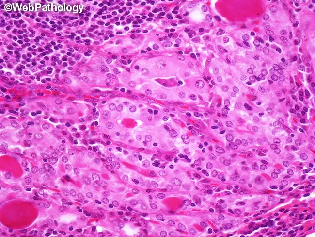
Bright-field light microscopy of thyroid tissue stained with Hematoxylin and Eosin demonstrates features of Hashimoto thyroiditis. The image shows diffuse lymphocytic infiltration with formation of large lymphoid follicles and well-demarcated germinal centers, consistent with chronic autoimmune thyroiditis. Residual thyroid follicles are small and atrophic, and follicular epithelial cells display prominent oxyphilic (Hurthle cell) change with abundant eosinophilic cytoplasm. Foci of squamous metaplasia and fibrous stromal expansion may be present, reflecting chronicity. The parenchymal architecture is disrupted by mononuclear cell infiltrates, including lymphocytes and plasma cells, with occasional residual colloid-containing follicles. These histologic features correlate with hypothyroid or fluctuating thyroid function and aid in distinguishing autoimmune thyroiditis from other thyroid disorders. Clinically, Hashimoto thyroiditis carries a risk for development of thyroid lymphoma, particularly extranodal marginal zone lymphoma; hence vigilance and multidisciplinary assessment are advised. This slide is a representative teaching example for pathology residents, endocrinology trainees, and head-and-neck oncologic surgeons. It is useful for differential diagnosis, immunohistochemical workup planning, and correlating histology with serology, imaging, and clinical presentation in autoimmune thyroid disease. This description supports researchers seeking biomarkers, educators designing case-based curricula, and clinicians integrating histology with ultrasound, fine-needle aspiration results, and serum antibodies to optimize patient management and therapeutic decisions.
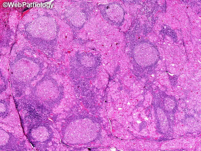
Light microscopy of thyroid tissue prepared with Hematoxylin and Eosin (H&E) staining shows features consistent with Hashimoto thyroiditis. The specimen arises from the thyroid gland in the cervical region. The slide demonstrates diffuse lymphocytic infiltration with formation of enlarged lymphoid follicles bearing prominent germinal centers, a hallmark of autoimmune thyroiditis. Interspersed are residual thyroid follicles that are small and atrophic, many lined by oncocytic (Hürthle) cells with abundant granular eosinophilic cytoplasm. The architectural disruption of normal thyroid parenchyma and the presence of germinal center-rich lymphoid infiltrates reflect chronic inflammation. These histologic features are classic for Hashimoto thyroiditis and are associated with an increased risk of secondary thyroid lymphoma, notably diffuse large B-cell lymphoma (DLBCL) comprising the majority of lymphomas in this setting, with mucosa-associated lymphoid tissue (MALT) lymphoma accounting for a smaller but meaningful fraction. The image highlights autoimmune etiologies and potential malignant transformation. Clinically, such histology supports autoimmune hypothyroidism in many patients and warrants surveillance for lymphoma development. Diagnostic significance includes correlation with serology (anti-thyroid peroxidase antibodies) and thyroid function tests, and may guide therapeutic decisions, including consideration of targeted biopsy in suspicious cases. These features aid educational teaching and differential diagnosis for pathology trainees in practice and research.
Thyroid gland pathology - MBBS 3rd year
- Follicular cells make thyroid hormones: T3 and T4
- Follicle lumen contains colloid, mainly thyroglobulin
- Parafollicular or C cells make calcitonin
Normal axis
Hypothalamus (TRH) -> Pituitary (TSH) -> Thyroid gland (T3, T4)Main thyroid disorders
- Hyperthyroidism
- Hypothyroidism
- Thyroiditis
- Hashimoto thyroiditis
- Subacute granulomatous thyroiditis
- Painless/postpartum thyroiditis
- Goitre
- Thyroid tumors
- Follicular adenoma
- Papillary carcinoma
- Follicular carcinoma
- Medullary carcinoma
- Anaplastic carcinoma
1. Hyperthyroidism / Thyrotoxicosis
Definition
Common causes
- Graves disease - most common
- Toxic multinodular goitre
- Toxic adenoma
- Thyroiditis, where preformed hormone leaks from damaged follicles
- Rarely, TSH-secreting pituitary adenoma
Clinical features
- Weight loss despite increased appetite
- Heat intolerance
- Sweating
- Warm, moist skin
- Palpitations and tachycardia
- Tremor
- Anxiety, irritability
- Hyperreflexia
- Diarrhea
- Proximal muscle weakness
- Menstrual disturbances
- In elderly patients: atrial fibrillation or heart failure may occur
T3/T4 high + TSH lowThyroid storm
- High fever
- Severe tachycardia/arrhythmia
- Delirium or coma
- Heart failure
Robbins & Kumar Basic Pathology, pp. 729-730.
2. Graves disease
Definition
Pathogenesis
TSH receptor antibody -> continuous stimulation of thyroid -> diffuse hyperplasia -> increased T3/T4Clinical triad
- Hyperthyroidism
- Diffuse goitre
- Ophthalmopathy
Clinical features
- Diffuse, soft thyroid enlargement
- Exophthalmos/proptosis
- Lid lag and staring look
- Diplopia due to eye muscle involvement
- Pretibial myxedema: thickened skin over shins
- Thyroid acropachy, rarely: clubbing and swelling of fingers
Why does exophthalmos occur?
- Glycosaminoglycan deposition
- Edema
- Fibrosis
- Enlargement of extraocular muscles and retro-orbital tissue
Morphology
Gross
- Diffuse, symmetrical enlargement of thyroid
- Soft, fleshy, red-brown gland
Microscopy
- Diffuse hypertrophy and hyperplasia of follicular cells
- Follicular lining cells become tall and crowded
- Papillary infoldings project into colloid
- Colloid becomes pale and shows scalloped margins due to active resorption
- Lymphoid infiltrate may be present
Important differentiation
| Graves disease | Toxic multinodular goitre |
|---|---|
| Autoimmune | Usually due to long-standing nodular goitre |
| Diffuse enlargement | Multiple nodules |
| TSH receptor antibodies present | Antibodies usually absent |
| Ophthalmopathy common | Ophthalmopathy absent |
| Pretibial myxedema can occur | Absent |
3. Hypothyroidism
Definition
Causes
Primary hypothyroidism: problem is in thyroid gland
- Hashimoto thyroiditis
- Iodine deficiency
- Surgical removal of thyroid
- Radioiodine therapy
- Drugs such as lithium
- Congenital thyroid dysgenesis
- Dyshormonogenesis
Secondary hypothyroidism: pituitary failure
- Low TSH due to pituitary disease
Tertiary hypothyroidism: hypothalamic failure
- Low TRH
Laboratory pattern
| Type | T3/T4 | TSH |
|---|---|---|
| Primary hypothyroidism | Low | High |
| Secondary/tertiary hypothyroidism | Low | Low or inappropriately normal |
Clinical features in adults: myxedema
- Fatigue and lethargy
- Weight gain
- Cold intolerance
- Constipation
- Dry, coarse skin
- Puffy face
- Hair loss
- Slow speech and thinking
- Depression
- Bradycardia
- Reduced cardiac output
- Raised cholesterol
- Menstrual irregularities
- Hoarse voice
Congenital hypothyroidism
- Severe intellectual disability
- Growth retardation
- Short stature
- Coarse facial features
- Large protruding tongue
- Umbilical hernia
Robbins & Kumar Basic Pathology, pp. 730-731.
4. Thyroiditis
A. Hashimoto thyroiditis
Definition
Pathogenesis
- Anti-thyroid peroxidase antibodies: anti-TPO
- Anti-thyroglobulin antibodies
- CD8+ cytotoxic T-cell-mediated injury
- Cytokine-mediated damage
- Antibody/complement-related injury
Autoimmune response -> follicular destruction -> reduced T3/T4 -> raised TSH -> hypothyroidismMorphology
Gross
- Usually diffuse, painless, symmetrical thyroid enlargement initially
- In late stages, gland may become small and fibrotic
Microscopy - very important
- Dense lymphocytic and plasma-cell infiltration
- Lymphoid follicles with germinal centres
- Atrophic thyroid follicles
- Hürthle cell change
- Interstitial fibrosis
Hürthle cells
- Large size
- Abundant granular eosinophilic cytoplasm
- Many mitochondria
Clinical features
- Painless goitre
- Gradually developing hypothyroidism
- Fatigue and weight gain
- Cold intolerance
- Constipation
- Raised anti-TPO antibody levels
- May have a short early thyrotoxic phase due to leakage of stored hormones
Complications
- Permanent hypothyroidism
- Increased risk of B-cell lymphoma, especially MALT lymphoma of thyroid
One-line exam answer: Hashimoto thyroiditis is an autoimmune destruction of the thyroid characterized by lymphoid follicles with germinal centres, Hürthle-cell metaplasia, follicular atrophy, and fibrosis.
B. Subacute granulomatous thyroiditis
Cause
Clinical features
- Painful, tender thyroid swelling
- Fever and malaise
- Neck pain may radiate to jaw or ear
- Initially transient hyperthyroidism due to release of stored hormone
- Later temporary hypothyroidism may occur
- Usually recovers completely
Microscopy
- Disruption of thyroid follicles
- Colloid leakage
- Granulomatous inflammation
- Multinucleated giant cells surrounding colloid
Key difference from Hashimoto: de Quervain thyroiditis is painful and shows granulomas with giant cells. Hashimoto is painless and shows lymphoid follicles with Hürthle cells.
C. Painless/postpartum thyroiditis
- Autoimmune thyroiditis
- Painless thyroid enlargement
- Often occurs postpartum in women
- Transient hyperthyroid phase followed by hypothyroid phase
- Lymphocytic infiltration but little/no Hürthle cell change compared with Hashimoto
5. Goitre
Definition
A. Diffuse nontoxic goitre
Mechanism
Low thyroid hormone production -> increased TSH -> thyroid hyperplasia and enlargementB. Multinodular goitre
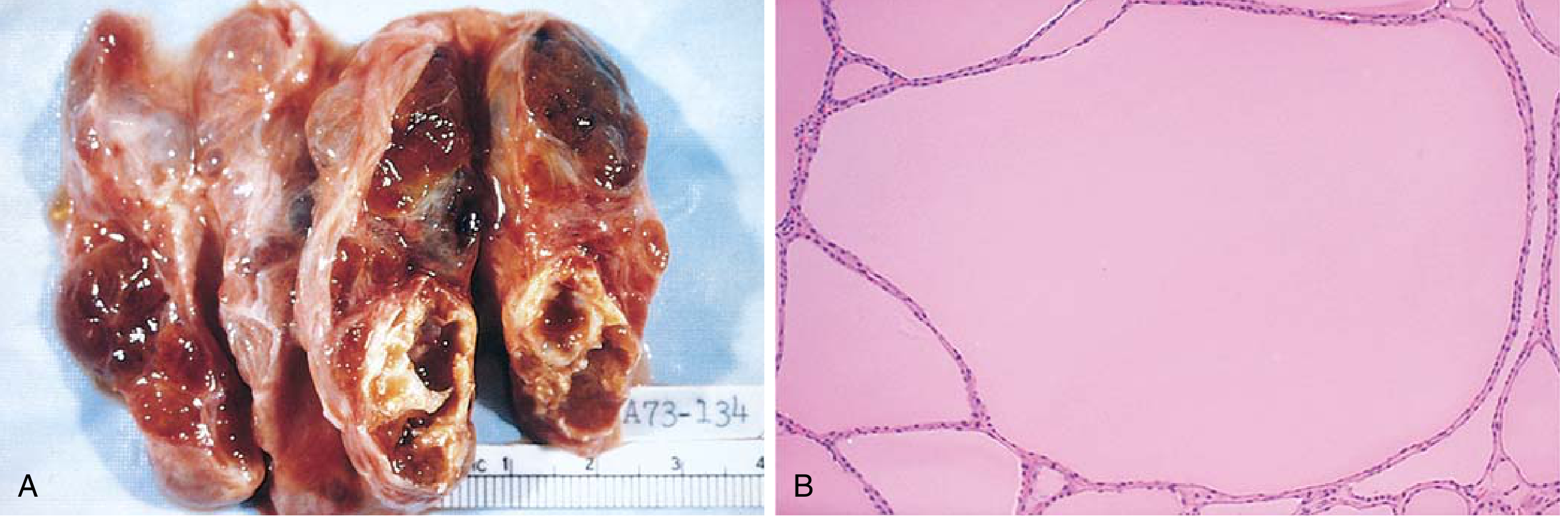
Morphology
Gross
- Enlarged gland with multiple nodules
- Areas of hemorrhage, cystic degeneration, fibrosis, and calcification may occur
Microscopy
- Follicles vary in size
- Some follicles are dilated and filled with colloid
- Others are hyperplastic
- Fibrosis, hemorrhage, and cystic changes may be seen
Clinical features
- Slowly enlarging neck mass
- Usually euthyroid initially
- Cosmetic concern
- Compression symptoms in large goitre:
- Dysphagia
- Dyspnea
- Hoarseness
- Superior vena cava obstruction, rarely
- May become toxic and cause hyperthyroidism: toxic multinodular goitre
6. Thyroid neoplasms
Important clinical clues suggesting malignancy in thyroid nodule
- Solitary nodule
- Nodule in male patient
- Nodule in child or young person
- History of neck irradiation
- Rapid increase in size
- Hoarseness or vocal cord palsy
- Cervical lymph node enlargement
- Cold nodule on radioactive iodine scan
Robbins & Kumar Basic Pathology, pp. 734-735.
A. Follicular adenoma
Definition
Morphology
- Solitary, well-circumscribed, encapsulated lesion
- Uniform follicles containing colloid
- Compresses adjacent thyroid tissue
- No capsular invasion
- No vascular invasion
Important point
7. Thyroid carcinomas
| Type | Cell of origin | Main spread | Key clue |
|---|---|---|---|
| Papillary carcinoma | Follicular cells | Lymphatics | Orphan Annie eye nuclei, nuclear grooves, psammoma bodies |
| Follicular carcinoma | Follicular cells | Blood | Capsular/vascular invasion |
| Medullary carcinoma | C cells | Lymph nodes/blood | Calcitonin, amyloid stroma, RET/MEN 2 |
| Anaplastic carcinoma | Follicular cells | Local invasion/metastasis | Very aggressive tumor in elderly |
A. Papillary thyroid carcinoma
Risk factors
- Ionizing radiation, especially in childhood
- Genetic alterations involving:
- BRAF
- RET/PTC rearrangements
- NTRK fusions
- RAS mutations
Morphology
Gross
- Solitary or multifocal tumor
- May be well circumscribed or infiltrative
Microscopy - very important
- Papillae with fibrovascular cores
- Orphan Annie eye nuclei or ground-glass nuclei
- Nuclear grooves
- Intranuclear cytoplasmic inclusions
- Psammoma bodies: laminated calcified bodies
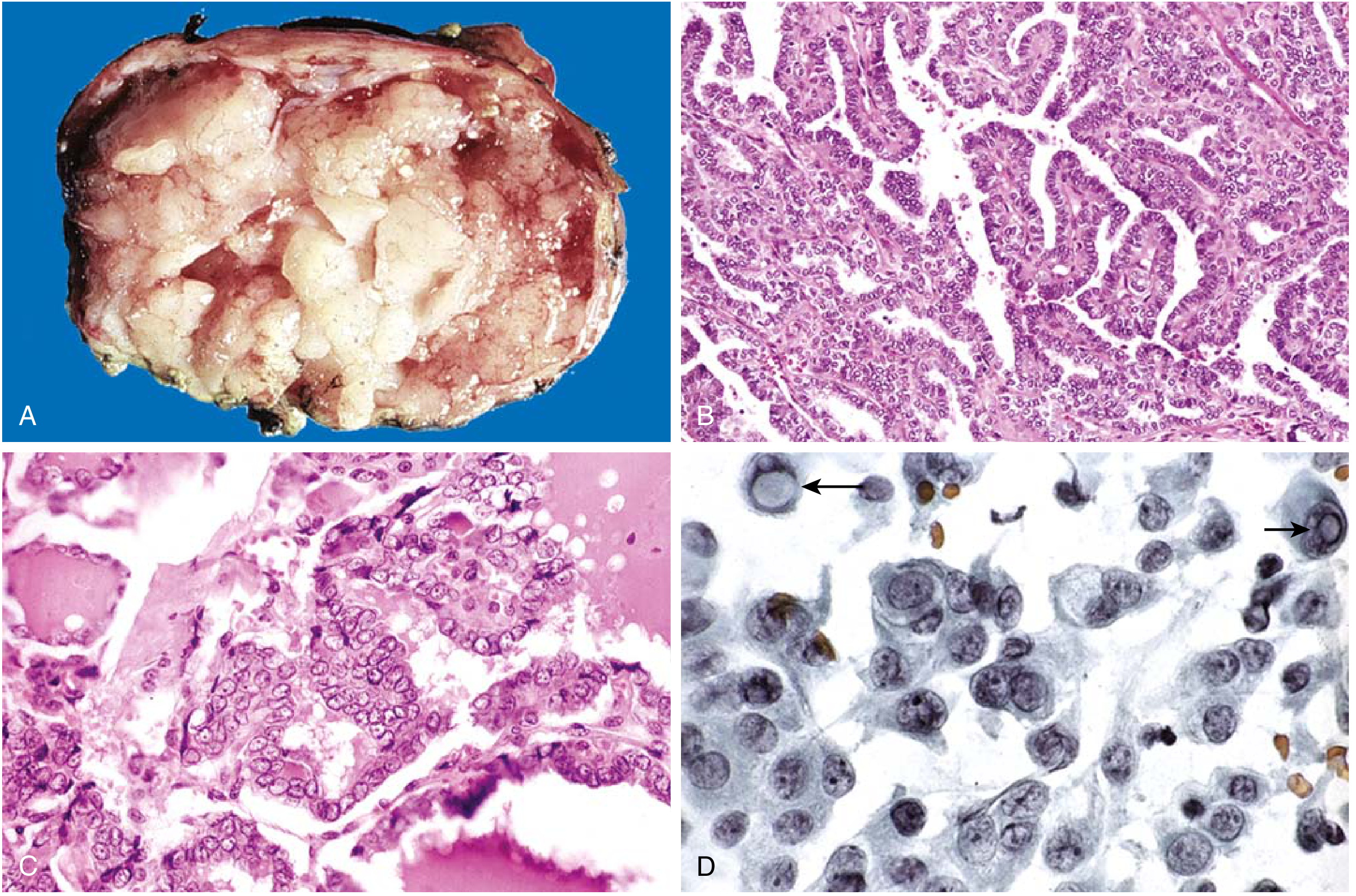
Clinical features
- Painless thyroid nodule
- Usually euthyroid
- Cervical lymph-node metastasis may be present
- Spreads mainly by lymphatics
Prognosis
Most important diagnostic feature: nuclear characteristics, not necessarily papillary architecture.
B. Follicular thyroid carcinoma
Definition
Pathogenesis
- RAS mutations
- PI3K/AKT pathway mutations
- PAX8-PPARG rearrangement
Morphology
- Capsular invasion
- Vascular invasion
Spread
- Bone
- Lungs
- Liver
Viva point: Papillary carcinoma spreads through lymphatics. Follicular carcinoma spreads hematogenously.
C. Medullary thyroid carcinoma
Origin
Types
- Sporadic
- Familial, associated with MEN 2A and MEN 2B
Morphology
- Nests, trabeculae, or sheets of tumor cells
- Amyloid deposits in stroma, derived from altered calcitonin molecules
- Amyloid is Congo-red positive
Clinical features
- Thyroid mass
- Raised calcitonin
- Diarrhea or flushing may occur due to hormone secretion
- In MEN 2, evaluate for pheochromocytoma and hyperparathyroidism, depending on subtype
High-yield line: Medullary carcinoma arises from C cells, secretes calcitonin, shows amyloid in stroma, and may be associated with RET mutation/MEN 2.
D. Anaplastic thyroid carcinoma
Definition
Clinical features
- Rapidly enlarging neck mass
- Dyspnea
- Dysphagia
- Hoarseness
- Early local invasion
- Distant metastasis
Morphology
- Highly pleomorphic cells
- Spindle cells and giant cells
- Numerous mitoses
- Necrosis
- No useful thyroid differentiation
Prognosis
Quick revision: thyroid pathology
| Disease | Key cause/pathogenesis | Key pathology finding | Important clue |
|---|---|---|---|
| Graves disease | TSH receptor-stimulating antibodies | Diffuse hyperplasia, scalloped colloid | Hyperthyroidism + eye signs |
| Hashimoto thyroiditis | Autoimmune follicular destruction | Lymphoid follicles, Hürthle cells | Painless goitre + hypothyroidism |
| de Quervain thyroiditis | Viral/postviral inflammation | Granulomas with giant cells | Painful tender thyroid |
| Multinodular goitre | Repeated TSH-driven hyperplasia/involution | Multiple nodules, colloid, hemorrhage, fibrosis | Large irregular thyroid |
| Follicular adenoma | Benign follicular neoplasm | Encapsulated follicular lesion | No invasion |
| Papillary carcinoma | BRAF/RET/NTRK pathway abnormalities | Orphan Annie nuclei, grooves, psammoma bodies | Lymphatic spread |
| Follicular carcinoma | RAS/PI3K pathway changes | Capsular/vascular invasion | Blood spread to bone/lung |
| Medullary carcinoma | C-cell tumor, RET in familial cases | Amyloid stroma | Calcitonin, MEN 2 |
| Anaplastic carcinoma | Highly undifferentiated malignancy | Pleomorphism, giant cells, necrosis | Elderly, rapidly enlarging mass |
Most asked viva questions
-
Most common thyroid carcinoma?
Papillary thyroid carcinoma. -
Most common cause of hypothyroidism in iodine-sufficient areas?
Hashimoto thyroiditis. -
Most common cause of hypothyroidism worldwide?
Iodine deficiency. -
Characteristic cells of Hashimoto thyroiditis?
Hürthle cells. -
Histological hallmark of papillary carcinoma?
Orphan Annie eye nuclei, nuclear grooves, intranuclear inclusions, and psammoma bodies. -
How is follicular carcinoma distinguished from follicular adenoma?
By capsular and/or vascular invasion. -
Which thyroid cancer spreads by blood?
Follicular carcinoma. -
Which thyroid cancer is associated with calcitonin and amyloid?
Medullary thyroid carcinoma. -
Which thyroid cancer is associated with MEN 2?
Medullary thyroid carcinoma. -
Which thyroiditis is painful?
Subacute granulomatous/de Quervain thyroiditis.
3
hyperparathyroidism pathology
hyperparathyroidism hypoparathyroidism pathology Merck Manual Professional
parathyroid adenoma histology normal rim pathology
osteitis fibrosa cystica brown tumor hyperparathyroidism bone pathology
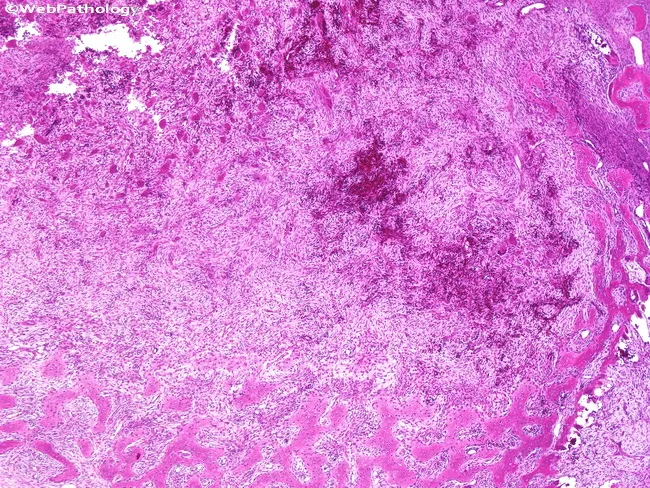
This histopathology image depicts an osteitis fibrosa cystica (brown tumor) within bone in the setting of hyperparathyroidism. The center shows a zone of bone resorption with loss of normal trabecular architecture, replaced by a fibrous, cellular stroma. The lesion is filled with fibrous tissue containing spindle-shaped fibroblasts and occasional osteoclast-type giant cells and hemorrhagic debris; the periphery demonstrates osteoid production forming an immature woven bone matrix with a fibrous dysplasia-like appearance. The overall pattern includes a mixture of radiating fibrous tissue and newly formed osteoid at the margins, which mimics fibrous dysplasia. The histologic spectrum reflects exuberant bone remodeling driven by excess parathyroid hormone, with interspersed areas of osteoclast resorption and fibrous tissue that may produce cystic spaces. Clinically, such brown tumors are part of osteitis fibrosa cystica and signal advanced hyperparathyroidism; identification on biopsy prompts evaluation of parathyroid function and calcium-phosphate homeostasis. Differential considerations include fibrous dysplasia, giant cell-rich lesions, and osseous metastases; however, the association with systemic hyperparathyroidism and parathyroid dysfunction supports the diagnosis. This image is relevant for medical students, pathology residents, and clinicians assessing metabolic bone disease, surgical planning, and educational case reviews.
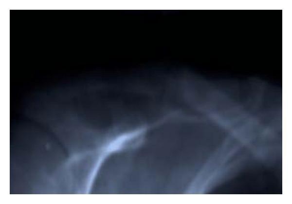
This anteroposterior (AP) radiograph of the right shoulder region demonstrates significant skeletal pathology of the clavicle. The image reveals a focal, expansile cystic lesion characterized by a well-defined area of radiolucency (decreased bone density) within the shaft of the clavicle. This lesion, consistent with osteitis fibrosa cystica (brown tumor), has caused marked thinning of the overlying cortical bone. A clear discontinuity in the cortical margin is visible through the lesion, signifying a pathologic fracture. The surrounding bone shows generalized osteopenia. This finding is a classic skeletal manifestation of primary hyperparathyroidism, where excessive parathyroid hormone leads to increased osteoclastic activity and the replacement of bone marrow with fibrous tissue and cystic vascular spaces.
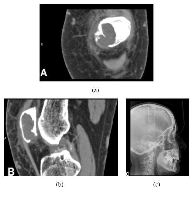
This composite of diagnostic images illustrates systemic skeletal manifestations of primary hyperparathyroidism. Images (a) and (b) are axial and sagittal CT scans of the left knee, revealing a prominent osteolytic, cyst-like lesion within the patella. The lesion exhibits a polylobate internal structure, regular borders, significant cortical thinning, and localized erosion, characteristic of a brown tumor. Image (c) is a lateral skull radiograph (X-ray) demonstrating the classic 'salt-and-pepper' appearance of the cranial vault. This sign is produced by multiple small, diffuse, punctate lucencies caused by trabecular resorption and replacement with fibrous tissue. Together, these findings highlight the generalized demineralization and focal bone lesions associated with hyperparathyroid-induced osteitis fibrosa cystica. The educational focus is on identifying systemic radiologic correlates of parathyroid pathology beyond common oral manifestations.
Parathyroid gland pathology - MBBS 3rd year
Main function of PTH
Low calcium -> increased PTH -> blood calcium rises- Bone: increases bone resorption, releasing calcium.
- Kidney: increases calcium reabsorption, so less calcium is lost in urine.
- Kidney: increases phosphate excretion, so serum phosphate falls.
- Vitamin D activation: activates vitamin D in kidney, increasing intestinal calcium absorption.
Net effect of PTH
| Effect | Result |
|---|---|
| Serum calcium | Increased |
| Serum phosphate | Decreased |
| Urinary phosphate | Increased |
| Bone resorption | Increased |
1. Hyperparathyroidism
Definition
High PTH -> hypercalcemia + hypophosphatemia + increased bone resorption- Primary hyperparathyroidism
- Secondary hyperparathyroidism
- Tertiary hyperparathyroidism
2. Primary hyperparathyroidism
Definition
Causes
| Cause | Approximate frequency |
|---|---|
| Parathyroid adenoma | 85% to 95% |
| Parathyroid hyperplasia | 5% to 10% |
| Parathyroid carcinoma | About 1% |
A. Parathyroid adenoma
Pathogenesis
- Cyclin D1 overexpression
- MEN1 mutation
Morphology
Gross
- Solitary
- Well-circumscribed
- Soft, tan or reddish-brown nodule
- Usually surrounded by a thin capsule
- Other parathyroid glands are normal or atrophic due to suppression by high calcium
Microscopy
- Mainly proliferation of chief cells
- Little or absent stromal fat
- A rim of compressed normal parathyroid tissue may be present at the edge
- Mild nuclear pleomorphism, called endocrine atypia, may occur but does not mean cancer
- Mitoses are rare
High-yield difference: Parathyroid adenoma is usually a single-gland lesion, while parathyroid hyperplasia usually involves multiple glands.
B. Parathyroid hyperplasia
Morphology
- Usually all four glands are enlarged
- May be diffuse or nodular
- Chief-cell hyperplasia is common
- Stromal fat is reduced
Association
- MEN 1
- MEN 2A
- Familial hyperparathyroidism
C. Parathyroid carcinoma
Clinical clue
Morphology
- Large, firm gray-white mass
- Thick fibrous capsule
- Cells may look rather uniform, so cytology alone cannot reliably diagnose malignancy
Definitive evidence of carcinoma
- Invasion into surrounding tissue
- Vascular invasion
- Metastasis
Do not diagnose parathyroid carcinoma only by pleomorphism. Invasion or metastasis is needed.
Clinical features of primary hyperparathyroidism
Stones, bones, abdominal groans, and psychic moans
1. Renal features: stones
- Hypercalciuria
- Renal stones: nephrolithiasis
- Calcium deposition in kidney: nephrocalcinosis
- Renal impairment in severe disease
2. Skeletal features: bones
- Bone pain
- Osteoporosis
- Fractures
- Osteitis fibrosa cystica
- Brown tumors
3. Gastrointestinal features: abdominal groans
- Constipation
- Abdominal pain
- Peptic ulcer disease
- Pancreatitis, occasionally
4. Neuropsychiatric features: psychic moans
- Fatigue
- Weakness
- Depression
- Poor concentration
- Confusion, especially in severe hypercalcemia
Laboratory findings
| Investigation | Finding in primary hyperparathyroidism |
|---|---|
| PTH | High |
| Serum calcium | High |
| Serum phosphate | Low |
| Urinary calcium | Usually high |
| Alkaline phosphatase | May be high if bone disease is active |
Bone pathology in hyperparathyroidism
Osteitis fibrosa cystica
- Cortical bone thinning
- Increased osteoclastic resorption
- Marrow replaced by fibrous tissue
- Hemorrhage and cyst-like spaces
- Bone becomes weak and prone to fracture
Brown tumor
- Osteoclast-type giant cells
- Fibrous tissue
- Hemorrhage
- Hemosiderin pigment, which gives the brown color
One-line answer: Brown tumors are giant-cell-rich, hemorrhagic lesions of osteitis fibrosa cystica caused by prolonged hyperparathyroidism.
3. Secondary hyperparathyroidism
Definition
Most common cause
Pathogenesis in chronic renal failure
-
Kidney cannot excrete phosphate properly
CKD -> phosphate retention -> high serum phosphate -
Phosphate binds calcium
High phosphate -> low free calcium -
Diseased kidneys cannot activate vitamin D adequately
Low active vitamin D -> reduced intestinal calcium absorption -
Low calcium stimulates parathyroid glands
Low calcium -> increased PTH -> parathyroid hyperplasia
Simple flowchart
Chronic renal failure -> phosphate retention + low active vitamin D -> hypocalcemia -> increased PTH -> secondary hyperparathyroidismOther causes
- Vitamin D deficiency
- Intestinal malabsorption
- Low dietary calcium
Laboratory findings in renal secondary hyperparathyroidism
| Investigation | Finding |
|---|---|
| PTH | High |
| Calcium | Low or normal |
| Phosphate | High in CKD |
| Vitamin D | Low |
| Alkaline phosphatase | Often high |
Morphology
- Usually involves all parathyroid glands
- Diffuse or nodular chief-cell hyperplasia
- Reduced fat in glands
Skeletal lesion
- Osteitis fibrosa cystica
- Osteomalacia due to vitamin D deficiency
- Osteosclerosis
- Growth retardation in children
4. Tertiary hyperparathyroidism
Definition
Flowchart
Long-standing CKD -> secondary hyperparathyroidism -> persistent gland hyperplasia -> autonomous PTH secretion -> tertiary hyperparathyroidismLaboratory findings
| Condition | PTH | Calcium | Phosphate |
|---|---|---|---|
| Primary hyperparathyroidism | High | High | Low |
| Secondary HPT due to CKD | High | Low/normal | High |
| Tertiary hyperparathyroidism | Very high | High | Often high in CKD |
5. Hypoparathyroidism
Definition
Low PTH -> hypocalcemia + hyperphosphatemiaCauses
-
Accidental removal or damage during thyroid surgery
Most common acquired cause. -
Autoimmune destruction of parathyroid glands
-
Congenital absence or underdevelopment
Example: DiGeorge syndrome -
Radiation injury
-
Severe magnesium deficiency can impair PTH secretion/action
Clinical features
- Tingling around mouth
- Tingling in fingers and toes
- Muscle cramps
- Carpopedal spasm
- Tetany
- Seizures
- Laryngospasm in severe cases
Clinical signs
Chvostek sign
Trousseau sign
Laboratory findings
| Investigation | Finding in hypoparathyroidism |
|---|---|
| PTH | Low |
| Serum calcium | Low |
| Serum phosphate | High |
| Alkaline phosphatase | Usually normal |
Primary versus secondary hyperparathyroidism
| Feature | Primary HPT | Secondary HPT |
|---|---|---|
| Basic problem | Autonomous parathyroid disease | Chronic low calcium stimulus |
| Common cause | Single parathyroid adenoma | Chronic kidney disease |
| PTH | High | High |
| Calcium | High | Low or normal |
| Phosphate | Low | High in CKD |
| Gland involvement | Usually one gland in adenoma | Usually all glands hyperplastic |
| Bone lesion | Osteitis fibrosa cystica, brown tumors | Renal osteodystrophy |
Short notes for exams
Write a short note on parathyroid adenoma
Write a short note on secondary hyperparathyroidism
Viva questions
-
What is the main function of PTH?
To increase ionized serum calcium. -
Commonest cause of primary hyperparathyroidism?
Parathyroid adenoma. -
Commonest cause of secondary hyperparathyroidism?
Chronic kidney disease. -
What is the difference between primary and tertiary hyperparathyroidism?
Primary is autonomous PTH secretion due to primary gland disease. Tertiary occurs when long-standing secondary hyperparathyroidism becomes autonomous. -
What are brown tumors?
Giant-cell-rich, hemorrhagic lesions in severe hyperparathyroidism. They are not true tumors. -
Which parathyroid cells produce PTH?
Chief cells. -
Most common cause of acquired hypoparathyroidism?
Accidental damage/removal during thyroid surgery. -
Laboratory findings in hypoparathyroidism?
Low PTH, low calcium, high phosphate. -
Which gene is associated with familial parathyroid tumors?
MEN1. -
What confirms parathyroid carcinoma?
Invasion of adjacent tissues or metastasis.