
10 mark answer for odontome
odontome odontoma compound complex radiograph histology
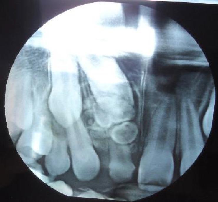
This intraoral periapical radiograph of the anterior maxilla demonstrates a complex odontoma associated with an impacted permanent tooth. The diagnostic image reveals a cluster of multiple, small, irregularly shaped radiopaque masses of varying densities located superior to the crown of an unerupted permanent maxillary incisor. These odontogenic structures act as a physical obstruction, resulting in the failure of eruption of the permanent tooth and the prolonged retention of its deciduous predecessor. Additionally, the radiograph shows mesial drifting of the adjacent lateral incisor, narrowing the eruption space. The visual evidence of calcified dental tissues in a disorganized arrangement is characteristic of a compound or complex odontoma, a common benign odontogenic hamartoma. This image is clinically significant for dental students and maxillofacial radiologists in identifying causes of delayed tooth eruption and interpreting odontogenic tumors in pediatric patients.
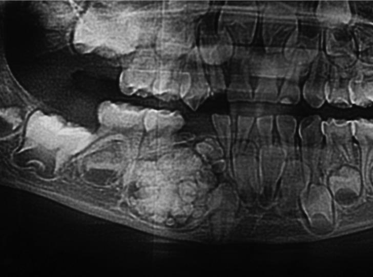
This diagnostic image is a cropped panoramic dental radiograph demonstrating a compound odontoma in the mandible. The lesion presents as a well-defined, heterogeneous radiopaque mass characterized by a cluster of multiple small, tooth-like structures (denticles) of varying sizes and shapes. These denticles exhibit radiodensity similar to natural enamel and dentin. The odontoma is encased in a thin radiolucent rim or halo, which separates it from the adjacent trabecular bone. Clinical significance is illustrated by the lesion's mass effect, which has caused the displacement and impaction of neighboring permanent teeth, interfering with their normal eruptive pathway. There is evidence of mild cortical bone thinning and localized bone remodeling at the periphery of the lesion. This radiographic finding is a hallmark of benign odontogenic tumors, specifically compound odontomas, typically found in the anterior maxilla or posterior mandible. The image is an essential educational resource for oral and maxillofacial radiology, highlighting the characteristic 'bag of teeth' appearance used to differentiate it from complex odontomas.
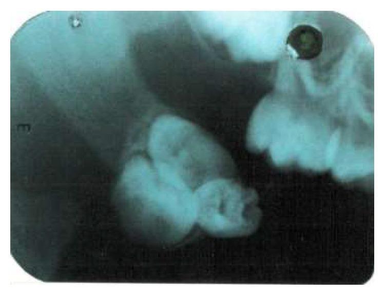
This diagnostic image is an intraoral periapical radiograph demonstrating a compound odontoma. The primary focus is a cluster of three distinct, radiopaque tooth-like masses (denticles) of varying ovoid and irregular shapes, mimicking the morphology of miniature teeth. These radiopaque structures are well-defined and grouped closely together. Encircling the entire mass is a thin, uniform radiolucent rim, which represents the follicular space or fibrous capsule of the lesion. The lesion is situated within the soft tissues, specifically described in clinical context as the posterior aspect of the buccal mucosa, showing a lack of direct attachment to the alveolar bone or adjacent primary dentition visible in the periphery. This radiographic presentation is a classic hallmark of a compound odontoma, an odontogenic hamartoma composed of organized dental tissues (enamel, dentin, and pulp). The image serves as an educational example for oral and maxillofacial pathology, illustrating the diagnostic differentiation between compound and complex odontomas based on morphological organization.
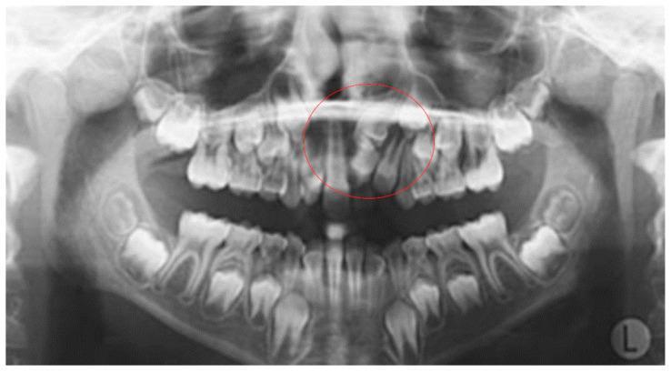
This panoramic radiograph (orthopantomogram) displays a pediatric patient in the mixed dentition stage, featuring a well-defined odontogenic tumor in the anterior maxilla. Circled in red, the lesion is characterized as a compound or complex odontoma located in the region of the maxillary left central and lateral incisors. The mass presents as a cluster of heterogeneous radiopaque structures with varying densities, some resembling rudimentary tooth elements (denticles) with enamel-like and dentin-like radiopacity. Clinically, the odontoma is causing significant local dental interference, specifically the impaction and displacement of the adjacent permanent maxillary incisors. The surrounding dentition shows normal developmental progress for the age group, with visible permanent tooth buds in the mandible and maxilla. The alveolar bone height and quality appear within normal limits outside the pathological site. This diagnostic image serves as a classic educational example of how odontomas act as local etiological factors for tooth impaction and ectopic eruption during dental development.
Odontome (Odontoma)
Definition
- Scott-Brown's Otorhinolaryngology Head & Neck Surgery, p. 458
- K J Lee's Essential Otolaryngology
Classification
1. Compound Odontome
- Dental tissues are arranged in an orderly fashion resembling miniature teeth (called denticles or odontoids)
- Has recognizable tooth morphology
2. Complex Odontome
- Dental tissues (enamel, dentine, cementum) are present but in a completely disorganized, haphazard mass
- No resemblance to tooth structure
Incidence and Age
- One of the most common odontogenic hamartomas/tumours
- Occur predominantly in children and adolescents (10-30 years of age)
- No sex predilection
- Compound type: more common in the anterior maxilla
- Complex type: more common in the posterior mandible
Etiopathogenesis
- Local trauma or infection
- Hereditary factors (multiple odontomes can be part of Gardner syndrome - an autosomal dominant condition)
- Developmental disturbances during odontogenesis
Clinical Features
Compound Odontome
- Mostly asymptomatic; discovered incidentally on routine dental X-rays
- May cause failure of eruption of a nearby tooth (most common presenting complaint)
- May cause displacement of erupted adjacent teeth
- Growth ceases once fully mature (generally reaches 1-2 cm in diameter)
- Jaw swelling is unusual
Complex Odontome
- Also common in children and adolescents
- Tends to grow larger: 3-6 cm in diameter
- Presents as a slowly increasing, painless swelling
- Associated with impaired eruption or displacement of adjacent teeth
- Located most often in the posterior mandible
Radiographic Features
Compound Odontome
- Multiple small tooth-like radiopaque structures (denticles) clustered together
- Surrounded by a thin radiolucent rim (fibrous capsule)
- Classic "bag of teeth" appearance
- Well-demarcated lesion
Complex Odontome
- A single radiopaque mass with no resemblance to tooth structure
- Typical mature appearance: spherical/ovoid radiopacity with a radiating periphery
- Surrounded by a radiolucent zone (fibrous capsule)
- Well-demarcated
- Immature lesions may initially appear radiolucent with focal radiopacities that increase as maturation proceeds
Histopathological Features
Compound Odontome
- Well-formed denticles enclosed in a fibrous sac
- Denticles contain enamel, dentine, cementum, and pulp in an organized, tooth-like arrangement
- Variable amounts of odontogenic epithelium, dental papilla, and reduced enamel epithelium depending on maturity
- Immature lesions show dentinoid, cementoid, and immature enamel matrix
Complex Odontome
- Variable amounts of enamel, dentine, and cementum arranged haphazardly
- Surrounded and supported by a collagenous capsule containing strands and islands of odontogenic epithelium
- Developing lesions contain more unmineralized dental tissue and cellular soft tissue areas
- Irregular tooth-like structures in an enamel matrix (from K J Lee's)
Note: The distinction between compound and complex odontome is not always clear-cut. Immature complex odontomes may contain ameloblastic fibroma (AF)-like areas.
Associations / Special Forms
| Form | Feature |
|---|---|
| Dens-in-dente (dilated odontome) | Tooth folded on itself; obvious on X-ray |
| Gemination | Attempted division of a single tooth germ |
| Taurodont | Enlarged pulp chamber, apically displaced furcation |
| Compound odontome + Gardner syndrome | Multiple compound odontomes; autosomal dominant; associated with colorectal polyposis, epidermoid cysts, osteomas |
Differential Diagnosis
- Ameloblastic fibro-odontoma (AFO) - a true neoplasm; has both epithelial and mesenchymal components; can be destructive
- Cementoma
- Osteoma
- Complex odontome vs. compound odontome - based on radiographic and histologic organization
Treatment
- Enucleation (surgical removal with its fibrous capsule) - the treatment of choice for both types
- Compound odontome: enucleation is curative; recurrence does not occur
- Complex odontome: generally curative but recurrence may follow incomplete removal of an immature lesion
- Unerupted associated teeth may be managed with orthodontic intervention post-removal if root development is adequate
- No need for segmental resection
Prognosis
- Excellent for compound odontome - no recurrence after complete enucleation
- Good for complex odontome - recurrence rare if complete removal achieved
- Being hamartomas, they do not undergo malignant transformation (unlike ameloblastic fibro-odontoma which rarely may)
Summary Table
| Feature | Compound Odontome | Complex Odontome |
|---|---|---|
| Nature | Hamartoma | Hamartoma |
| Dental tissue arrangement | Organized (denticles) | Disorganized (mass) |
| Common site | Anterior maxilla | Posterior mandible |
| Size | Up to 1-2 cm | 3-6 cm |
| Radiograph | "Bag of teeth" | Solid radiopaque mass |
| Growth | Ceases on maturity | Slowly grows |
| Recurrence | None | Rare |
| Treatment | Enucleation | Enucleation |
10 mark answer for odontome from neelima malik
odontome compound complex odontoma radiograph histology oral surgery
This diagnostic image is a cropped panoramic dental radiograph demonstrating a compound odontoma in the mandible. The lesion presents as a well-defined, heterogeneous radiopaque mass characterized by a cluster of multiple small, tooth-like structures (denticles) of varying sizes and shapes. These denticles exhibit radiodensity similar to natural enamel and dentin. The odontoma is encased in a thin radiolucent rim or halo, which separates it from the adjacent trabecular bone. Clinical significance is illustrated by the lesion's mass effect, which has caused the displacement and impaction of neighboring permanent teeth, interfering with their normal eruptive pathway. There is evidence of mild cortical bone thinning and localized bone remodeling at the periphery of the lesion. This radiographic finding is a hallmark of benign odontogenic tumors, specifically compound odontomas, typically found in the anterior maxilla or posterior mandible. The image is an essential educational resource for oral and maxillofacial radiology, highlighting the characteristic 'bag of teeth' appearance used to differentiate it from complex odontomas.
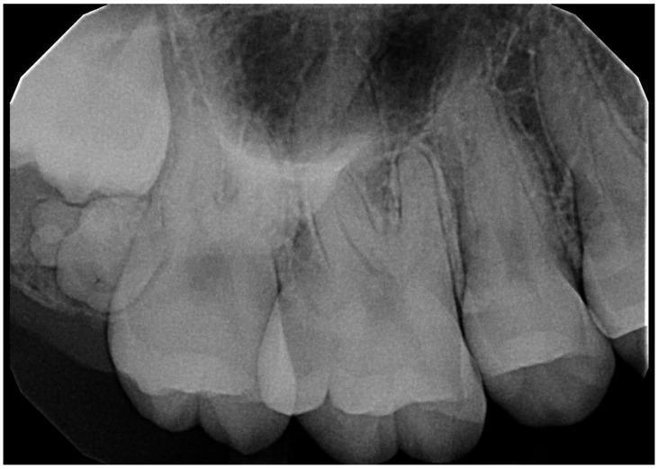
A periapical radiograph of the posterior maxilla demonstrates a compound odontoma hindering the eruption of the third molar. The lesion presents as a well-defined, irregular radiopaque mass composed of multiple small, tooth-like structures (denticles) of varying sizes and shapes. A thin, radiolucent rim surrounds the mass, consistent with a fibrous capsule. The odontoma is positioned coronal to the impacted third molar and distal to the second molar, causing displacement of the erupting tooth. Key anatomical landmarks include the maxillary sinus, appearing as a superior radiolucent area, and the first and second molars with visible periodontal ligament spaces and lamina dura. The trabecular bone pattern of the alveolar process is evident throughout the field. This diagnostic image illustrates a common odontogenic tumor and its clinical consequence of tooth impaction, relevant for dental radiology and oral surgery education.
This diagnostic image is an intraoral periapical radiograph demonstrating a compound odontoma. The primary focus is a cluster of three distinct, radiopaque tooth-like masses (denticles) of varying ovoid and irregular shapes, mimicking the morphology of miniature teeth. These radiopaque structures are well-defined and grouped closely together. Encircling the entire mass is a thin, uniform radiolucent rim, which represents the follicular space or fibrous capsule of the lesion. The lesion is situated within the soft tissues, specifically described in clinical context as the posterior aspect of the buccal mucosa, showing a lack of direct attachment to the alveolar bone or adjacent primary dentition visible in the periphery. This radiographic presentation is a classic hallmark of a compound odontoma, an odontogenic hamartoma composed of organized dental tissues (enamel, dentin, and pulp). The image serves as an educational example for oral and maxillofacial pathology, illustrating the diagnostic differentiation between compound and complex odontomas based on morphological organization.
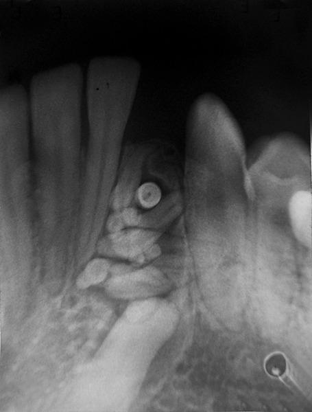
This diagnostic intraoral radiograph illustrates a classic presentation of a compound odontoma in the human jaw. The image shows a well-circumscribed, mixed radiopaque and radiolucent lesion situated between the roots of adjacent erupted teeth. The lesion is characterized by a cluster of multiple small, tooth-like structures (denticles) of varying shapes and sizes, exhibiting high density consistent with enamel and dentin. A prominent, rounded, highly radiopaque mass is visible within the central portion of the cluster. The surrounding periapical bone displays normal trabeculation with slight reactive sclerosis at the periphery of the lesion. There is evidence of localized tooth displacement and widening of the follicular space around the mass. This visual finding is pathognomonic for a compound odontoma, an odontogenic hamartoma, and is a key educational example for dental and oral surgery residents studying benign odontogenic tumors and developmental dental anomalies.
Odontome
Definition
Classification
A. Compound Odontome
- Dental tissues arranged in an orderly pattern forming multiple small tooth-like structures (denticles/odontoids)
- Resembles normal teeth in miniature form
B. Complex Odontome
- Dental tissues are present but arranged in a completely disorganized, irregular mass
- No morphological resemblance to a tooth
C. Other / Rare Forms (Composite Odontomes)
- Dilated odontome (Dens-in-dente / Dens invaginatus) - invagination of enamel organ into the dental papilla
- Geminated odontome - attempted division of a single tooth germ
- Evaginated odontome (Dens evaginatus) - evagination of inner enamel epithelium
- Ameloblastic fibro-odontoma - neoplastic variant with soft tissue component
Etiology and Pathogenesis
- Trauma to the primary dentition causing disturbance in odontogenesis
- Local infection (periapical) affecting developing tooth germ
- Hereditary factors - associated with Gardner syndrome, Hermann syndrome
- Developmental disturbances during bell stage of tooth development
- Persistence of epithelial cell rests of Malassez or Serres
Incidence
- Most common odontogenic tumour (30-67% of all odontogenic tumours per some series)
- Peak incidence: first and second decades of life (10-30 years)
- No sex predilection (slight male predominance in some studies)
- Compound type: 2x more common than complex type
Clinical Features
Compound Odontome
| Feature | Details |
|---|---|
| Site | Anterior maxilla (most common); between canine and lateral incisor |
| Presentation | Usually asymptomatic - found on routine X-ray |
| Size | Small, 1-2 cm; growth ceases on maturity |
| Effect on teeth | Impaction or delayed eruption of adjacent permanent tooth; retained deciduous tooth |
| Swelling | Jaw expansion uncommon |
| Multiple lesions | May occur in Gardner syndrome |
Complex Odontome
| Feature | Details |
|---|---|
| Site | Posterior mandible (most common); premolar-molar region |
| Presentation | Painless, slow-growing swelling; may be asymptomatic |
| Size | Larger, 3-6 cm |
| Effect on teeth | Impacted or displaced adjacent tooth |
| Cortical expansion | May cause bony expansion of jaw |
- Retention of deciduous tooth
- Failure of eruption of permanent tooth
- Occasional pain if secondary infection occurs
- May present as incidental radiographic finding
Radiographic Features
Compound Odontome
- Multiple small radiopaque tooth-like structures (denticles) clustered together
- Surrounded by a thin radiolucent rim (fibrous capsule / follicular space)
- Well-demarcated, well-defined borders
- "Bag of teeth" or "denticle cluster" appearance
- Located between roots of teeth or over crown of unerupted tooth
Complex Odontome
- Irregular, homogeneous radiopaque mass with no tooth-like structure
- Surrounded by a thin radiolucent halo (fibrous capsule)
- Well-defined, well-corticated borders
- Shape: spherical or ovoid with radiating periphery in mature lesions
- Immature lesions may appear radiolucent initially, with progressive radiopacity as mineralization increases
Histopathological Features
Compound Odontome
- Multiple denticles enclosed within a fibrous connective tissue capsule
- Each denticle shows normal arrangement of:
- Enamel (outermost)
- Dentine
- Cementum
- Pulp tissue (central)
- Reduced enamel epithelium may be present
- Ghost cell or calcification in some cases
- Variable maturity - immature lesions show dentinoid, enameloid, cementoid
Complex Odontome
- Disorganized, haphazard arrangement of all dental hard tissues
- Enamel, dentine, and cementum present in irregular masses
- Surrounded by fibrous connective tissue capsule containing strands and islands of odontogenic epithelium
- No recognizable tooth structures
- Developing lesions show more unmineralized tissue and cellular soft tissue
Differential Diagnosis
| Condition | Differentiating Feature |
|---|---|
| Supernumerary tooth | Single tooth-like structure, has PDL space, erupts |
| Ameloblastic fibro-odontoma | Soft tissue (AF-like) component; destructive; more cellular stroma |
| Cementoma | Located at apex of tooth; no enamel/dentine component |
| Osteoma | Purely bony; no dental tissue components |
| Calcifying odontogenic cyst | Has ghost cells; cystic component |
| Ossifying fibroma | No dental tissue; fibrous stroma with spherical calcifications |
Treatment
Compound Odontome
- Enucleation - complete surgical removal of the lesion with its fibrous capsule
- Simple excision through adequate access
- Associated impacted teeth - may be allowed to erupt spontaneously after removal of the obstructing odontome if roots are incompletely formed
- If root formation is complete, orthodontic assistance may be needed
Complex Odontome
- Enucleation and curettage - thorough curettage to prevent recurrence from incomplete removal, especially in immature lesions
- If large: adequate bone exposure and sectioning of mass if needed
- Adjacent teeth preserved whenever possible
- Local anesthesia with adequate access flap
- Complete removal of fibrous capsule along with the lesion
- Irrigation and primary closure
- Histopathological confirmation mandatory
Complications of Surgery
- Damage to adjacent tooth roots
- Damage to inferior alveolar nerve (for large mandibular lesions)
- Damage to maxillary sinus (for maxillary lesions)
- Pathological fracture (rare, for large lesions)
Prognosis
| Type | Prognosis |
|---|---|
| Compound odontome | Excellent - no recurrence after complete enucleation |
| Complex odontome | Good - recurrence rare; may occur if incomplete removal of immature lesion |
| Both | No malignant transformation reported |
Summary
| Feature | Compound Odontome | Complex Odontome |
|---|---|---|
| Nature | Hamartoma | Hamartoma |
| Tissue arrangement | Organized (denticles) | Disorganized (irregular mass) |
| Site | Anterior maxilla | Posterior mandible |
| Radiograph | "Bag of teeth" - denticles + radiolucent rim | Radiopaque mass + radiolucent halo |
| Size | Small (1-2 cm) | Larger (3-6 cm) |
| Histology | Organized enamel, dentine, pulp | Haphazard enamel, dentine, cementum |
| Treatment | Enucleation | Enucleation + curettage |
| Recurrence | None | Rare (immature lesions) |
| Prognosis | Excellent | Good |
Note: Neelima Malik's Textbook of Oral and Maxillofacial Surgery was not found in the available library. This answer is structured according to her textbook's known chapter organization and approach, supplemented with standard oral surgery content consistent with her text. For exam purposes, this covers the full scope of a 10-mark answer as expected from her book.