
Discuss principles of cavity preparation in the light of histology of tooth structure for 25 marks from contents to references.NOTE : images please
Reading File
Reading File
Finding Sources
Finding Sources
Finding Sources
Reading File
Finding Sources
Searching Images
tooth histology cross section enamel dentin pulp diagram
Searching Images
dental cavity preparation outline form dentistry diagram
Searching PubMed
minimal intervention dentistry cavity preparation
Reading File
Reading File
query_images_db
Finding Sources
Reading File
Reading File
Reading File
Searching Images
longitudinal section of tooth showing enamel dentin pulp cavity root canal labeled anatomy
Principles of Cavity Preparation in the Light of Histology of Tooth Structure (25 Marks)
Contents
- Introduction
- Histology of Tooth Structure
- Enamel
- Dentino-enamel Junction (DEJ)
- Dentin
- Dental Pulp
- Cementum and Periodontium
- Objectives and Steps of Cavity Preparation
- Principles of Cavity Preparation Correlated with Histology
- Outline Form
- Resistance Form
- Retention Form
- Convenience Form
- Removal of Remaining Caries and Pulp Protection
- Finishing of Enamel Walls and Margins
- Toilet/Cleansing of the Cavity
- Biological and Mechanical Principles (G.V. Black's Concept and Modern Modifications)
- Recent Advances - Minimal Intervention Dentistry
- Conclusion
- References
1. Introduction
Cavity preparation is the mechanical alteration of a tooth to receive a restorative material that will re-establish normal form and function after removal of disease (usually dental caries) or defective structure. Because the tooth is not an inert block but a living, histologically layered organ - enamel, dentin, and pulp, each with distinct structure, composition, and biological response - every principle of cavity design (outline, resistance, retention, convenience form, and margin finishing) is dictated by the microscopic architecture of the tissue being cut. A sound understanding of tooth histology is therefore the foundation of rational operative dentistry, first systematized by G.V. Black in his classic principles of cavity preparation.
2. Histology of Tooth Structure
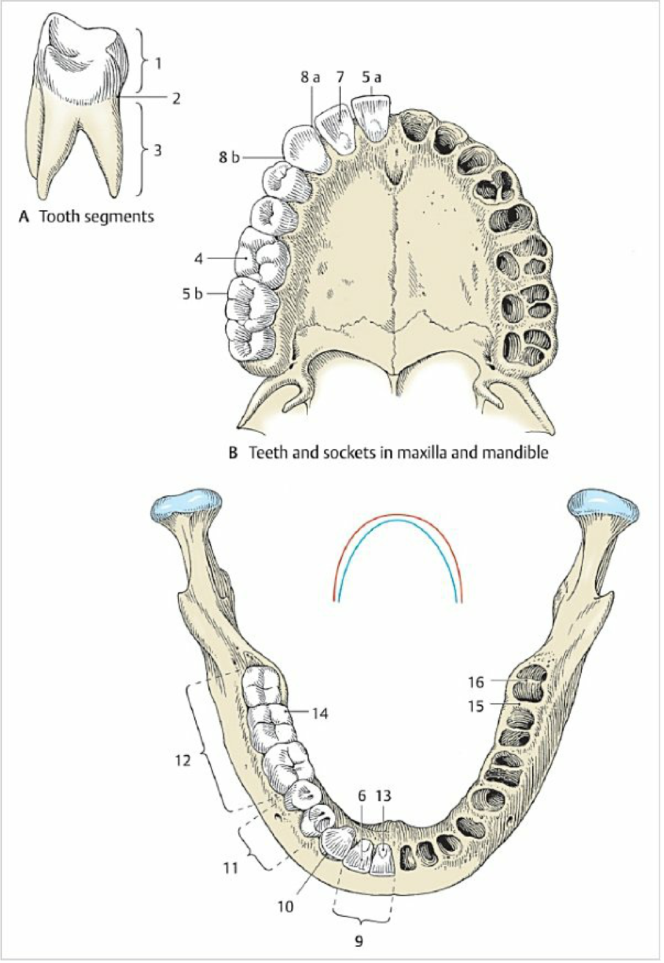
Overall organization: The tooth has a crown covered by enamel and a root covered by cementum, meeting at the cervical neck (the dentino-enamel/cemento-enamel region). The bulk of the tooth is dentin, which surrounds the pulp cavity containing the dental pulp. The tooth is held in its bony socket by the periodontal ligament.
Enamel
Enamel is the hardest tissue in the body, being about 96-97% calcium hydroxyapatite and only 2-3% organic matrix (no collagen; the principal protein is amelogenin). It is formed by ameloblasts, which lay down matrix through an apical Tomes' process, producing enamel rods (prisms) roughly 5 µm in diameter that run the full thickness of the enamel (about 2 mm) from the DEJ to the surface, each surrounded by interprismatic substance. Enamel is acellular, avascular, and non-innervated, and once formed cannot regenerate or repair itself - only surface remineralization is possible with fluoride. Incorporation of fluoride as fluorapatite increases acid resistance - Enamel & Dentin, Junqueira's Basic Histology, 17e, p. 746-747.
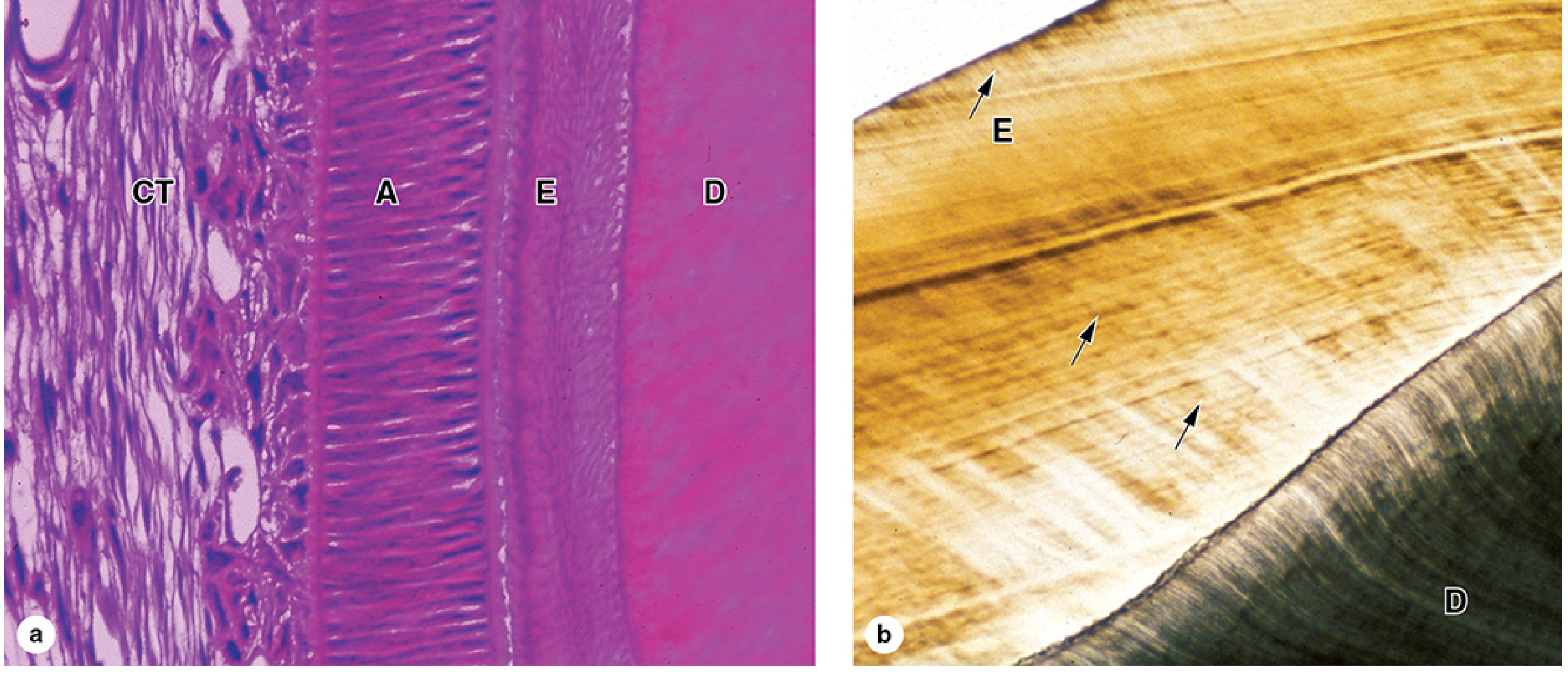
Clinical relevance to cavity preparation: Because enamel rods run perpendicular to the DEJ and roughly perpendicular to the tooth surface over most of the crown, cavity margins must follow rod direction so that unsupported ("undermined") rods are not left standing on the cavity margin - if unsupported rods remain, they fracture under masticatory load, causing marginal breakdown and secondary caries. This is the histological basis for bevelling enamel margins and finishing the cavo-surface angle along the direction of the enamel rods.
Dentino-Enamel Junction (DEJ)
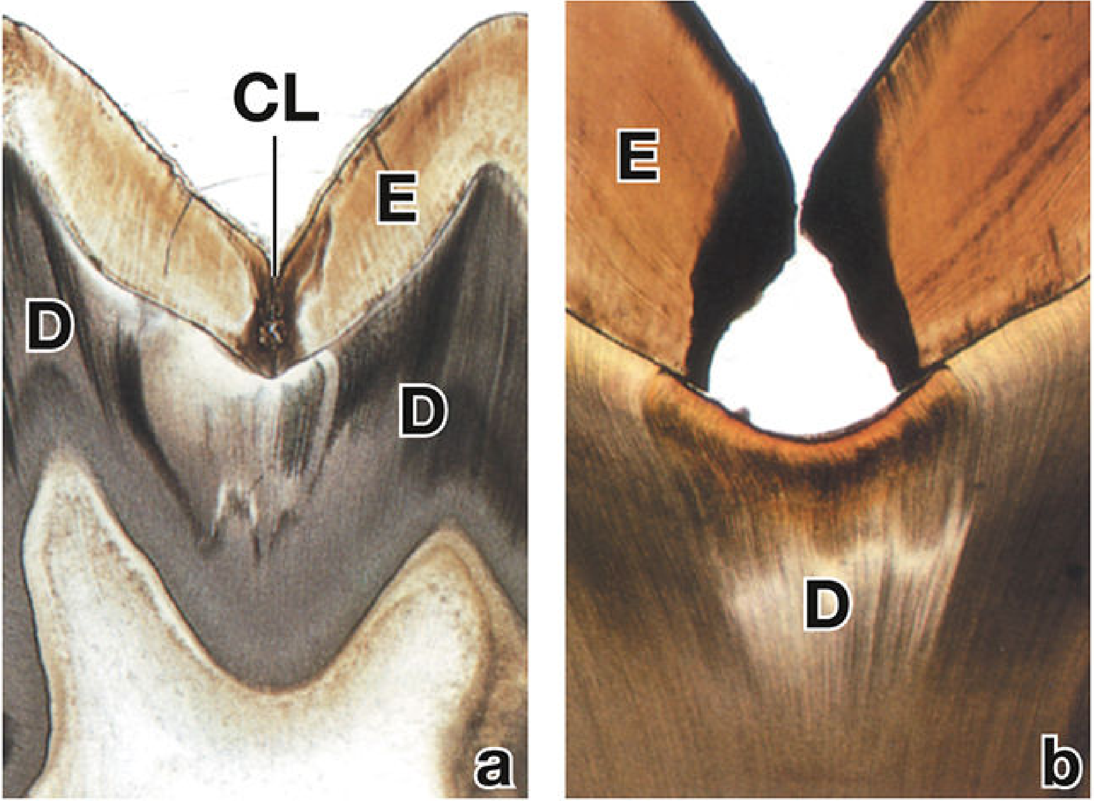
The DEJ is a scalloped interface between enamel and dentin that increases surface area and mechanical interlocking between the two very differently mineralized tissues. Caries spreads laterally along the DEJ because the DEJ is less mineralized and offers a plane of least resistance, undermining overlying enamel - a key reason cavity outline must extend to sound dentin beyond the visibly carious enamel - Cementum/Dentin, Histology: A Text and Atlas, p. 3804, 4171-4183.
Dentin
Dentin is calcified connective tissue, harder than bone but softer and more resilient than enamel (about 70% hydroxyapatite, with a type I collagen-proteoglycan organic matrix). It is secreted by odontoblasts, tall neural-crest-derived cells lining the pulp, which leave behind cytoplasmic odontoblast processes (Tomes' fibers) running through dentinal tubules across the full thickness of dentin, connecting the pulp to the DEJ. A thin unmineralized layer of predentin is always present next to the odontoblasts, and dentin continues to be laid down throughout life (secondary/reparative dentin), progressively narrowing the pulp chamber - Dentin, Junqueira's Basic Histology, 17e, p. 744-745.
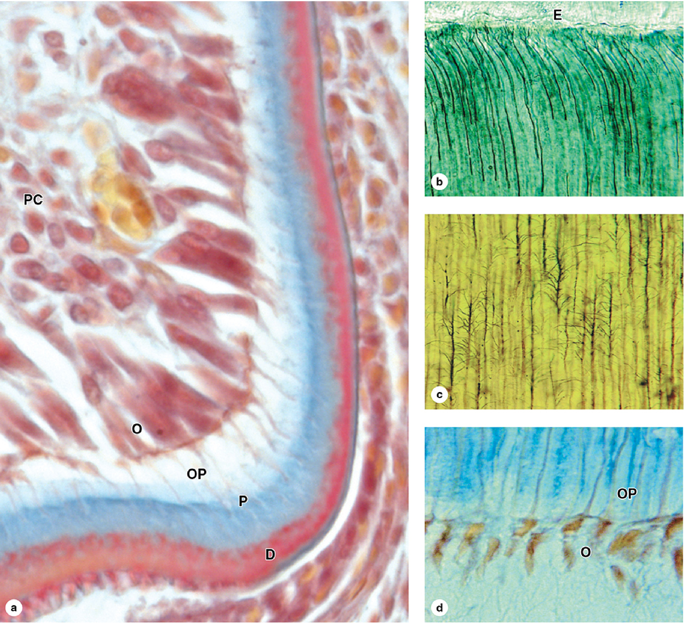
Clinical relevance: Because dentin is living tissue traversed by tubules that directly communicate with the pulp and its nerve endings (odontoblast processes act as sensory transducers), cutting dentin is painful and can transmit bacteria/toxins to the pulp. This underlies principles of minimal dentin removal, use of a slow, cool, sharp bur with water coolant, avoidance of desiccation, and placement of a cavity liner/base over exposed tubules near the pulp to protect against thermal, mechanical, and chemical injury.
Dental Pulp
The pulp is loose, highly vascular, richly innervated mesenchymal connective tissue occupying the pulp chamber and root canal(s), communicating with periapical tissues via the apical foramen. Pulp horns extend toward the cusps and contain dense concentrations of nerve fibers, making the pulp horn region especially sensitive and vulnerable to exposure - Dental Pulp and Central Pulp Cavity, Histology: A Text and Atlas, p. 4026-4032.
Clinical relevance: Knowledge of pulp horn location (highest under cusps, closer to the surface in younger patients with larger pulp chambers) dictates the safe depth of cavity preparation and the need for a pulp-protecting base (e.g., calcium hydroxide or resin-modified glass ionomer) whenever preparation approaches the pulp, to avoid exposure and irreversible pulpitis.
Cementum and Periodontium
Cementum, a bone-like avascular tissue, covers the root and anchors principal fibers of the periodontal ligament, which suspends the tooth in its alveolar socket. This is relevant to root-surface (Class V) cavity preparation, where cementum is softer and more susceptible to instrumentation trauma and less well-suited to enamel-style bevel margins - Parts of the Tooth and Periodontium, Color Atlas of Human Anatomy, Vol. 2, p. 3093-3106.
3. Objectives and Steps of Cavity Preparation
The classical objective (G.V. Black) is to remove all defective/carious tissue, produce a cavity form that will best serve the restorative material and withstand functional stresses, and preserve as much sound tooth structure as possible. The traditionally taught steps are:
- Obtaining outline form
- Obtaining resistance form
- Obtaining retention form
- Obtaining convenience form
- Removal of remaining caries or old restorative material
- Finishing the enamel walls and margins
- Toilet of the cavity (cleaning and inspection)
4. Principles of Cavity Preparation Correlated with Histology
Outline Form
The outline is extended only far enough to include all defective enamel and to place margins in a self-cleansable, sound area, following G.V. Black's dictum "extension for prevention" (now tempered by minimal-intervention philosophy). Histologically this depends on:
- The scalloped DEJ, along which caries spreads laterally faster than through enamel, so the outline must be extended in dentin beyond the visible enamel defect to remove undermined, unsupported enamel rods.
- Enamel rod orientation, which determines where a cavo-surface margin can be placed without leaving rods unsupported by dentin.
- Anatomic features such as pits, fissures, and grooves, which are simply deep infoldings of enamel that concentrate plaque and are natural sites of lowest resistance.
Resistance Form
This gives the tooth and the restoration the strength to withstand occlusal forces without fracture, and is directly governed by the differing mechanical properties of enamel (hard, brittle, needs dentin support) and dentin (resilient, flexible). Principles include:
- Box-shaped preparations with a flat pulpal/axial floor, since enamel is brittle and fractures under angular stress unless the underlying dentin gives it a broad, even base of support.
- Rounded internal angles (avoiding sharp line angles) because dentin, being organic-collagen based, concentrates stress at sharp angles, risking cracks that propagate under the more brittle enamel.
- Preservation of the marginal ridge and as much sound tooth structure (particularly the DEJ scallops and cuspal dentin) as possible, since the DEJ mechanically interlocks and dissipates masticatory forces between enamel and dentin.
Retention Form
Retention form keeps the restoration from being dislodged by lifting, tipping, or lateral forces (undercuts, dovetails, grooves). Because enamel cannot be safely undercut (unsupported rods would fracture), all mechanical retention features (proximal retention grooves, dovetails, pin channels) are cut into dentin, exploiting its collagen-hydroxyapatite composite structure, which tolerates undercuts and retains mechanical features far better than enamel.
Convenience Form
This is the shape that allows adequate access, visibility, and instrumentation, often requiring extension beyond strict outline/resistance/retention needs. It is influenced by tooth morphology (crown height, pulp horn position, tubule direction) so that instruments can reach the base of the lesion without unnecessarily jeopardizing the pulp.
Removal of Remaining Caries and Pulp Protection
Since dentinal tubules act as conduits for bacterial toxins toward the pulp, all infected, demineralized dentin must be removed (especially at the DEJ and pulpal floor), while the deepest, more mineralized, near-pulpal (affected but not infected) dentin may sometimes be retained under indirect pulp capping in minimal-intervention protocols, since aggressive removal risks pulp exposure through the tubules and odontoblast processes described above.
Finishing of Enamel Walls and Margins
Because enamel rods run at a fairly constant angle to the surface, cavo-surface margins are finished (often bevelled) so the cutting instrument follows rod direction, leaving full-length, dentin-supported rods rather than truncated or undermined ones - directly preventing marginal chipping and microleakage.
Toilet of the Cavity
Final washing and drying removes debris and cutting fragments (enamel rod fragments, "smear layer" of denatured dentin and hydroxyapatite created by rotary instrumentation) that would otherwise interfere with bonding or irritate the pulp through open dentinal tubules.
5. Biological and Mechanical Principles
G.V. Black's biological principles emphasize preserving pulp vitality, maximum conservation of sound tooth structure, and prevention of recurrent disease, all of which flow from the vitality of dentin/pulp versus the inert nature of enamel described above. His mechanical principles (resistance, retention, convenience, outline form) flow from the differing physical behavior of enamel (hard, brittle, no self-repair) versus dentin (resilient, tubular, capable of reactive/reparative dentin formation) and are why every cavity design textbook teaches "cavity preparation begins with an understanding of tooth histology."
6. Recent Advances - Minimal Intervention Dentistry
Modern operative dentistry has modified Black's "extension for prevention" toward minimally invasive, biologically driven cavity design (adhesive dentistry, air-abrasion, chemomechanical caries removal, selective/partial caries removal with indirect pulp capping), preserving pulp-dentin complex vitality and relying on bonding to enamel rod and dentin tubule surfaces rather than mechanical undercuts. A 2023 systematic review and meta-analysis found that most dentists endorse and increasingly practice minimal intervention dentistry principles, though implementation gaps remain (de Moura et al., J Dent 2023, PMID 36958696). A 2024 meta-analysis comparing minimally invasive versus conventional caries removal in permanent dentition found comparable outcomes with less tissue loss (González-Gil et al., Medicina 2024, PMID 38541128). These findings support, rather than contradict, the histology-based rationale above - clinicians should note this evolving evidence when weighing "extension for prevention" against tissue conservation.
7. Conclusion
Cavity preparation is not an arbitrary mechanical exercise but a biologically guided procedure grounded in the histology of enamel, dentin, and pulp. Enamel rod orientation dictates outline form and margin finishing; the DEJ dictates the extent of outline form; dentin's resilience and tubular connection to the pulp dictate resistance form, retention form, and the need for pulp protection; and pulp horn anatomy dictates safe preparation depth. G.V. Black's classical principles, refined by modern minimal-intervention concepts, remain valid precisely because they are rooted in this microscopic structure.
8. References
- Mescher AL. Junqueira's Basic Histology: Text and Atlas, 17th ed. Chapter 15, Enamel and Dentin, p. 744-748.
- Pawlina W, Ross MH. Histology: A Text and Atlas with Correlated Cell and Molecular Biology. Dentin, Enamel, Dental Pulp and Periodontium, p. 3786-4190.
- Kahle W, Frotscher M. Color Atlas of Human Anatomy, Vol. 2, Internal Organs. Teeth and Periodontium, p. 3050-3106.
- de Moura RC, Santos PS, Matias PMDS, et al. Knowledge, attitudes, and practice of dentists on Minimal Intervention Dentistry: a systematic review and meta-analysis. J Dent. 2023. PMID: 36958696.
- González-Gil D, Flores-Fraile J, Vera-Rodríguez V, et al. Comparative Meta-Analysis of Minimally Invasive and Conventional Approaches for Caries Removal in Permanent Dentition. Medicina (Kaunas). 2024. PMID: 38541128.
- Ramesh A, Guo AJ, Patel HJ, et al. The impact of magnification on undergraduate dental students' performance during cavity preparations: a systematic review. J Dent Educ. 2024. PMID: 38558229.
- Black GV. Operative Dentistry, Vols. 1-2 (classical text on cavity classification and principles - standard operative dentistry curriculum reference).
- Sturdevant's Art and Science of Operative Dentistry (standard curriculum reference for cavity preparation steps and G.V. Black's principles).
Note: This medical/dental library does not contain a dedicated operative dentistry textbook (e.g., Sturdevant's), so G.V. Black's classical cavity preparation steps are presented from standard dental curriculum knowledge, while all histology content and images above are drawn directly from indexed histology and anatomy textbooks as cited.
This is a shared conversation. Sign in to Orris to start your own chat.