
Hepatopulmonary syndrome 10 mark answer
Reading File
Now let me search the medical library for hepatopulmonary syndrome content in parallel.
Finding Sources
Finding Sources
Searching PubMed
"hepatopulmonary syndrome"[MeSH Terms]
Excellent. I have two strong textbook sources. Let me read the main sections from Sleisenger & Fordtran and Murray & Nadel in parallel.
Reading File
Reading File
Searching Images
hepatopulmonary syndrome pathophysiology pulmonary vascular dilation
Great content found. Let me read the clinical features, diagnosis, and treatment sections.
Reading File
Loading Image
Excellent content and the pathophysiology diagram is perfect. Let me now read the treatment/management and prognosis sections.
Reading File
Loading Image
I now have comprehensive content for a complete 10-mark answer. Let me compile the full structured answer.
Hepatopulmonary Syndrome (HPS)
Definition
Hepatopulmonary syndrome (HPS) is defined as a triad of:
- Advanced liver disease or portal hypertension
- Intrapulmonary vascular dilatation (IPVD)
- Abnormal arterial oxygenation - specifically a widened age-corrected alveolar-arterial oxygen gradient (AaPO2 >15 mmHg, or >20 mmHg in patients ≥64 years) on room air, with or without frank hypoxemia
The term was coined by Kennedy and Knudson in 1977. It occurs in 5-35% of cirrhotic patients evaluated for liver transplantation (LT).
Pathophysiology
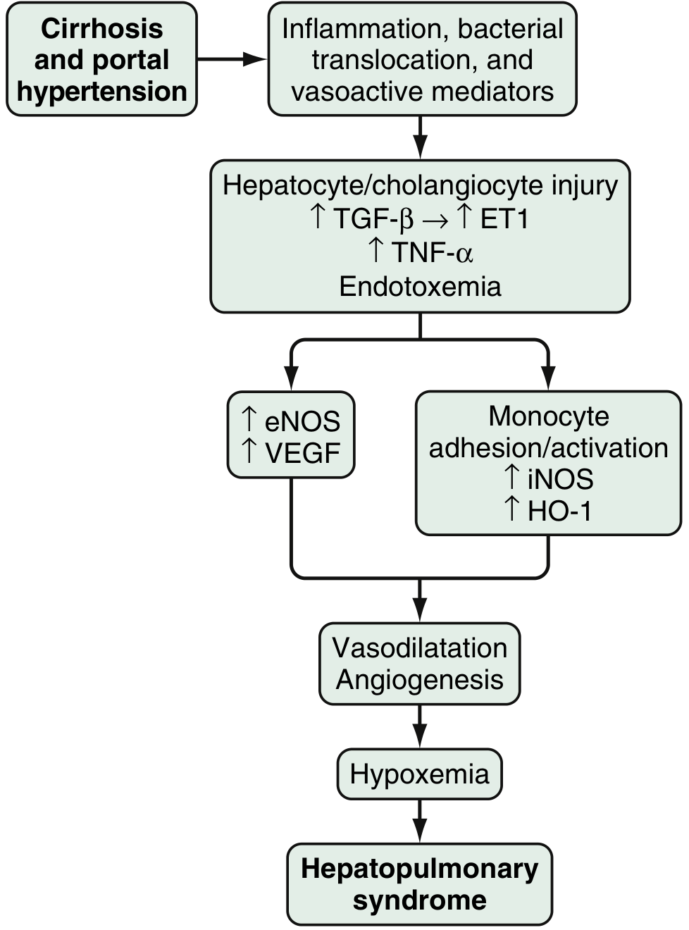
The central abnormality is intrapulmonary vascular dilatation - dilation of pulmonary capillaries and precapillary vessels from normal <8 µm to 15-100 µm. This is driven by a cascade triggered by cirrhosis and portal hypertension:
Key mediators:
- Cirrhosis/portal hypertension → inflammation, bacterial translocation, vasoactive mediator release
- Hepatocyte/cholangiocyte injury → ↑TGF-β → ↑Endothelin-1 (ET-1); ↑TNF-α; endotoxemia
- ET-1 acts on overexpressed pulmonary endothelin-B receptors → activates eNOS → ↑Nitric oxide (NO)
- TNF-α and fractalkine drive pulmonary monocyte adhesion/activation → ↑iNOS-derived NO, ↑heme oxygenase-1 (HO-1, producing CO), ↑VEGF-mediated angiogenesis
- Net result: vasodilatation + angiogenesis → hypoxemia
Mechanisms of Hypoxemia
Three mechanisms operate in HPS, often in combination:
| Mechanism | Explanation |
|---|---|
| Low V/Q mismatch (predominant) | Dilated vessels at lung bases receive excess perfusion relative to ventilation; worsens upright (explains orthodeoxia) |
| Diffusion-perfusion impairment | High cardiac output + vasodilation → reduced erythrocyte transit time; O2 can't diffuse to RBCs in center of dilated capillaries |
| True intrapulmonary shunt | Arteriovenous communications allow venous blood to bypass ventilated alveoli; predominates in severe HPS (PaO2 35-67 mmHg) |
The "shunt" in HPS is atypical - even when macroaggregated albumin suggests 32% shunt fraction, 100% O2 can partially reverse it (measured shunt by O2 method only 19%), because many "shunts" behave like low-V/Q units at high FiO2.
Clinical Features
Symptoms:
- Dyspnea - most common; insidious onset, progressive
- Platypnea - dyspnea worsening in the upright position, relieved by lying down (pathognomonic but uncommon)
- Orthodeoxia - fall in PaO2 >4 mmHg or SpO2 >5% in upright vs supine position (caused by increased perfusion of dilated basal vessels when upright)
- Cough
Signs:
- Digital clubbing
- Peripheral cyanosis
- Spider angiomata (common association)
- Signs of chronic liver disease and portal hypertension
- Up to 70% develop marked hypoxemia during sleep (nocturnal desaturation)
Note: Severity of HPS does not clearly correlate with the degree of hepatic dysfunction (Child-Pugh or MELD score).
Severity Grading (ERS/ATS Task Force Consensus)
| Grade | PaO2 (room air) |
|---|---|
| Mild | ≥80 mmHg |
| Moderate | 60-80 mmHg |
| Severe | 50-60 mmHg |
| Very severe | <50 mmHg |
Diagnosis
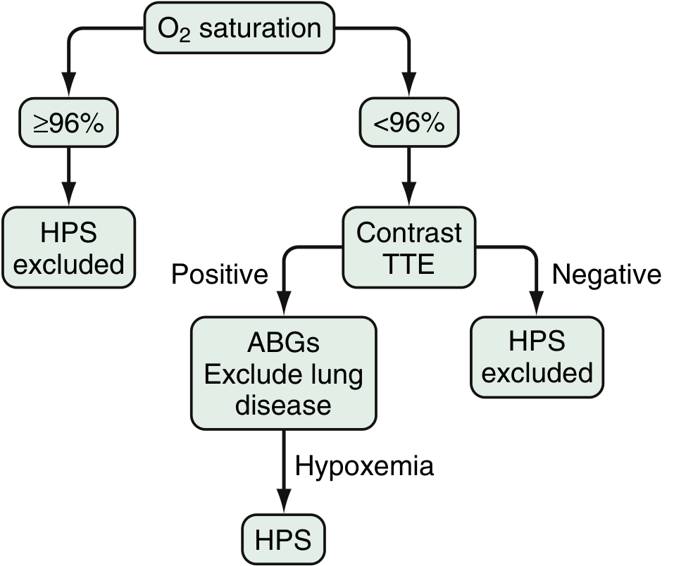
Diagnosis requires:
- Clinical suspicion + evidence of liver disease/portal hypertension
- Arterial blood gas (ABG) - demonstrating widened AaPO2 ± hypoxemia (pulse oximetry is insensitive and unreliable)
- Detection of intrapulmonary shunting
- Exclusion of intrinsic cardiopulmonary disease
Screening algorithm:
Investigations
1. Contrast-enhanced transthoracic echocardiography (CE-TTE) - most sensitive test
- Agitated saline ("bubble study") injected IV: normally, microbubbles are trapped in pulmonary capillaries
- In HPS: bubbles appear in left heart after 3-6 cardiac cycles (delayed appearance = intrapulmonary origin, distinguishing from intracardiac shunt where appearance is immediate at ≤3 cycles)
2. Macroaggregated albumin (MAA) lung perfusion scan (99mTc-MAA)
- Radiolabeled albumin macroaggregates (>20 µm) pass through dilated pulmonary vessels and appear in brain/kidney
- Shunt fraction >6% is diagnostic; also quantifies shunt severity
- Useful when CE-TTE is equivocal
3. Arterial blood gas - gold standard for quantifying hypoxemia; measures PaO2 and AaPO2
4. High-resolution CT chest - may identify anatomic AV shunts amenable to embolization; not routine
5. Pulmonary angiography - only if CT identifies large AV shunts potentially amenable to coil embolization
Treatment
Medical Therapy
- No proven effective medical therapy exists
- Supplemental oxygen is used for resting or exercise-induced hypoxemia (PaO2 <60 mmHg); improves symptoms but no RCT evidence for clinical benefit
- Garlic preparations - two small uncontrolled studies + one RCT reported improved oxygenation; considered experimental
- Pentoxifylline - conflicting results in small case series
- Agents targeting NO, ET-1, VEGF pathways have shown benefit in animal models but not established in humans
Radiological/Interventional
- TIPS (Transjugular intrahepatic portosystemic shunt) - case reports/series show improvement; a 2025 systematic review (PMID 40562306) evaluated TIPS for HPS; not routinely recommended
- Pulmonary angiography + embolization - for large AV communications detected on CT; not standard practice
Liver Transplantation (LT) - Definitive Treatment
- LT reverses HPS in up to 80% of patients
- Resolution can take >12 months post-LT (even longer in severe cases)
- Mortality post-LT is higher in HPS vs non-HPS patients, especially with:
- PaO2 <50 mmHg (very severe)
- Shunt fraction >20%
- MELD exception points are allocated for HPS with resting PaO2 <60 mmHg to increase transplant priority
- Unique post-LT complications: transient paradoxical worsening of hypoxemia, development of pulmonary hypertension, embolic cerebrovascular events
Prognosis
- Without LT: median survival 10.6-24 months after diagnosis; HPS significantly increases mortality in cirrhosis
- HPS tends to worsen over time - mild-moderate disease can progress to severe
- With LT: improved survival; best outcomes when transplanted before PaO2 drops below 50 mmHg
- Presence of HPS does not generally resolve spontaneously
HPS vs Portopulmonary Hypertension (POPH) - Key Distinction
| Feature | HPS | POPH |
|---|---|---|
| Pulmonary vessels | Dilated | Constricted/remodeled |
| PaO2 | Decreased | Usually normal/mildly decreased |
| Response to O2 | Improves (partial) | Minimal response |
| LT | Curative | Often contraindicated (moderate-severe) |
| PAP | Normal or low | Elevated (>25 mmHg) |
Sources: Sleisenger and Fordtran's Gastrointestinal and Liver Disease, Chapter 94; Murray & Nadel's Textbook of Respiratory Medicine, Chapter 126
This is a shared conversation. Sign in to Orris to start your own chat.