
How to remember changes in breast in a primi gravida with duration
Reading File
The question is about a mnemonic to remember the changes in the breast during pregnancy (primigravida) with their timing. Let me search the textbook library for this.
Finding Sources
Finding Sources
Reading File
Reading File
I now have all the information needed. Here is a complete, structured answer with a mnemonic:
Breast Changes in Primigravida - With Timing & Mnemonic
Mnemonic: "TENSE SCAN MA" (or read as "Tense Scan Ma" - the breast feels tense, scan it!)
| Letter | Change | Timing |
|---|---|---|
| T | Tenderness + Tingling | Early weeks (1st month) |
| E | Enlargement (size increases, nodular due to hypertrophy of mammary alveoli) | After 2nd month |
| N | Nipple - deeply pigmented + erectile | 2nd month onwards |
| S | Superficial veins engorged/distinct | 2nd month onwards |
| E | Areola - pink → dark brown (Montgomery's tubercles appear) | End of 2nd month |
| C | Colostrum secreted (thin, yellowish; fat globules + large phagocytic cells) | 3rd month |
| S | Striae (silvery lines on skin) | After 6 months |
Alternate Memory Hook: "2-2-2-3-6"
A simpler timing-based anchor:
- Early weeks - Tenseness + Tingling
- 2nd month - size ↑, veins enlarge, nipple darkens & becomes erectile
- End of 2nd month - Areola darkens (pink → brown), Montgomery's tubercles form
- 3rd month - Colostrum expressed
- After 6 months - Striae appear (primipara)
Why Important in Primigravida?
These breast changes are highly significant in a primigravida but are of less diagnostic value in multiparas, because multiparas may already have permanently altered breast anatomy from previous pregnancies. This is a key point in Forensic Medicine (signs of pregnancy/delivery examination).
Complete Summary of Changes (Sequence)
- Sense of tenseness and tingling - early weeks
- Increase in size + nodularity (hypertrophy of mammary alveoli) - after 2nd month
- Superficial veins become distinct and engorged - 2nd month
- Nipple - deeply pigmented, more erectile - 2nd month
- Areola - changes from pink (in virgin) → gradually dark brown - 2nd month
- Montgomery's tubercles - sebaceous glands enlarge to form small, round, dark-colored tubercles around nipple - end of 2nd month
- Colostrum secreted (thin, yellowish fluid with fat globules + large phagocytic cells) - 3rd month
- Striae (stretch marks) - after 6 months, especially in primipara
Quick mnemonic summary:
"Tingling starts EARLY, Veins & Nipple darken at 2, Montgomery's at 2-end, Colostrum at 3, Striae at 6"
Sources: The Essentials of Forensic Medicine and Toxicology 36th ed. (2026), p. 8215-8231; P C Dikshit Textbook of Forensic Medicine and Toxicology, p. 3244-3254
Will linea nigra appear first or striae gravidarum
Linea nigra appears first. It is a pigmentation change that occurs early in pregnancy (around the 2nd-3rd month), while striae gravidarum appear later as the abdomen stretches (usually after 5-6 months). Let me verify this from the textbooks.
Searching sources...
Finding Sources
Finding Sources
Reading File
Finding Sources
The textbook data confirms it clearly. Here is the answer:
Linea Nigra Appears FIRST
Timing Comparison
| Sign | Timing | Category |
|---|---|---|
| Linea nigra | ~2nd-3rd month (part of early skin pigmentation changes) | Presumptive sign |
| Striae gravidarum | 6th-7th month (late pregnancy) | Probable sign |
Why Linea Nigra Comes First
Linea nigra is a pigmentation change - it results from hormonal (MSH/estrogen) stimulation of melanocytes, which occurs early in the 1st trimester. It is a dark line extending from the pubis upward to/beyond the umbilicus, and appears alongside other pigmentary changes (darkening of vulva, areola, axillae).
Striae gravidarum are mechanically caused - they result from rupture of subcuticular elastic fibers due to the gradual distension (stretching) of the abdomen. The abdomen doesn't stretch significantly until the uterus rises out of the pelvis (after 3rd month) and becomes large enough to cause skin stress, which only happens in the 6th-7th month.
What Happens to Striae After Delivery?
- During pregnancy: pinkish or slightly bluish, curved, irregular, depressed lines arranged concentrically around the umbilicus
- After delivery: become paler with time
- After ~1 year: appear as white scars called Lineae albicantes
Memory Hook
"Pigment before Stretch" Hormones act early (linea nigra ~ 2-3 months) → Skin stretches late (striae ~ 6-7 months)
Source: The Essentials of Forensic Medicine and Toxicology 36th ed. (2026), p. 8244-8291
Difference between boy and girl foetus in ultrasonography
Fetal sex determination on USG relies on visualization of the genitalia, typically reliable after 16-18 weeks. Male shows scrotal sac/penis, female shows labia. Let me get the detailed textbook content.
Searching sources...
Finding Sources
Finding Sources
Finding Sources
Finding Sources
Finding Sources
Finding Sources
Finding Sources
Finding Sources
Reading File
Finding Sources
Loading Image
Finding Sources
Here is a complete answer with the textbook USG image:
Fetal Sex Determination on Ultrasonography
When Can It Be Determined?
| Stage | Reliability |
|---|---|
| < 12 weeks | Not possible - external genitalia are morphologically indifferent |
| 12 weeks | Final differentiation of external genitalia occurs - earliest possible |
| 16-18 weeks | Reasonably reliable if fetus is in good position |
| After 20 weeks | Highly reliable (>95% accuracy) |
| 33 weeks (3rd trimester) | Definitive - clear structures visible |
USG Features: Male vs Female Fetus
| Feature | Male (Boy) | Female (Girl) |
|---|---|---|
| Primary structure | Penis - echogenic linear/cylindrical structure | Labia majora - two parallel echogenic lines ("hamburger sign") |
| Scrotum | Visible as rounded echogenic sac posterior to penis | Absent |
| Testes | Visible in scrotum (especially 3rd trimester) | Absent |
| Urethral view | Single midline linear structure | Three parallel lines (labia + clitoris) |
| "Turtle sign" | Penis + scrotum resemble a turtle head emerging from shell | Absent |
| Sagittal view (Nub/Angle sign) | Genital tubercle angled >30° above horizontal spine | Genital tubercle angled <30° (nearly parallel to spine) |
| Transverse view | Three dots - two testes + penis | Three lines - "three-line sign" or "fork sign" |
The Classic USG Image - 33-Week Male Fetus
The image below (from the textbook) shows a 33-week male fetus with clearly visible external genitalia:
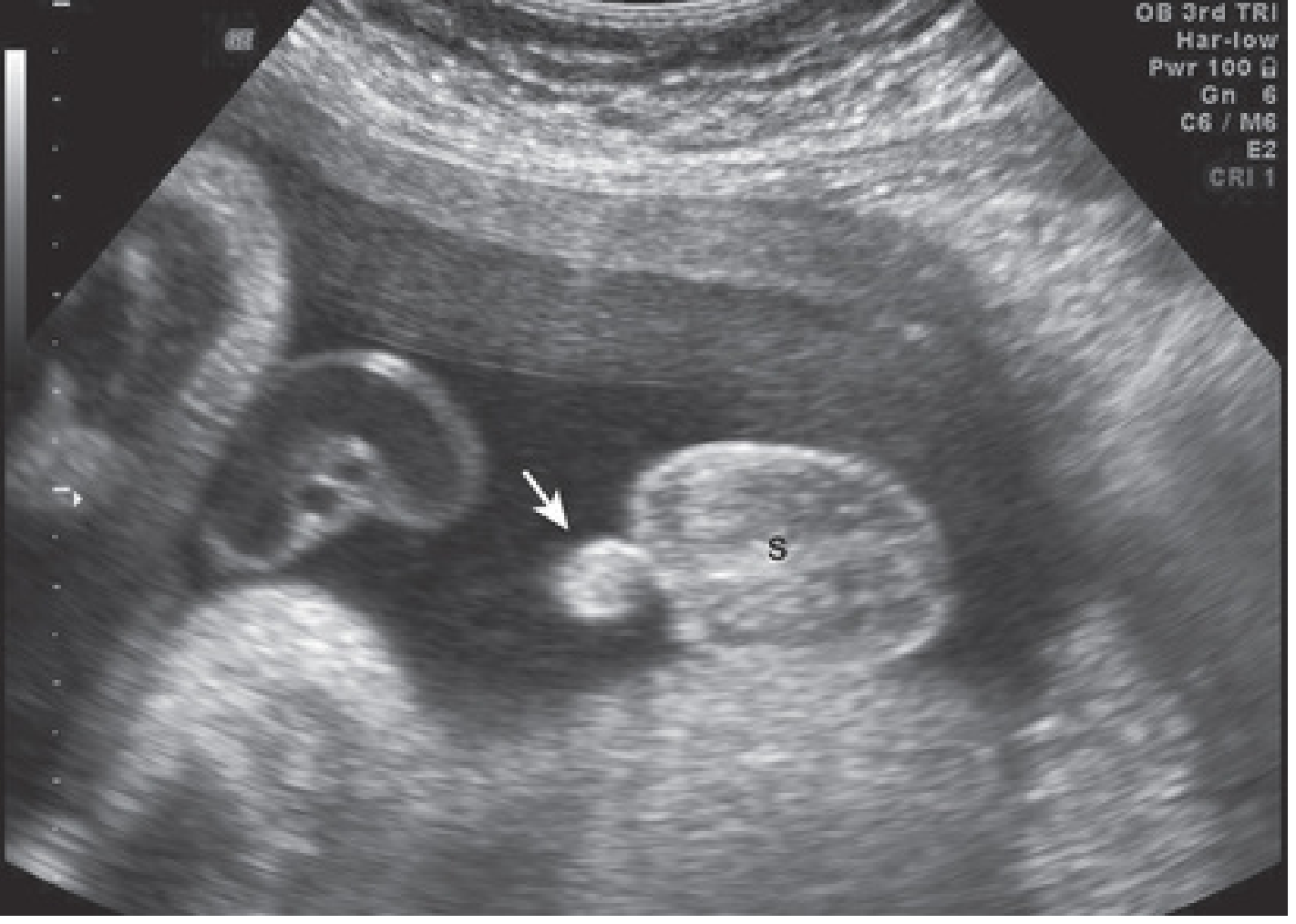
Arrow = Penis, S = Scrotum with testes visible inside
Key Signs to Remember
Male:
- "Turtle sign" - penis resembles a turtle's head
- "Three-dot sign" on transverse view (two testes + penis)
- Nub angle >30° in sagittal view
Female:
- "Hamburger sign" - labia on either side of central clitoris resembling a burger bun
- "Three-line sign" on sagittal/transverse view
- Nub angle <30° (lying flat, parallel to spine)
Important Legal Note (Forensic Relevance)
In India, fetal sex determination by USG is prohibited under the Pre-Conception and Pre-Natal Diagnostic Techniques (PCPNDT) Act, 1994 (amended 2003). Disclosure of the sex of the fetus to any person is a punishable offense, to prevent female foeticide. This is a high-yield forensic medicine exam topic.
Memory Hook
"Boys point UP (>30°, turtle sticks out), Girls lie FLAT (<30°, hamburger lies down)"
Source: The Developing Human - Clinically Oriented Embryology, p. 3251; Color Atlas of Human Anatomy Vol. 2, p. 1104
With pictures detailed USG differentiation of male and female
Searching sources...
Finding Sources
Searching the Web
USG differentiation male female fetus ultrasound signs turtle sign hamburger sign nub theory images
Finding Sources
Searching the Web
fetal sex determination ultrasound nub theory angle sign 12 weeks sagittal view boy girl
Loading Image
Loading Image
Searching the Web
hamburger sign three line sign female fetus ultrasound image vs turtle sign male fetus USG comparison
Reading Web Page
https://radiopaedia.org/cases/turtle-sign-ultrasound?lang=us
Loading Image
Here is the complete, picture-supported guide to USG differentiation of male vs. female fetus:
USG Differentiation: Male vs Female Fetus - With Images
1. EMBRYOLOGICAL BASIS
At < 12 weeks, both sexes have an identical genital tubercle - it is impossible to differentiate. From week 12 onward, testosterone in males causes the tubercle to elongate into a penis, while in females it remains as the clitoris. This is the foundation of all USG sex determination.
2. NUB THEORY / ANGLE SIGN (11-14 Weeks) - Sagittal View
This is the earliest method of sex determination on USG.
How It Works:
Measure the angle of the genital tubercle (nub) relative to the fetal spine in a mid-sagittal view:
Girl - Nub angle <30° (horizontal/flat):
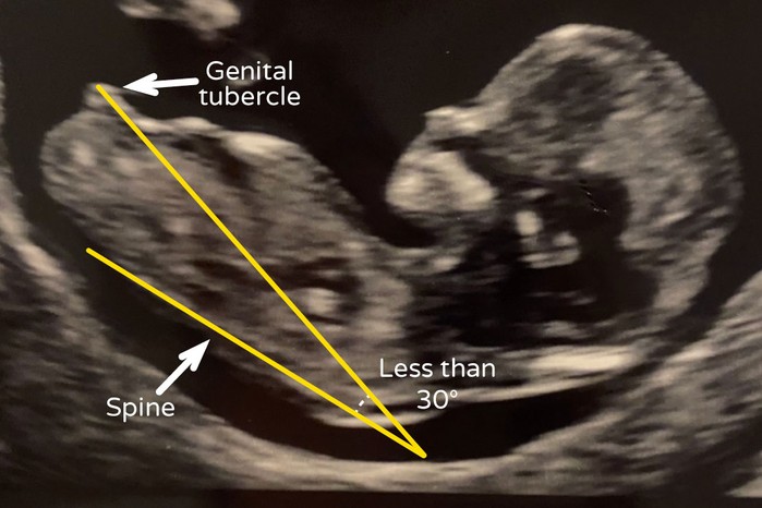
Boy vs Girl nub comparison at 12 weeks:
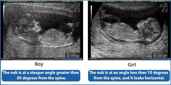
| MALE (Boy) | FEMALE (Girl) | |
|---|---|---|
| Nub angle | >30° from spine | <30° (parallel/flat) |
| Nub tip | Pointed, cranial notch upward | Rounded/forked, caudal notch downward |
| Appearance | Steeply pointing up | Lies along the spine |
| Accuracy at 12 weeks | ~96-99% (trained observer) | ~96-99% (trained observer) |
3. TURTLE SIGN - Male (2nd & 3rd Trimester)
Seen when the penis protrudes in front of the scrotum, like a turtle's head emerging from its shell.
Textbook USG - 33-week MALE fetus (3rd trimester):
Arrow = Penis | S = Scrotum | Testes visible inside scrotum
What to look for:
- Penis - echogenic cylindrical linear structure projecting forward
- Scrotum - rounded echogenic sac posterior to penis
- Testes - visible as oval structures inside scrotum (especially after 28 weeks as they descend)
- On transverse view: "Three-dot sign" - two testes on either side + penis in the middle
4. HAMBURGER SIGN / THREE-LINE SIGN - Female (2nd & 3rd Trimester)
What to look for:
- Three parallel echogenic lines between the legs
- Central line = clitoris/labia minora
- Two outer lines = labia majora
- Resembles a hamburger bun with a filling in the middle
- Also called the "fork sign" in sagittal view
| View | Female Appearance |
|---|---|
| Sagittal | 3 parallel lines, nub flat/horizontal |
| Transverse/axial | Two echogenic labia flanking central cleft |
| Coronal | "Fork" or "trident" appearance |
5. COMPLETE COMPARISON TABLE
| Feature | MALE | FEMALE |
|---|---|---|
| Nub angle (12 wks) | >30° from spine | <30°, parallel to spine |
| Nub tip | Cranial notch (upward) | Caudal notch (forked/downward) |
| Classic sign | Turtle sign | Hamburger / 3-line sign |
| Primary structure | Penis + scrotum | Labia majora + clitoris |
| Transverse view | Three dots (2 testes + penis) | Three parallel lines |
| Testes | Visible in scrotum (esp. 3rd trimester) | Absent |
| Uterus/ovaries | Not seen | Occasionally visible in 3rd trimester |
| Accuracy (>16 wks) | >95% | >95% |
| Accuracy (3rd tri) | ~99% | ~99% |
6. OPTIMAL TIMING FOR SEX DETERMINATION
| Gestational Age | Method | Accuracy |
|---|---|---|
| 11-12 weeks | Nub theory (sagittal angle) | ~70-80% |
| 13-14 weeks | Nub theory | ~96-99% |
| 16-20 weeks | Direct visualization of genitalia | >90% |
| 20-28 weeks | Turtle / Hamburger sign | >95% |
| 28+ weeks (3rd trimester) | All signs + testes visible in males | ~99% |
7. PITFALLS & ERRORS
- Crossed legs - most common cause of wrong sex determination
- Hypospadias in males can mimic female genitalia (3-line appearance)
- Clitoromegaly (CAH) in females can resemble a small penis
- Cord between legs can be mistaken for penis
- Suboptimal fetal position reduces accuracy significantly
Memory Hook
Boys POINT UP (nub >30°, turtle sticks out) Girls LIE FLAT (nub <30°, hamburger lies down)
⚠️ Legal Note (India): Disclosure of fetal sex is prohibited under the PCPNDT Act, 1994 - punishable by imprisonment and fine. This is a high-yield Forensic Medicine exam topic.
Sources: The Developing Human - Clinically Oriented Embryology (textbook, p.3251); PMC study on USG at 11-12 weeks; Nub Theory - Baby-Hunch
This is a shared conversation. Sign in to Orris to start your own chat.