
Give me the best photo of a unrooted teeth, single-rooted teeth.
Reading File
Searching the Web
single rooted teeth anatomy photo diagram
Now let me search the medical library for dental anatomy images and fetch the best image from the web results simultaneously.
Finding Sources
Loading Image
Loading Image
Finding Sources
Loading Image
Loading Image
Let me search for better, more specific single-rooted tooth images from the library and web.
Finding Sources
Searching the Web
single rooted teeth incisor canine labeled anatomy photo
Now let me fetch the best images from these results and also pull the textbook content:
Loading Image
Loading Image
Reading File
Let me now find the actual textbook images for teeth and fetch a better clinical photo:
Reading File
Loading Image
I found a textbook image URL. Let me now fetch it along with another detailed image:
Loading Image
I now have excellent textbook and anatomical images. Here is the full answer:
Single-Rooted Teeth - Anatomy & Best Images
Here are the best images available, sourced from authoritative medical textbooks and anatomical references:
1. Textbook Overview - Teeth and Sockets in the Jaws (Color Atlas of Human Anatomy)
This illustration from the Color Atlas of Human Anatomy, Vol. 2 (Fig. 4.9) shows the full dental arcade of the maxilla and mandible. The incisors (labeled 9) and canines (labeled 10) at the front of the arch are the primary single-rooted teeth in humans. Their sockets are small, round, and simple - contrasting with the larger multi-chambered sockets of molars visible at the back:
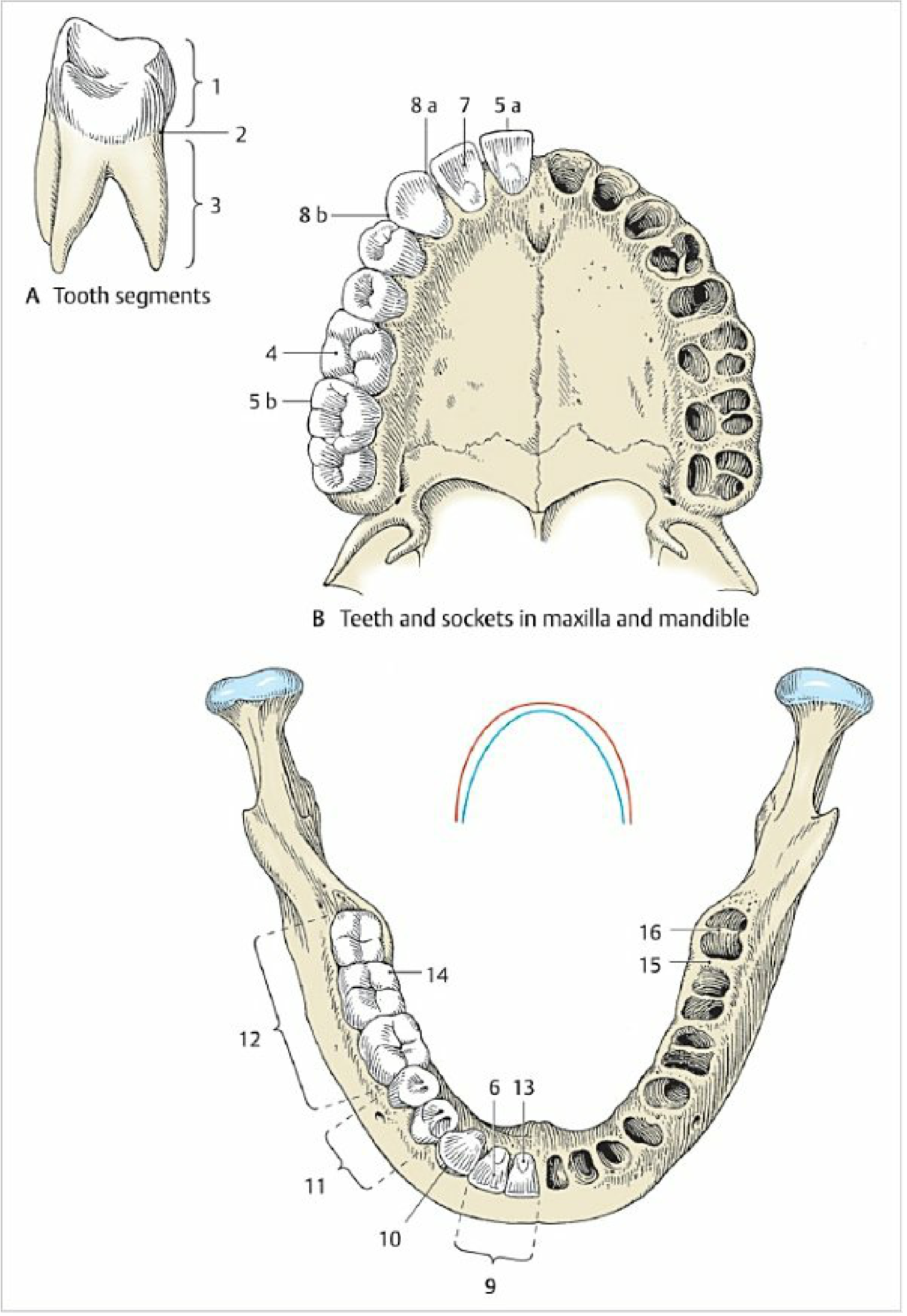
Color Atlas of Human Anatomy, Vol. 2 - Fig. 4.9
2. Labeled Diagram - Single-Rooted vs Multi-Rooted Tooth Comparison
This classic veterinary anatomy diagram shows a single-rooted incisor/canine (left column) clearly labeled with Crown and Root thirds (Incisal, Middle, Cervical, Apical), compared side-by-side with a multi-rooted molar (right). This is among the clearest labeled depictions available:

3. Cross-Section of a Single-Rooted Tooth (Endodontic Anatomy)
This clean clinical diagram shows the internal structure of a single-rooted tooth in cross-section, clearly showing the single root canal, pulp chamber, cementum, bone, periodontal ligament, and apical foramen - the defining feature of single-rooted teeth:

Key Facts: Which Teeth Are Single-Rooted?
Based on Color Atlas of Human Anatomy, Vol. 2 and Gray's Anatomy for Students:
| Tooth Type | Root Count | Notes |
|---|---|---|
| Central incisors | 1 | Single long, conical root |
| Lateral incisors | 1 | Single root (may be peg-shaped) |
| Canines (cuspids) | 1 | Longest single root in the mouth |
| Lower premolars | 1 | Simple single root |
| Upper premolars | 2 | Roots are divided |
| Lower molars | 2 | Two roots |
| Upper molars | 3 | Three roots |
The canine tooth is anatomically the archetypal single-rooted tooth - it has the longest, strongest single root in the entire dentition, making it a key anchor tooth in restorative dentistry. The incisor root is described as "single, long, and conical." - Color Atlas of Human Anatomy, Vol. 2, p. 267
This is a shared conversation. Sign in to Orris to start your own chat.