Now I have comprehensive data from the textbooks. Let me compile the full viva guide.
🧠 POSTERIOR FOSSA TUMOURS — COMPLETE NEUROSURGERY VIVA GUIDE
Sources: Bradley & Daroff's Neurology in Clinical Practice, Adams & Victor's Principles of Neurology (12th ed.), Robbins & Kumar Basic Pathology, Harrison's Internal Medicine 22E
📊 QUICK DIFFERENTIAL DIAGNOSIS AT A GLANCE
| Feature | Medulloblastoma | Ependymoma | Pilocytic Astrocytoma | Hemangioblastoma |
|---|
| Age | Children 4–8 yr | Children <3 yr (infratentorial) | Children & young adults | Adults 15–50 yr (peak 40) |
| WHO Grade | IV | II–III | I | I |
| Location | Cerebellar vermis / 4th ventricle roof | 4th ventricle floor | Cerebellar hemisphere (cyst + nodule) | Cerebellar hemisphere (cyst + nodule) |
| MRI hallmark | Solid, midline, restricted diffusion | Plastic/toothpaste extension through foramina | Large cyst + enhancing mural nodule | Cyst + intensely enhancing nodule (vascular) |
| Key molecule | WNT/SHH/Group 3/4 | ZFTA fusion, H3K27 loss (PF-A) | BRAF-KIAA1549 fusion | VHL gene mutation |
| Metastasis | CSF seeding (leptomeningeal) | CSF drop mets (~5%) | Rare | Rare (retina, spinal cord) |
| Prognosis | 70–80% 5-yr survival | Poor in <3 yr | Excellent (>90% 5-yr after GTR) | Excellent if complete excision |
🔵 TUMOR 1: MEDULLOBLASTOMA
1. DEMOGRAPHY
- Most common malignant brain tumor of childhood — accounts for ~20% of all childhood brain tumors, ~9.3% of all pediatric brain tumors
- Males > Females — 3:1 to 3:2 (varies by molecular subtype; SHH-activated has equal sex distribution)
- Peak age: 4–8 years; second smaller peak 18–25 years (adults rare)
- >50% occur in children under age 10
- Incidence stable over 20 years (SEER registry data)
2. GENETICS / ETIOLOGY
Molecular Subgroups (WHO 2021):
| Subgroup | Key Mutation | Age | Location | Prognosis |
|---|
| WNT-activated | CTNNB1 (β-catenin) mutations | Older children/adults | Cerebellar hemisphere | Best — >95% 5-yr survival |
| SHH-activated, TP53 wildtype | PTCH1, SMO, SUFU mutations | Infants / Adults | Lateral cerebellum | Intermediate |
| SHH-activated, TP53 mutant | TP53 + SHH pathway | Adolescents | Vermis/lateral | Worst in SHH |
| Non-WNT/Non-SHH Group 3 | MYC amplification | Infants/young children | Vermis | Poor |
| Non-WNT/Non-SHH Group 4 | MYCN, CDK6 amplification | Older children | Vermis | Intermediate |
Chromosome 17:
- Isochromosome 17q (i17q) — most common cytogenetic abnormality (>40%)
Familial Syndromes:
- Gorlin syndrome (Nevoid Basal Cell Carcinoma Syndrome) — PTCH gene mutation on 9q → desmoplastic/nodular variant
- Turcot syndrome — APC/WNT pathway mutations (colorectal polyps + brain tumor)
- Li-Fraumeni syndrome — TP53 mutation
Histological Variants:
| Variant | Features | Prognosis |
|---|
| Classic | Small blue cells, Homer Wright rosettes | Intermediate |
| Desmoplastic/Nodular | Pale nodular areas (neuronal diff), reticulin-rich stroma | Better (adults, lateral) |
| Extensively Nodular (MBEN) | Marked pale nodule expansion, neuropil-like tissue | Best (infants) |
| Large cell/Anaplastic | Widespread cellular anaplasia, high mitoses | Worst |
3. CLINICAL FEATURES
Symptoms (from hydrocephalus — 4th ventricle obstruction):
- Morning headaches awakening child from sleep (key feature)
- Projectile vomiting (often misdiagnosed as GI disease initially)
- Papilledema (due to raised ICP)
- Ataxia of gait + frequent falls → truncal ataxia (vermis involvement)
- 6th nerve palsy (false localizing sign from raised ICP)
- Diplopia, strabismus
- Mental torpor, deteriorating school performance
Duration before diagnosis: typically 1–5 months
Metastatic Spread:
- CSF/Leptomeningeal seeding — most common (drop metastases to spine)
- Bone mets — rare
- Lymph node — very rare
- Hematogenous — rare
Chang Staging (M-stage for metastasis):
| Stage | Definition |
|---|
| M0 | No metastasis |
| M1 | Tumor cells in CSF only |
| M2 | Nodular seeding in cerebellar/cerebral subarachnoid space |
| M3 | Nodular seeding in spinal subarachnoid space |
| M4 | Metastasis outside CNS |
4. RADIOLOGY
CT:
- Hyperdense (small blue cells, high N:C ratio) midline cerebellar mass
- Homogeneous contrast enhancement
- Hydrocephalus (dilated lateral + 3rd ventricles)
- Calcification in ~20%
MRI (Investigation of Choice):
When to do: Any child with signs of raised ICP, ataxia, morning vomiting
| Sequence | Finding |
|---|
| T1 | Hypointense to isointense |
| T2 | Isointense to hyperintense (heterogeneous) |
| FLAIR | Hyperintense |
| DWI/ADC | Restricted diffusion (low ADC) — KEY FEATURE: hypercellular small blue cells restrict water |
| T1+Gd | Heterogeneous enhancement |
| SWI | Calcification or hemorrhage blooming |
| MR Spectroscopy | High Cho/Cr, reduced NAA, taurine peak |
When NOT to do MRI: Pacemaker, cochlear implant (do CT instead)
Radiological Classification (Chang T-staging):
| T Stage | Description |
|---|
| T1 | <3 cm, limited to cerebellum |
| T2 | ≥3 cm, fills 4th ventricle |
| T3a | Fills 4th ventricle, exits through foramina |
| T3b | Fills 4th ventricle, invades brainstem |
| T4 | Extends through sylvian aqueduct or cervical canal |
Whole Spine MRI: MANDATORY for staging before surgery (M-staging for drop mets)
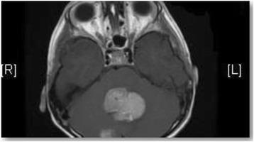
Axial T1+Gd MRI: large midline enhancing medulloblastoma with secondary obstructive hydrocephalus. Note dilation of temporal horns.
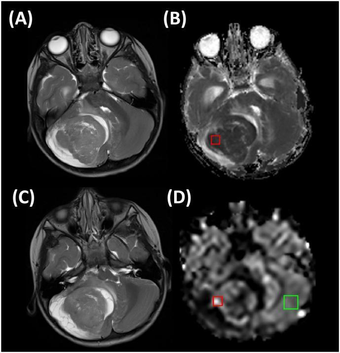
Multimodal MRI: T2 (A,C), ADC map (B — hypointense = restricted diffusion), CBF map (D — high perfusion = hypercellular tumor)
Medulloblastoma with Extensive Nodularity (MBEN): sagittal T2 shows "grape-like" hyperintense nodules — characteristic of infants, favorable prognosis
5. TUMOR MARKERS
- No specific serum tumor marker for medulloblastoma
- CSF cytology: malignant cells may be found (confirm M1 staging)
- LDH: elevated in CSF (nonspecific)
- Molecular markers (tissue): β-catenin nuclear staining (WNT), GLI2/PTCH (SHH)
6. BIOPSY & PATHOLOGY
Cell of Origin:
- Pluripotential stem cells of the external granular layer OR subependymal matrix of 4th ventricle
How/When to Take Biopsy:
- Preferred: Gross total surgical resection (primary) — tissue obtained at surgery
- Stereotactic biopsy if unresectable (rare for medulloblastoma)
- Avoid CSF sampling before surgery if herniation risk is high
Histology (H&E):
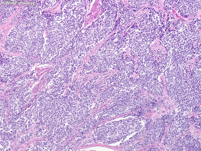
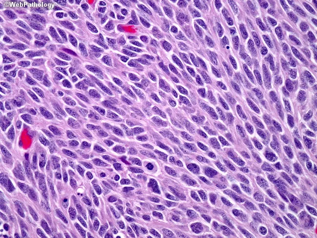
- Small round blue cells — scant cytoplasm, hyperchromatic round/carrot-shaped nuclei
- Homer Wright rosettes (neuroblastic rosettes) — tumor cells around neuropil core (no true lumen) — in ~50%
- Numerous mitoses and apoptotic bodies
- Synaptophysin-positive areas (neuronal differentiation)
Classic medulloblastoma: densely packed small blue cells, Homer Wright rosettes (neuropil core, no true lumen), high mitotic rate
Large cell/anaplastic variant: pronounced nuclear atypia, marked mitoses, geographic necrosis — worst prognosis
IHC Panel:
| Marker | Result | Significance |
|---|
| Synaptophysin | + | Neuronal differentiation |
| NSE | + | Neuroectodermal origin |
| GFAP | Variable/+ | Glial differentiation |
| β-catenin (nuclear) | + (WNT) | Diagnostic of WNT subgroup |
| INI1/SMARCB1 | Retained | Excludes ATRT |
| Ki-67 | High (>20%) | High proliferation |
| p53 | + in TP53 mutant SHH | Poor prognosis |
7. MANAGEMENT
🔪 Surgery
Indications: All fit patients — surgery is the cornerstone
Goal: Gross Total Resection (GTR) — <1.5 cm² residual tumor = acceptable
Approach: Posterior fossa craniotomy / suboccipital craniectomy
Surgical Steps:
- Position: prone, head fixed in Mayfield clamp, neck flexed
- Suboccipital midline incision
- Craniectomy/craniotomy at foramen magnum level
- Dural opening
- Identify tumor in vermis/4th ventricle
- Microsurgical resection with ultrasonic aspirator (CUSA)
- Identify and protect brainstem floor of 4th ventricle
- Confirm hemostasis, duraplasty if needed
- Temporary EVD (external ventricular drain) if hydrocephalus pre-op
Limitations: Floor of 4th ventricle — close relationship to brainstem (facial colliculus, vagal triangle)
Contraindications (relative): Severe brainstem invasion, distant M4 disease
Complications:
- Posterior fossa syndrome (cerebellar mutism — 25% of patients)
- CSF leak
- Cranial nerve palsies (6th, 7th)
- Bleeding from vermian veins
☢️ Radiotherapy
- Craniospinal irradiation (CSI) is MANDATORY — medulloblastoma is radiosensitive
- Standard risk: CSI 23.4 Gy + posterior fossa boost 54–55.8 Gy
- High risk: CSI 36–39.6 Gy + PF boost 55.8 Gy
- Infants <3 yr: Avoid RT (neurocognitive effects) → chemotherapy first, delay RT
- Proton beam therapy preferred to reduce integral dose
💊 Chemotherapy
| Regimen | Drugs | Use |
|---|
| Packer (Standard) | Vincristine (during RT) + CCNU + Cisplatin (post-RT) | Standard risk post-RT |
| High risk | Cisplatin + Etoposide + Cyclophosphamide | High risk post-RT |
| Infant | Carboplatin + VP-16 + Cyclophosphamide (Baby Brain protocols) | Delay RT in infants |
Key Drug Doses & Mechanisms:
| Drug | Dose | MOA | Side Effects |
|---|
| Cisplatin | 75 mg/m² q3-4 weeks | DNA crosslinks (alkylating) | Nephrotoxicity, ototoxicity, peripheral neuropathy |
| Carboplatin | AUC 5–7 | DNA crosslinks | Myelosuppression (less nephrotoxic than cisplatin) |
| Vincristine | 1.5 mg/m² weekly | Vinca alkaloid — spindle poison (anti-tubulin) | Peripheral neuropathy, SIADH |
| CCNU (Lomustine) | 75 mg/m² | Alkylating (nitrosourea) | Myelosuppression, secondary leukemia |
| Etoposide | 100 mg/m²/day × 3 | Topoisomerase II inhibitor | Myelosuppression, secondary AML |
| Cyclophosphamide | 1000–1500 mg/m² | Alkylating | Hemorrhagic cystitis (prevent with MESNA), alopecia |
🎯 Targeted Therapy
- Vismodegib (GDC-0449) — Hedgehog pathway (SMO inhibitor) for SHH-activated tumors
- Dose: 150 mg/day orally
- MOA: Inhibits Smoothened → blocks SHH signaling
- Side effects: Muscle cramps, alopecia, teratogenicity, premature growth plate fusion in children
- BET bromodomain inhibitors and CDK4/6 inhibitors — investigational for Group 3/4
🔑 MANAGEMENT CRUX FLOWCHART — MEDULLOBLASTOMA
Suspected Medulloblastoma (child, morning headache, vomiting, ataxia)
          ↓
MRI Brain + Whole Spine + CSF cytology (post-op)
          ↓
Staging: Standard risk vs High risk
   Standard: M0, GTR or <1.5cm² residual, no LCA histology
   High risk: M1–M4, or subtotal resection, or LCA/anaplastic, or age <3
          ↓
SURGERY (Posterior Fossa Craniotomy + GTR)
          ↓
CSF cytology (lumbar puncture ≥2 weeks post-op)
          ↓
┌─────────────────────────────────────┐
│ STANDARD RISK (>3 yr, M0, GTR)     │ HIGH RISK / <3 yr
│ CSI 23.4 Gy + PF boost 54 Gy      │ CSI 36 Gy + PF boost
│ + Vincristine (concurrent)         │ + Intensive chemo
│ + CCNU + Cisplatin (adjuvant)      │ (if <3 yr → chemo only first)
└─────────────────────────────────────┘
          ↓
Follow-up MRI every 3–6 months × 5 yr, then annually
8. FOLLOW-UP & PROGNOSIS
- 5-year survival: 70–80% overall; WNT subgroup >95%, SHH-TP53 mutant worst
- Standard risk: 80% 5-year EFS
- High risk: 60–65% 5-year EFS
- MRI brain + spine every 3 months for 2 years, then every 6 months, then annually
- Neuropsychological testing (IQ, memory) — radiation effects
- Audiogram (cisplatin ototoxicity)
- Endocrine surveillance (GH deficiency, hypothyroidism from CSI)
🟢 TUMOR 2: PILOCYTIC ASTROCYTOMA (PA)
1. DEMOGRAPHY
- Most common glioma in children — ~17.6% of all pediatric brain tumors
- Most common pediatric cerebellar tumor overall
- WHO Grade I — benign, potentially curable
- No clear gender predilection
- Predominantly first two decades of life (but can occur in adults)
- NF1 (Neurofibromatosis Type 1) associated — bilateral optic nerve gliomas; germline NF1 mutation
2. GENETICS / ETIOLOGY
Molecular Hallmark:
- BRAF-KIAA1549 fusion (~70% of sporadic cases) — constitutively active BRAF → MAPK/ERK signaling
- BRAF V600E mutation — lower percentage; more common in cerebral/diencephalic PAs
- FGFR1 mutations, NTRK2 fusions — rare subsets
- NF1 gene inactivation — NF1-associated cases
- No IDH mutation (distinguishes from diffuse astrocytomas)
Predisposing Conditions:
- NF1 — most important; optic pathway gliomas often indolent in NF1
- No other definite environmental factors
3. CLINICAL FEATURES
Locations (in order of frequency):
- Cerebellum (most common) — cerebellar hemisphere
- Optic pathways (optic nerve, chiasm) — especially in NF1
- Hypothalamus / 3rd ventricle
- Brainstem (dorsal exophytic)
- Cerebral hemispheres
- Spinal cord (rare)
Symptoms by Location:
- Cerebellum: Raised ICP (headache, vomiting), limb ataxia, papilledema
- Optic pathway: Visual loss, proptosis, nystagmus
- Hypothalamus: Diencephalic syndrome (in infants) — emaciation despite normal linear growth, hyperemesis, hyperkinesis, nystagmus (classic viva question!)
- Brainstem: Obstructive hydrocephalus, cranial nerve palsies
4. RADIOLOGY
MRI — Investigation of Choice:
Classic Appearance (cerebellum): Large cyst + intensely enhancing mural nodule
| Feature | Finding |
|---|
| T1 cyst | Hypointense (CSF-like) |
| T2 cyst | Hyperintense |
| Mural nodule | Iso/hypointense T1 → intense enhancement post-Gd |
| T2 nodule | Hyperintense |
| Edema | Minimal |
| DWI | No restricted diffusion (low cellularity — key differentiator from medulloblastoma) |
| Spectroscopy | Elevated Cho, decreased NAA, reduced Cho/Cr ratio vs GBM |
When to do: All children with posterior fossa symptoms, ataxia, or raised ICP
When NOT to do: Active hemorrhage risk or claustrophobia (use sedation for young children)
Note: Hypothalamic/optic gliomas in NF1 are usually solid (not cystic)
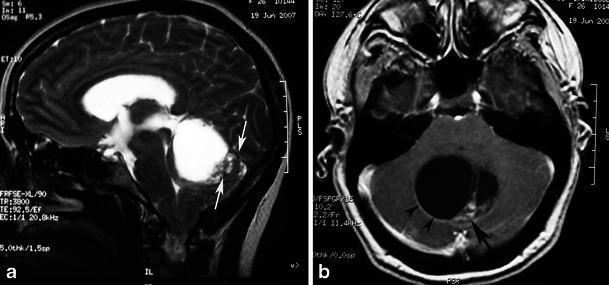
Sagittal T2 (a): large cystic cerebellar PA with posterior mural nodule (arrows). Axial T1+Gd (b): intense nodule enhancement with cyst wall enhancement (arrowheads) — classic PA in a 26-year-old female
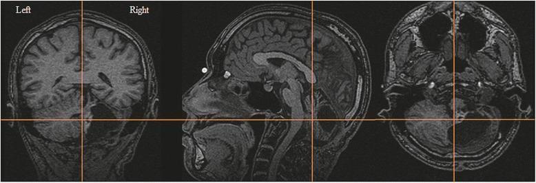
Coronal, sagittal, axial T1 MRI of cerebellar PA: large hypointense cyst with enhancing mural nodule in vermis area, significant hemispheric displacement
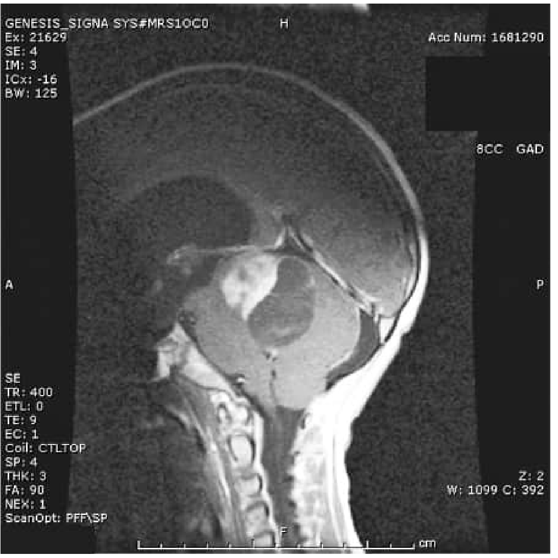
Sagittal contrast MRI in 9-year-old boy: anterior posterior fossa solid component + posterior cyst. Resection confirmed pilocytic astrocytoma. (Bradley & Daroff)
5. TUMOR MARKERS
- No specific serum marker
- Molecular testing (tissue): BRAF-KIAA1549 fusion (FISH or RT-PCR), BRAF V600E (IHC or sequencing)
6. BIOPSY & PATHOLOGY
Cell of Origin: Astrocytes (GFAP-positive glial cells)
When to Biopsy:
- Tissue diagnosis should always be attempted (except NF1 patients with typical optic pathway glioma — may observe)
- Resection specimen is standard; stereotactic biopsy for inaccessible lesions (e.g., hypothalamus)
Histology — KEY FEATURES:
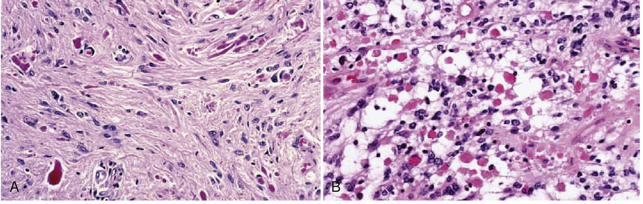
- Biphasic pattern — dense compact pilocytic areas + loose microcystic spongy areas (most important histologic feature)
- Piloid cells — bipolar astrocytes with long hair-like processes
- Rosenthal fibers — bright red/pink, fusiform or corkscrew-shaped intracellular aggregates of GFAP (pathognomonic of low-grade/indolent process)
- Eosinophilic granular bodies (EGBs) — mulberry-shaped pink inclusions
- Low mitotic rate, no necrosis, no microvascular proliferation
Fig. 72.8 (Bradley & Daroff): Biphasic PA — Dense (A) with Rosenthal fibers (bright red corkscrews) and loose (B) areas with eosinophilic granular bodies (hematoxylin-eosin ×200)
IHC:
| Marker | Result |
|---|
| GFAP | Strongly positive |
| S100 | + |
| BRAF V600E (clone VE1) | + (if V600E mutant) |
| IDH1/2 | Negative (key distinguisher from diffuse astrocytoma) |
| Ki-67 | Low (<5%) |
| p53 | Negative/rare |
WHO 2021 Classification:
- Pilocytic astrocytoma, WHO Grade I — all subtypes
- Pilomyxoid astrocytoma — variant (historically Grade II, now ungraded; age <3 yr, hypothalamic)
7. MANAGEMENT
🔪 Surgery
Indication: All accessible cerebellar PAs → Gross Total Resection is curative
Surgical Steps (cerebellar PA):
- Prone position, Mayfield head clamp
- Posterior fossa craniotomy (suboccipital)
- Identify cyst — evacuate cystic fluid
- Locate mural nodule (wall of cyst — does NOT need full cyst wall excision as cyst wall is not tumor)
- Excise the mural nodule completely — this is the actual tumor (cyst wall = reactive gliosis, benign)
- Cyst wall alone does not recur — key surgical principle
- Duraplasty and closure
Limitations: Hypothalamic, optic chiasmatic, brainstem location — risks of visual loss, hormone dysfunction
Contraindications (relative): NF1 with bilateral optic gliomas — observe if asymptomatic
☢️ Radiotherapy
- Avoid in children — neurocognitive effects, secondary high-grade gliomas (especially NF1!)
- Radiation is a relative contraindication in NF1 (risk of vasculopathy, moyamoya, secondary tumors)
- Reserved for progressive unresectable disease in adults or refractory cases
💊 Chemotherapy (for unresectable/progressive disease)
| Regimen | Drugs | Notes |
|---|
| First line | Carboplatin + Vincristine | Carboplatin: AUC 6.5, Vincristine: 1.5 mg/m² |
| Alternative | Vinblastine monotherapy | Weekly, 6 mg/m² |
| TPCV | Thioguanine + Procarbazine + CCNU + Vincristine | Marginally superior EFS but worse safety → not first line |
🎯 Targeted Therapy (Emerging — Key for Viva!)
- BRAF + MEK inhibitors — powerful second-line options:
- Dabrafenib + Trametinib (BRAF V600E + MEK inhibitor) — impressive responses
- Selumetinib (MEK inhibitor) — for BRAF-fused tumors, especially NF1 (FDA approved for NF1 plexiform neurofibromas)
- Mechanism: MEK inhibition → blocks MAPK/ERK pathway → tumor regression
🔑 MANAGEMENT CRUX FLOWCHART — PILOCYTIC ASTROCYTOMA
Suspected PA (child, cerebellar symptoms, MRI cyst + nodule)
          ↓
MRI Brain (± Spine if symptomatic)
Molecular testing (BRAF status)
          ↓
          ↓
┌──────────────────────────────────────────┐
│ ACCESSIBLE (cerebellum, cortical)        │ INACCESSIBLE / UNRESECTABLE
│ → GTR = CURATIVE                        │ (hypothalamus, optic pathway, BS)
│ → >90% recurrence-free at 5 yr          │ → Observe (NF1 optic glioma)
│ → No adjuvant therapy needed            │ → Chemo: Carboplatin + Vincristine
└──────────────────────────────────────────┘ → Targeted: BRAF/MEK inhibitor
          ↓
FOLLOW-UP MRI every 6 months × 3 yr
then annually
(Visual testing if optic pathway)
8. FOLLOW-UP & PROGNOSIS
- Excellent — 80% 20-year survival (Bradley & Daroff)
- After GTR cerebellum: >90% 5-year recurrence-free survival (Shaw & Wisoff)
- Malignant transformation is rare
- Centrally located tumors (hypothalamus, brainstem) have reduced PFS
- MRI every 6 months × 2–3 years, then annually
🟡 TUMOR 3: EPENDYMOMA
1. DEMOGRAPHY
- 3rd most common pediatric brain tumor — ~4–10% of all brain tumors; 5.5% of pediatric brain tumors
- Most common in 1st decade of life (infratentorial)
- Males:Females ≈ 2:1 (infratentorial)
- Age <3 years → significantly worse prognosis
- 90% intracranial, 10% spinal (spinal ependymoma more common in adults)
- Of intracranial: 75% infratentorial (posterior fossa, 4th ventricle) in children; supratentorial more common in adults
2. GENETICS / ETIOLOGY
Molecular Classification (WHO 2021 — DNA methylation based):
Posterior Fossa:
| Subgroup | Age | Biology | Prognosis |
|---|
| PF-EPN-A | Infants/young children | H3K27 trimethylation loss (EZHIP/EZH2); bland genome; chr 1q gain (20%) | Poor |
| PF-EPN-B | Adolescents/adults | Chr gains/losses; H3K27 trimethylation retained | Better |
Supratentorial:
- ST-EPN-RELA (ZFTA-RELA fusion) — NF-κB pathway activation
- ST-EPN-YAP (YAP1-MAML2 fusion) — better prognosis
Spinal:
- NF2 mutations (22q deletion) — spinal ependymomas
- Myxopapillary, classic, subependymoma subtypes
Key Points:
- NF2 mutations / 22q deletion — spinal ependymomas
- Chr 22q deletion — common in all groups
- No IDH mutations (unlike adult diffuse gliomas)
3. CLINICAL FEATURES
Infratentorial Ependymoma (4th Ventricle):
- Headache, vomiting, papilledema (obstructive hydrocephalus)
- Ataxia (cerebellar compression)
- Torticollis / neck pain (extension through foramen of Magendie)
- Hoarseness, dysphagia, lower cranial nerve palsies (extension through foramina of Luschka → CPA cistern)
- Downward beating nystagmus (impending tonsillar herniation)
- Increased head circumference in infants
Symptoms present for 1–2 years before diagnosis (slower onset than medulloblastoma)
Spinal Ependymoma:
- Localized pain worse at night (recumbent → spinal venous congestion)
- Radiculopathy, weakness, sensory loss
Metastatic Spread:
- CSF seeding / leptomeningeal drop mets in ~5% (less than medulloblastoma)
- Associated with poor prognosis
4. RADIOLOGY
Classic MRI Appearance:
"Plastic" or "toothpaste" extension through foramina — the tumor squeezes out through foramina of Luschka (laterally) and Magendie (inferiorly) into cisterns — PATHOGNOMONIC
| Feature | Finding |
|---|
| Location | 4th ventricle floor → extends to foramina |
| T1 | Hypointense/isointense |
| T2 | Hyperintense, heterogeneous |
| Enhancement | Moderate, heterogeneous |
| Calcification | Common (seen on CT/SWI) — distinguishes from medulloblastoma |
| DWI/ADC | No significant restriction (unlike medulloblastoma) — higher ADC values |
| SWI | Blooming from calcification/hemorrhage |
| Cysts | Common |
CT: May show calcification (useful differentiator from medulloblastoma)
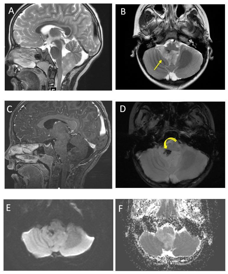
Posterior fossa ependymoma in 6-year-old: T2 (A,B) — heterogeneous 4th ventricular mass with classic "plastic" extension through right foramen of Luschka (yellow arrow). SWI (D) — calcification blooming. ADC (F) — high values (no significant restriction)
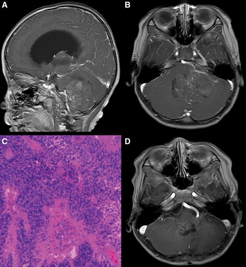
Ependymoma WHO Grade II: (A,B) Preop sagittal/coronal T1+Gd — large 4th ventricular mass with hydrocephalus. (C) H&E showing perivascular pseudorosettes. (D) Recurrence at 34 months on follow-up MRI
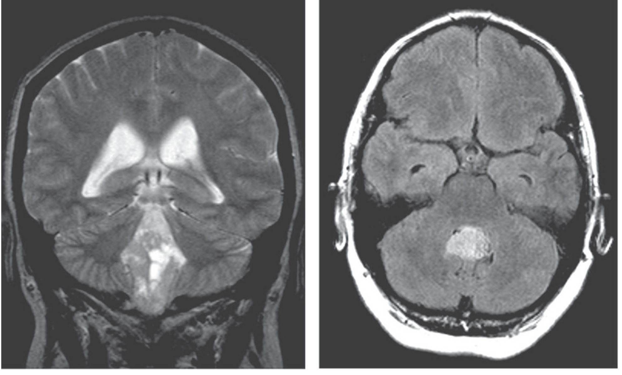
Fig. 30-12 (Adams & Victor): Coronal T2 — ependymoma growing from 4th ventricle floor. Axial FLAIR — mass obliterating 4th ventricle
Whole Spine MRI: Recommended for staging (drop metastases)
5. TUMOR MARKERS
- No specific serum markers
- CSF cytology — may detect malignant cells
- Molecular: ZFTA fusion (ST-RELA), H3K27me3 loss (PF-A — IHC loss = PF-A subtype)
6. BIOPSY & PATHOLOGY
Cell of Origin: Ependymal cells lining ventricles and central canal
Histology — KEY FEATURES:
- Perivascular pseudorosettes — cells radiating around blood vessels with anuclear zones (most common, most important — always present)
- True ependymal rosettes — cells around a central lumen (canal) — in ~10% only but more specific
- Ependymal canals — slit-like structures resembling miniature ventricles
- Well-circumscribed, compresses rather than infiltrates
- Uniform cells with round/oval nuclei, moderate chromatin
Electron Microscopy (if morphologically ambiguous):
- Cilia with basal bodies (blepharoplasts)
- Microvilli
- Zipper-like intercellular junctions
- Intracellular lumina
(EM is confirmatory when light microscopy is ambiguous)
Grading:
- WHO Grade II — ependymoma (classic)
- WHO Grade III — anaplastic ependymoma (hypercellularity, increased mitoses, microvascular proliferation, necrosis)
- Note: Histological grading may be phased out — molecular classification more prognostic
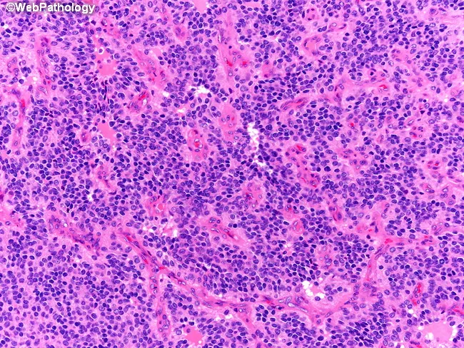
Classic ependymoma: perivascular pseudorosettes — tumor cells radiate around central blood vessels, creating anuclear halo zones
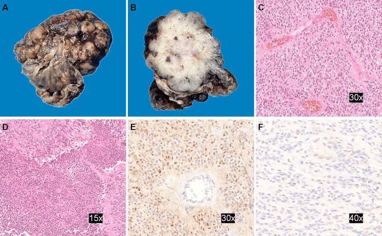
Ependymoma pathology: macroscopic solid-cystic tumor, H&E showing perivascular pseudorosettes, EMA immunohistochemistry showing characteristic dot-like/ring-like cytoplasmic positivity
IHC:
| Marker | Result | Note |
|---|
| GFAP | + (highlights pseudorosette processes) | Thin processes to vessels |
| EMA | + (dot-like / ring-like cytoplasmic) | Key distinguishing feature |
| CD99 | + | Dot-like |
| D2-40 | + | Dot-like |
| S100 | + | Nonspecific |
| H3K27me3 (IHC) | Loss = PF-EPN-A | Important prognostic marker |
| Synaptophysin | − | Excludes medulloblastoma |
| Ki-67 | Variable | |
7. MANAGEMENT
🔪 Surgery
Goal: GTR (Gross Total Resection) — extent of resection is the most important prognostic variable
Surgical Steps (4th ventricular ependymoma):
- Prone positioning, Mayfield head clamp
- Suboccipital craniotomy ± C1 laminectomy (if tumor extends into cervical canal)
- Dural opening, gentle retraction of cerebellar tonsils
- Identify tumor in 4th ventricle
- Microsurgical debulking with CUSA/bipolar
- Careful dissection from floor of 4th ventricle (brainstem)
- If tumor extends through foramina of Luschka → may need additional CPA approach
- EVD placement if hydrocephalus
Indications: All patients — attempt GTR
Limitation: Floor of 4th ventricle — brainstem attachment
Second-look surgery: Considered if residual tumor found on post-op MRI (<72 hours)
☢️ Radiotherapy
- Standard of care post-surgery for all children >1–3 years old
- Local conformal RT to tumor bed: 54–59.4 Gy (NOT craniospinal — unlike medulloblastoma, because CSF dissemination is rare)
- CSI only if metastatic disease confirmed
- Infants <1–3 yr: Chemotherapy first to delay RT
- Proton therapy preferred (posterior fossa — cochlea sparing)
💊 Chemotherapy
- Adjuvant chemo does NOT improve survival (unlike medulloblastoma) — chemotherapy role limited
- Used to delay RT in infants (Baby protocols)
- Regimens: Carboplatin + Vincristine, or Cisplatin + Etoposide
- Re-irradiation used at recurrence
🎯 Targeted/Future Therapy
- EZH2 inhibitors (tazemetostat) — for PF-EPN-A with EZHIP overexpression (investigational)
- VEGF pathway (bevacizumab) — investigational
🔑 MANAGEMENT CRUX FLOWCHART — EPENDYMOMA
Posterior fossa ependymoma (4th ventricular mass, "plastic" extension)
          ↓
MRI Brain + Spine + CSF cytology
          ↓
SURGERY (GTR primary goal)
If residual: Second-look surgery (<72 hr post-op MRI)
          ↓
Post-op MRI at 48–72 hours
          ↓
Molecular profiling (H3K27me3 IHC, ZFTA fusion, DNA methylation)
          ↓
┌────────────────────────────────────────┐
│ >3 yr, localized (M0)                 │ <3 yr OR M+ disease
│ Local conformal RT 54–59.4 Gy         │ Chemotherapy (delay RT)
│ (No CSI unless M+)                    │ CSI if M+ disease
└────────────────────────────────────────┘
          ↓
MRI every 3 months × 2 yr, then every 6 months
Recurrence → Re-irradiation ± chemo
8. FOLLOW-UP & PROGNOSIS
- Prognosis worse than pilocytic astrocytoma, better than medulloblastoma high-risk
- Age <3 years: significantly worse prognosis
- PF-EPN-A: poor prognosis regardless of grade
- PF-EPN-B: better prognosis
- GTR vs. subtotal resection: major survival difference
- 5-year OS: ~75% (GTR) vs ~30% (incomplete resection)
- MRI follow-up every 3 months for 2 years, then every 6 months
🔴 TUMOR 4: HEMANGIOBLASTOMA
1. DEMOGRAPHY
- Most common PRIMARY cerebellar neoplasm in adults
- WHO Grade I — benign
- Age range: 15–50 years, peak at 40 years
- Males > Females
- ~10% associated with Von Hippel-Lindau (VHL) disease (familial); rest sporadic
- Equally common in all races (blacks, whites, Asians)
- Rare in children
2. GENETICS / ETIOLOGY
VHL Disease (Autosomal Dominant):
- VHL gene on chromosome 3p25-26 — tumor suppressor gene
- Germline VHL mutation ("first hit") → second somatic hit → tumor formation
- VHL protein normally promotes HIF-α degradation → VHL loss → HIF-α accumulation → VEGF/EPO overexpression → angiogenesis
- Presents with CNS hemangioblastomas + renal cell carcinoma (clear cell) + pheochromocytoma + pancreatic cysts + retinal angiomas
Sporadic:
- Somatic VHL inactivation (both alleles)
Associations in VHL:
- CNS hemangioblastomas (cerebellum > brainstem > spinal cord)
- Retinal hemangioblastoma (first sign!)
- Clear cell renal cell carcinoma (major cause of morbidity/mortality)
- Pheochromocytoma
- Pancreatic neuroendocrine tumors/cysts
3. CLINICAL FEATURES
Symptoms:
- Dizziness and ataxia (unilateral cerebellar hemisphere involvement)
- Signs of raised ICP (hydrocephalus from 4th ventricle compression)
- Polycythemia — (viva pearl!) tumor elaborates erythropoietin → secondary erythrocytosis (Hb elevated)
- Retinal angioma — may cause visual symptoms (often the first finding in VHL)
- Spontaneous hemorrhage (tumor is very vascular)
VHL Syndrome Features:
- Hepatic/pancreatic cysts
- Renal cell carcinoma
- Pheochromocytoma (episodic hypertension, sweating, palpitations)
4. RADIOLOGY
MRI — Investigation of Choice:
Classic Appearance: Cyst + intensely enhancing mural nodule (similar to PA but in adults)
| Feature | Finding |
|---|
| T1 cyst | Hypointense |
| T2 cyst | Hyperintense |
| Mural nodule | Intense homogeneous enhancement (highly vascular) |
| Flow voids | Present in/around nodule (large feeding vessels) |
| Location | Cerebellar hemisphere (posterior pial surface) |
| Solid variant | ~40% are solid (multiple lesions in VHL) |
| SWI | May show hemosiderin from prior hemorrhage |
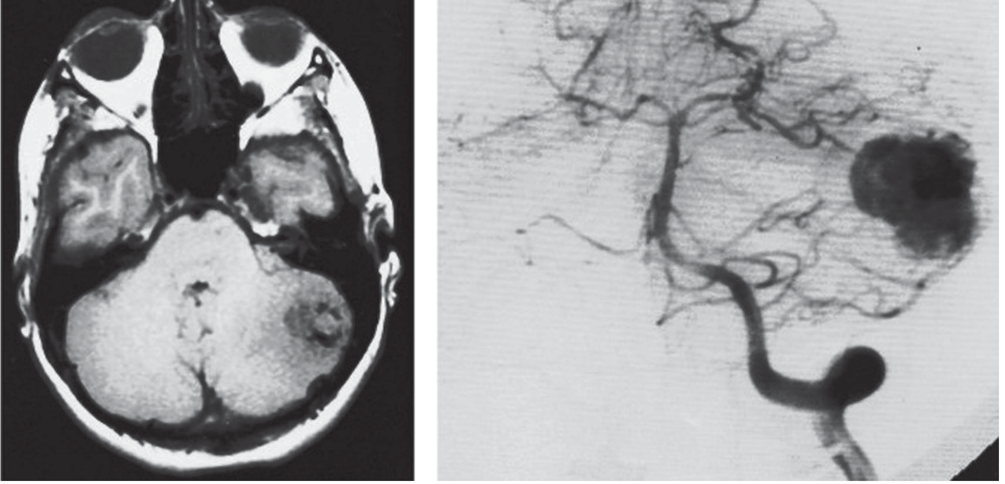
Angiography (DSA): Classic — cluster of small vessels forming a hypervascular nodule 1–2 cm diameter with dilated draining veins (Fig. 30-13, Adams & Victor)
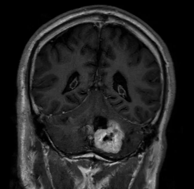
Cerebellar hemangioblastoma in 38-year-old male: MRI gadolinium — cystic mass with sharply enhancing mural nodule (high vascularity). VHL screen should be performed.
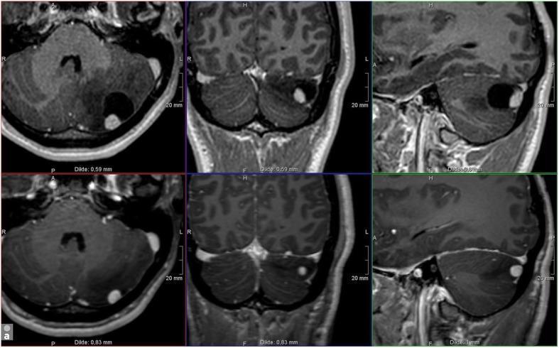
VHL disease — 20-year-old male: 3D-T1 Gd axial/coronal/sagittal (upper 2018 vs lower 2017): interval growth of enhancing nodule + cyst — supporting surveillance protocol in VHL
Fig. 30-13 (Adams & Victor): Axial contrast MRI — vascular left cerebellar tumor. Vertebral angiogram — hypervascular nodule with dilated draining veins
When to screen for VHL: Any cerebellar hemangioblastoma, any age → genetic testing especially if:
- Age <40 years
- Multiple lesions
- Family history
- Retinal angioma
5. TUMOR MARKERS
- Erythropoietin (EPO) — elevated serum EPO → polycythemia (Hb >18 g/dL, Hct >52%)
- Normal: Men 4.3–17.7 mIU/mL; Women 4.3–9.9 mIU/mL
- In hemangioblastoma: often >20–30 mIU/mL
- Secondary erythrocytosis resolves after tumor removal
- VHL genetic testing — germline mutation analysis
6. BIOPSY & PATHOLOGY
Cell of Origin: Stromal cells (neoplastic component) — uncertain histogenesis (mesenchymal origin)
Histology — KEY FEATURES:
- Abundant capillaries throughout tumor mass (rich vascular network)
- Foamy lipid-laden stromal cells — the actual neoplastic component (interspersed between capillaries)
- Solid portions are dark red (vascular)
- Sharply demarcated from surrounding tissue
IHC:
| Marker | Result | Note |
|---|
| Inhibin-α | Strongly + | Most specific marker |
| S100 | + | Stromal cells |
| NSE | + | Stromal cells |
| Brachyury | + | Recent finding |
| GFAP | Patchy/+ | Nonspecific |
| D2-40 | + | Stromal cells |
| EMA | − | Helps exclude metastatic RCC |
| CD10 | − | Helps exclude metastatic RCC |
| RCC antigen | − | Distinguishes from metastatic RCC |
Distinguishing from Metastatic Renal Cell Carcinoma (VHL context!):
- Inhibin-α +, RCC antigen −, CD10 − = hemangioblastoma
- RCC: EMA+, PAX8+, CD10+, inhibin-α −
7. MANAGEMENT
🔪 Surgery
Indication: Symptomatic hemangioblastomas — surgical excision is curative if complete
Key Surgical Principles:
- En-bloc excision (do NOT piecemeal excision — risk of torrential hemorrhage)
- Open the cerebellar cyst → evacuate fluid → identify mural nodule on wall
- Excise the nodule en-bloc — cyst wall alone does not need resection
- Pre-operative embolization (endovascular) — controversial; used for large vascular nodules; does not clearly reduce recurrence
- Complete removal = cure (high recurrence if nodule left)
Complications:
- Hemorrhage (highly vascular tumor)
- Cranial nerve injury
- Recurrence if incomplete
☢️ Radiosurgery
- Stereotactic radiosurgery (SRS/Gamma Knife) — for:
- Multiple lesions (VHL)
- Surgically inaccessible lesions
- Small asymptomatic lesions
- Recurrent lesions
- Series show good control rates (~90% at 5 years for small lesions)
No standard chemotherapy
🎯 Targeted Therapy (Investigational):
- Bevacizumab (anti-VEGF) — for VHL-associated hemangioblastomas (VHL pathway drives VEGF)
- HIF-2α inhibitors (belzutifan) — FDA approved for VHL-associated tumors (RCC, CNS hemangioblastomas)
- Dose: 120 mg orally daily
- MOA: Inhibits HIF-2α → reduces VEGF/EPO transcription
- Major breakthrough for VHL disease management (2021 FDA approval)
🔑 MANAGEMENT CRUX FLOWCHART — HEMANGIOBLASTOMA
Adult, cerebellar cyst + mural nodule, ataxia ± polycythemia
          ↓
MRI Brain + Spine (for multiple lesions)
CBC (polycythemia?), EPO level
VHL gene testing + ophthalmic exam (retinal angioma)
Abdominal MRI (renal, pancreatic involvement in VHL)
          ↓
┌──────────────────────────────────────┐
│ SYMPTOMATIC / GROWING               │ ASYMPTOMATIC / SMALL
│ → Surgical excision (en-bloc)       │ → Observe (VHL: serial MRI 6-12 mo)
│ → OR Gamma Knife SRS               │ → SRS if growing/asymptomatic
│ (if small/multiple/inaccessible)    │
└──────────────────────────────────────┘
          ↓
VHL disease? → Multidisciplinary:
Screen family, Renal surveillance,
Ophthalmology, Endocrine (pheo)
          ↓
Follow-up MRI every 6–12 months
New lesions: treat as they become symptomatic
8. FOLLOW-UP & PROGNOSIS
- Excellent with complete excision of nodule
- High recurrence if nodule not completely removed
- VHL patients: new lesions form throughout life → lifelong surveillance
- Children of affected parents should be tested for VHL mutation and screened for retinal angioma
- Retinal lesions cause blindness if untreated
- Polycythemia resolves after tumor removal
- MRI annually for VHL; every 1–2 years after sporadic complete excision
⚖️ DIFFERENTIAL DIAGNOSIS OF POSTERIOR FOSSA TUMORS
Comprehensive Comparison Table
| Feature | Medulloblastoma | Ependymoma | Pilocytic Astrocytoma | Hemangioblastoma | ATRT | Brainstem Glioma |
|---|
| Age | 4–8 yr | <3 yr | <15 yr | 30–50 yr | <3 yr | Any (peak 5–10 yr) |
| Grade | IV | II–III | I | I | IV | II–IV |
| Location | Vermis/4th ventricle roof | 4th ventricle floor | Cerebellar hemisphere | Cerebellar hemisphere | 4th ventricle / CP angle | Pons/brainstem |
| Onset | Rapid (1–5 mo) | Slower (1–2 yr) | Slow | Slow | Rapid | Gradual |
| MRI key | Midline, restricted diffusion, homogeneous | Plastic extension foramina, calcification | Cyst + nodule, no restriction | Cyst + intensely enhancing nodule, flow voids | Heterogeneous, hemorrhage | DIPG: pons expansion |
| ADC | Low (restricted) | High (no restriction) | High (no restriction) | High (no restriction) | Low | Variable |
| Calcification | 20% | Common | Rare | Rare | Present | Rare |
| Polycythemia | No | No | No | Yes (EPO) | No | No |
| Hydrocephalus | Early, severe | Early | When large | Late | Yes | From aqueduct |
| CSF dissemination | Common | 5% | Rare | Rare | Common | Rare |
| Key marker | CTNNB1/SHH | ZFTA fusion/H3K27 | BRAF-KIAA1549 | VHL mutation/inhibin-α | SMARCB1 loss | H3K27M (DIPG) |
| Best prognosis | WNT subtype | PF-EPN-B | ALL (especially GTR) | ALL (if complete excision) | Very poor | Very poor (DIPG) |
Quick Radiological Differentiators
POSTERIOR FOSSA MASS
        ↓
Is it MIDLINE (vermis/4th ventricle)?
├── YES → Medulloblastoma (child) OR Ependymoma
│   ├── Restricted diffusion (low ADC) → Medulloblastoma
│   ├── Plastic extension through foramina, calcification → Ependymoma
│   └── Floor attachment (brainstem floor) → Ependymoma
└── NO → Hemispheric mass
    ├── Cyst + mural nodule, CHILD → Pilocytic Astrocytoma
    ├── Cyst + mural nodule, ADULT ± polycythemia → Hemangioblastoma
    ├── Solid, enhancing, ADULT → Metastasis (lung, breast, melanoma)
    └── Multiple lesions → Metastases or VHL hemangioblastomas
Additional Posterior Fossa Tumors to Know
Atypical Teratoid/Rhabdoid Tumor (ATRT)
- Age: <3 years (most common CNS malignancy in infants)
- SMARCB1/INI1 deletion/mutation (chromosome 22q11.2) — IHC: INI1 loss
- Heterogeneous MRI, restricted diffusion, hemorrhage
- Extremely aggressive, poor prognosis
- Chemotherapy-intensive, RT if tolerated
Choroid Plexus Tumors
- Papilloma (WHO I) vs. Carcinoma (WHO III)
- Location: 4th ventricle (childhood) and lateral ventricle
- Presents with hydrocephalus (overproduction of CSF)
- MRI: frond-like enhancing mass in ventricle
- Surgery curative for papilloma
Brainstem Glioma / DIPG
- Diffuse Intrinsic Pontine Glioma (DIPG) — H3K27M mutation (histone)
- Expands the pons (>50% pons involved = diagnostic)
- Clinical: cranial nerve palsies (VI, VII), long tract signs, ataxia
- Biopsy: Now recommended (for molecular diagnosis, clinical trials)
- Treatment: RT (54 Gy) — palliation; ONC201/suramin (targeted for H3K27M)
Dermoid / Epidermoid Cysts
- Epidermoid: CPA angle, young adults; "pearly tumor"; DWI shows restriction (NOT enhancement)
- Dermoid: Midline, children; skin appendages; fat signal on T1
- Chemical meningitis if ruptured
📋 PEDIATRIC BRAIN TUMOR FREQUENCY TABLE (from Bradley & Daroff)
| Histology | % All Pediatric Brain Tumors | Median Age |
|---|
| Pilocytic Astrocytoma | 17.6% | 7 yr |
| Other low-grade glioma | 14.3% | 6 yr |
| High-grade glioma | 11.1% | 7 yr |
| Medulloblastoma | 9.3% | 6 yr |
| Ependymal tumors | 5.5% | 4 yr |
| ATRT | 2.3% | 1 yr |
🔬 OVERALL IHC COMPARISON CHEATSHEET
| IHC Marker | Medulloblastoma | Ependymoma | Pilocytic Astrocytoma | Hemangioblastoma |
|---|
| GFAP | Variable | + (pseudorosettes) | Strongly + | Patchy |
| Synaptophysin | + | − | − | − |
| EMA | − | + (dot-like) | − | − |
| Inhibin-α | − | − | − | Strongly + |
| IDH1/2 | − | − | − | − |
| H3K27me3 loss | − | + (PF-A only) | − | − |
| β-catenin (nuclear) | + (WNT) | − | − | − |
| Ki-67 | High | Moderate | Low | Low |
| INI1/SMARCB1 | Retained | Retained | Retained | Retained (loss = ATRT) |
| BRAF V600E | − | − | + (V600E subset) | − |
💉 DRUG SUMMARY CHEATSHEET
| Drug | Class | MOA | Key Side Effect |
|---|
| Cisplatin | Alkylating (platinum) | DNA crosslinks | Nephrotoxicity, ototoxicity, neuropathy |
| Carboplatin | Alkylating (platinum) | DNA crosslinks | Myelosuppression (less nephrotoxic) |
| Vincristine | Vinca alkaloid | Anti-tubulin (spindle poison) | Peripheral neuropathy, SIADH, constipation |
| CCNU (Lomustine) | Alkylating (nitrosourea) | DNA alkylation | Myelosuppression, pulmonary fibrosis |
| Etoposide (VP-16) | Topoisomerase II inhibitor | DNA strand breaks | Secondary AML, myelosuppression |
| Cyclophosphamide | Alkylating | DNA crosslinks | Hemorrhagic cystitis, alopecia |
| Vismodegib | Hedgehog (SMO) inhibitor | Blocks SHH signaling | Muscle cramps, alopecia, teratogenic |
| Dabrafenib | BRAF inhibitor | Blocks BRAF V600E kinase | Pyrexia, arthralgia, secondary cutaneous SCC |
| Selumetinib/Trametinib | MEK inhibitor | Blocks MAPK/ERK | Rash, GI, cardiomyopathy |
| Belzutifan | HIF-2α inhibitor | Blocks VHL-pathway VEGF/EPO | Anemia, fatigue (FDA approved VHL 2021) |
| Bevacizumab | Anti-VEGF | Blocks angiogenesis | HTN, proteinuria, bleeding |
🎯 HIGH-YIELD VIVA POINTS
| Question | Answer |
|---|
| Most common malignant pediatric brain tumor | Medulloblastoma |
| Most common overall pediatric brain tumor | Pilocytic astrocytoma |
| Most common primary cerebellar tumor in adults | Hemangioblastoma |
| Best prognosis medulloblastoma subgroup | WNT-activated |
| Tumor with polycythemia | Hemangioblastoma (EPO) |
| "Plastic toothpaste" extension | Ependymoma through foramina of Luschka/Magendie |
| Restricted diffusion in posterior fossa = | Medulloblastoma (small blue cells) |
| No restricted diffusion + cyst + nodule + adult | Hemangioblastoma |
| No restricted diffusion + cyst + nodule + child | Pilocytic astrocytoma |
| Rosenthal fibers + EGBs | Pilocytic astrocytoma |
| Homer Wright rosettes | Medulloblastoma (and neuroblastoma) |
| Perivascular pseudorosettes | Ependymoma |
| Inhibin-α positive cerebellar tumor | Hemangioblastoma |
| EMA dot-like positivity | Ependymoma |
| H3K27me3 loss = which subgroup | PF-EPN-A (poor prognosis ependymoma) |
| BRAF-KIAA1549 fusion | Pilocytic astrocytoma (~70%) |
| VHL gene on chromosome | 3p25-26 |
| Gorlin syndrome → which tumor | Medulloblastoma (desmoplastic/nodular, SHH) |
| Turcot syndrome → which tumor | Medulloblastoma (WNT/APC) or Glioblastoma |
| NF1 → which posterior fossa tumor | Pilocytic astrocytoma (optic pathway glioma, cerebellar PA) |
| Treatment of WNT medulloblastoma direction | De-escalation protocols (so good prognosis) |
| Why avoid RT in NF1 children with PA | Secondary high-grade glioma, vasculopathy/moyamoya |
| Cerebellar mutism occurs after | Posterior fossa surgery (medulloblastoma resection) — ~25% |
| Surgery type for hemangioblastoma nodule | En-bloc (piecemeal = hemorrhage) |
| Craniospinal RT for | Medulloblastoma (NOT ependymoma unless M+) |
Sources: Bradley & Daroff's Neurology in Clinical Practice | Adams & Victor's Principles of Neurology 12th ed. | Robbins & Kumar Basic Pathology | Harrison's Principles of Internal Medicine 22EThat's your complete posterior fossa tumour viva guide! Here's what's covered:
What's included:
4 Tumors in Full Detail:
- Medulloblastoma — most common malignant pediatric brain tumor, full WHO molecular subgrouping (WNT/SHH/Group 3-4), Chang staging, CSI protocol, Packer regimen doses
- Pilocytic Astrocytoma — most common pediatric glioma, BRAF-KIAA1549 fusion, biphasic histology with Rosenthal fibers/EGBs, BRAF/MEK targeted therapy, GTR is curative
- Ependymoma — plastic/toothpaste foramen extension, PF-EPN-A/B molecular classification, H3K27me3 IHC, local RT (not CSI), EZH2 inhibitors
- Hemangioblastoma — most common primary cerebellar tumor in adults, VHL/3p25-26, polycythemia from EPO, inhibin-α IHC, belzutifan (FDA 2021)
Every section has:
- Demography → Genetics → Clinical features → Radiology (with when to do/not do) → Tumor markers → Pathology/IHC → Full management with drug doses/MOA/side effects → Management flowchart → Follow-up & prognosis
Bonus tables:
- Full differential diagnosis comparison (including ATRT, DIPG, dermoid)
- IHC cheatsheet across all tumors
- Drug summary with doses, MOA, side effects
- High-yield viva Q&A at the end
All sourced from Bradley & Daroff, Adams & Victor, Robbins & Kumar, and Harrison's. Good luck with your viva! 🎯