
Teratoma of Testis - gross, microscopic features 2. Seminoma***** - gross and microscopic features; Labelled diagram; Classical Seminoma - morphology, modes of spread 3. Classify Testicular Tumors* 4. Teratoma - define, classify, Extragonadal sites of appearance 5. Testicular Germ Cell Tumors - ennumerate, describe any one answer in detail covering imp points as welll in exaam point of view 2mbbs pathology refer harsh moham add diagrams as well cover required points for muhs exam
Testicular Tumors - Complete MUHS 2nd MBBS Pathology Notes
3. CLASSIFY TESTICULAR TUMORS
A. GERM CELL TUMORS (GCTs) - 95%
- Germ cell neoplasia in situ (GCNIS)
- Gonadoblastoma
- Seminoma (most common, ~50% of GCTs)
- Embryonal carcinoma
- Yolk sac tumor (postpubertal type) - aka endodermal sinus tumor
- Choriocarcinoma
- Teratoma (postpubertal type)
- Teratoma with somatic-type malignancy
- Spermatocytic tumor (formerly spermatocytic seminoma)
- Teratoma, prepubertal type (dermoid cyst, epidermoid cyst)
- Yolk sac tumor, prepubertal type
B. SEX CORD-STROMAL TUMORS - 5%
- Leydig cell tumor
- Sertoli cell tumor
- Granulosa cell tumor
C. MISCELLANEOUS
- Lymphoma (most common testicular tumor in men >60 years)
- Metastatic tumors
Exam tip: GCTs = 95% of testicular tumors; affect males 15-45 years of age; most common cancer in this age group.
5. TESTICULAR GERM CELL TUMORS - ENUMERATION & DETAILED DESCRIPTION
Enumeration of GCTs:
- Seminoma
- Embryonal carcinoma
- Yolk sac tumor (endodermal sinus tumor)
- Choriocarcinoma
- Teratoma (mature/immature)
- Mixed germ cell tumors
1. SEMINOMA - Detailed Description (★★★★★)
Definition
GROSS FEATURES
- Bulky mass - sometimes 10 times the normal testis size
- Homogeneous, gray-white, lobulated cut surface
- Devoid of hemorrhage or necrosis (in contrast to NSGCT which shows hemorrhage)
- Tunica albuginea usually not penetrated
- Occasional extension to epididymis, spermatic cord, or scrotal sac
- Soft to firm consistency
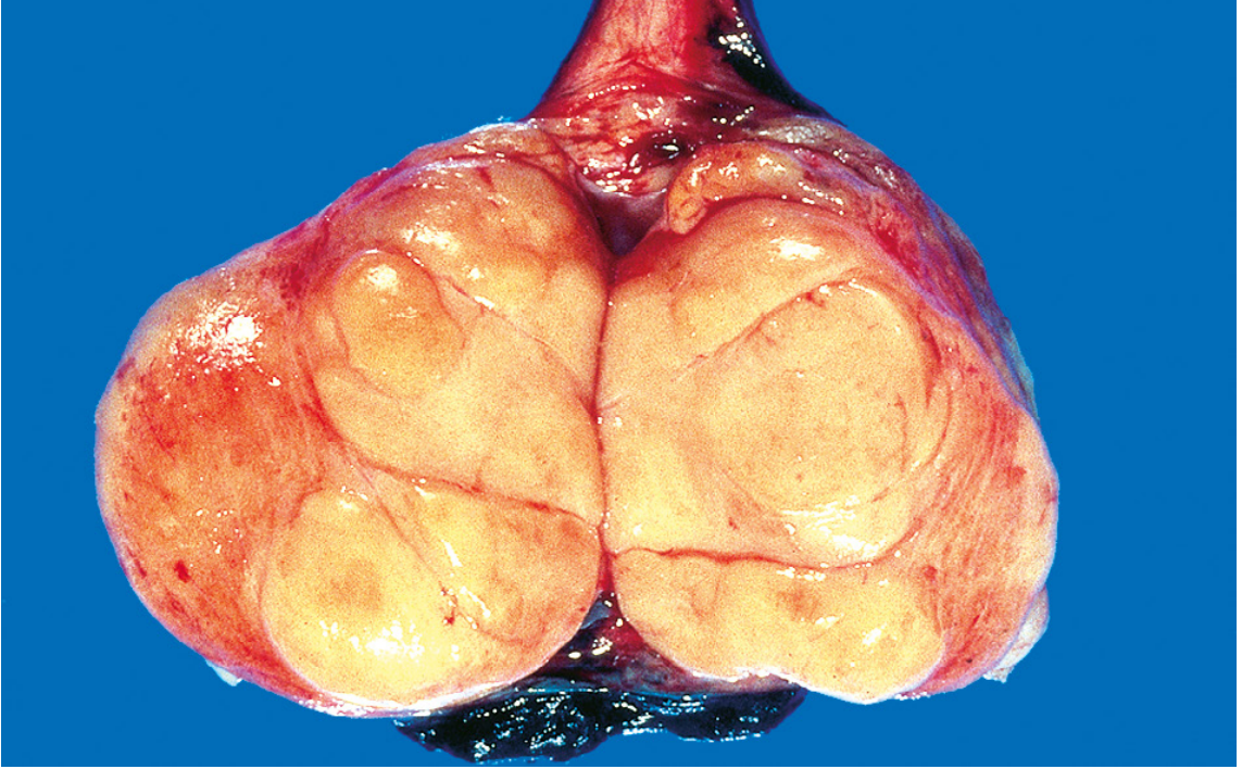
MICROSCOPIC FEATURES
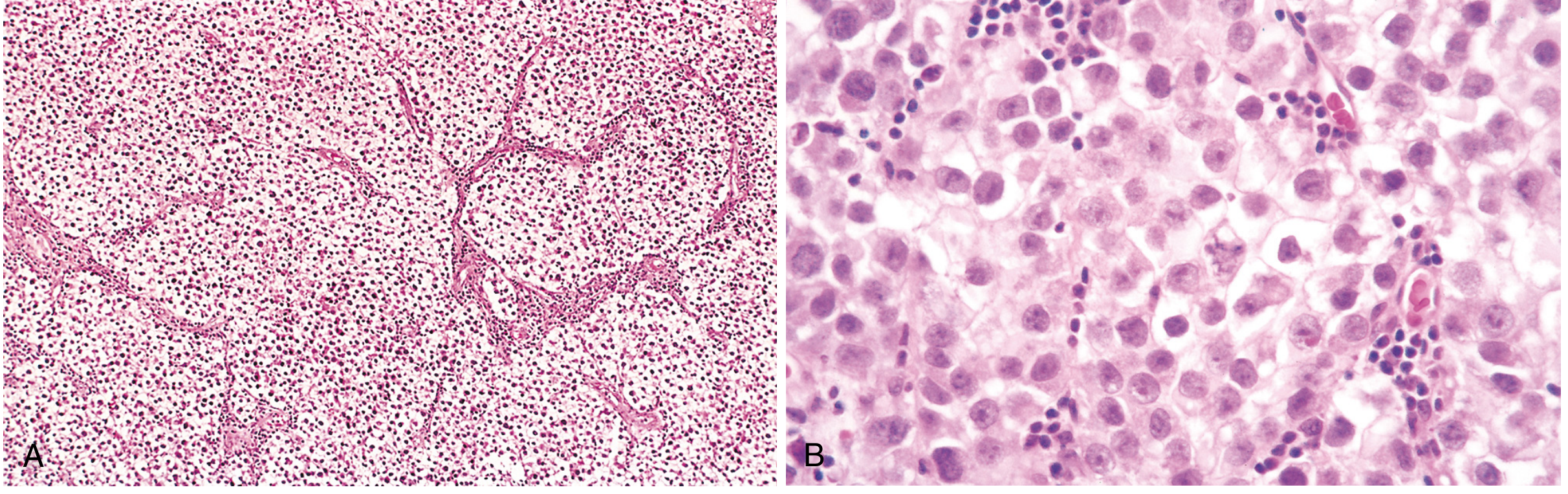
| Feature | Description |
|---|---|
| Architecture | Sheets of uniform cells divided into poorly demarcated lobules |
| Septa | Delicate fibrous septa containing lymphocytic infiltrate |
| Cells | Round to polyhedral, large, uniform |
| Cell membrane | Distinct (clear outline) |
| Cytoplasm | Clear/watery-appearing, contains glycogen (PAS +ve) |
| Nucleus | Large, central, with 1-2 prominent nucleoli |
| Granulomas | Ill-defined granulomas present (host response) |
| Syncytiotrophoblasts | Present in ~15% of cases - positive for hCG |
| Inflammatory infiltrate | Lymphocytes + granulomatous reaction (characteristic) |
- Positive: KIT (CD117), OCT3/4, PLAP (placental alkaline phosphatase), podoplanin, SALL4
- Negative: CD30, cytokeratin, AFP
LABELLED DIAGRAM OF SEMINOMA
                    SEMINOMA - MICROSCOPIC DIAGRAM
    ┌─────────────────────────────────────────────────────┐
    │                                                     │
    │    ████████     ░░░░░░░     ████████     ░░░░░     │
    │    ████████  ←──FIBROUS──→  ████████     ░░░░░     │
    │    ████████     SEPTUM      ████████               │
    │    ●●●●●●●●     ▲           ●●●●●●●●               │
    │    ● = Tumor    │           ● = Tumor               │
    │      cells    LYMPHO-         cells                 │
    │               CYTES                                 │
    │                                                     │
    │  [LOBULE 1]              [LOBULE 2]                 │
    │                                                     │
    │  Each cell:                                         │
    │  ┌──────────────────┐                               │
    │  │  Distinct cell   │                               │
    │  │  border          │                               │
    │  │  ○ = Large       │                               │
    │  │    central       │                               │
    │  │    nucleus with  │                               │
    │  │    prominent     │                               │
    │  │    nucleolus     │                               │
    │  │  Clear/watery    │                               │
    │  │  cytoplasm       │                               │
    │  └──────────────────┘                               │
    └─────────────────────────────────────────────────────┘
- Sheet of tumor cells (lobular arrangement)
- Fibrous fibrovascular septa
- Lymphocytic infiltrate within septa
- Granuloma (ill-defined)
- Tumor cell: distinct cell border, clear cytoplasm, central nucleus, prominent nucleolus
- Syncytiotrophoblast (present in 15%)
MODES OF SPREAD OF SEMINOMA
- First to retroperitoneal para-aortic lymph nodes (iliac nodes NOT involved first - important)
- Then mediastinal and supraclavicular nodes
- Follows lymphatics along the spermatic vessels (testicular lymphatics drain to paraaortic nodes, NOT inguinal nodes)
Exception: If scrotal skin is involved → inguinal lymph nodes
- Occurs later in course (contrast: NSGCTs spread hematogenously early)
- Sites: Lungs (most common), then liver, brain, bones
| Feature | Seminoma | NSGCT |
|---|---|---|
| Main route | Lymphatic | Hematogenous |
| Spread timing | Late hematogenous | Early hematogenous |
| First LN | Para-aortic | Para-aortic |
| Lung mets | Later | Earlier |
4. TERATOMA - Definition, Classification, Extragonadal Sites
DEFINITION
CLASSIFICATION OF TERATOMA
| Prepubertal Type | Postpubertal Type | |
|---|---|---|
| GCNIS association | Absent | Present |
| Isochromosome 12p | Absent | Present |
| Behavior | Benign | Malignant |
| Common in | Infants/children | Adults |
| Type | Features |
|---|---|
| Mature teratoma | Well-differentiated adult-type tissues; benign in ovary; malignant in testes (adults) |
| Immature teratoma | Contains fetal/embryonic-type tissues (especially immature neuroepithelium); malignant potential |
| Teratoma with somatic-type malignancy | Squamous cell carcinoma, adenocarcinoma, sarcoma arising within - chemoresistant |
- Dermoid cyst - contains hair, teeth, skin appendages
- Epidermoid cyst
- Well-differentiated neuroendocrine tumor (monodermal teratoma)
GROSS & MICROSCOPIC FEATURES OF TERATOMA
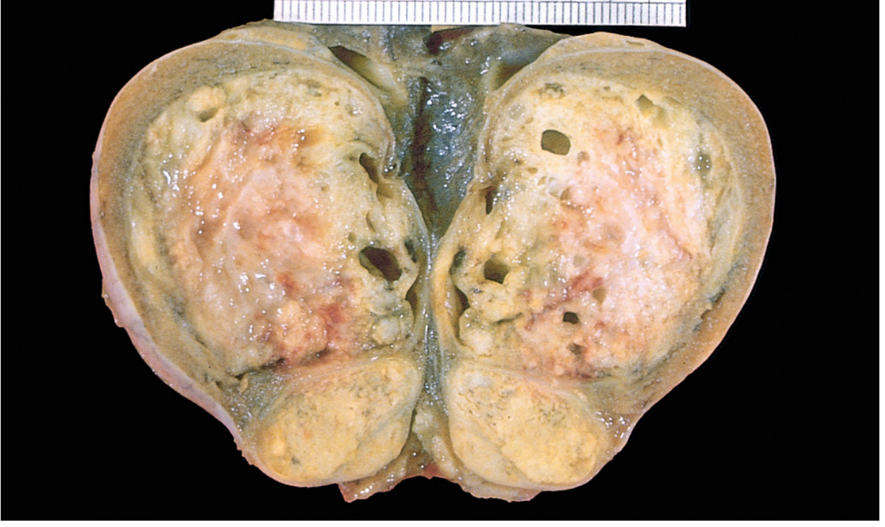
- Size: 5-10 cm in adult males
- Variegated/heterogeneous cut surface
- Solid + cystic areas
- May contain cartilaginous, fleshy, mucinous areas
- Cysts lined by various epithelia
- Collections of differentiated cells / organoid structures from multiple germ layers:
- Ectoderm: neural tissue, squamous epithelium, skin adnexa
- Mesoderm: muscle bundles, islands of cartilage, bone
- Endoderm: bronchial epithelium, intestinal wall, thyroid tissue
- Elements embedded in fibrous or myxoid stroma
- Elements may be mature (adult-like) or immature (fetal/embryonic-like)
EXTRAGONADAL SITES OF TERATOMA
| Site | Details |
|---|---|
| Sacrococcygeal region | Most common site overall; most common in newborns/infants |
| Anterior mediastinum | Most common site in adults; associated with Klinefelter syndrome |
| Pineal gland / CNS | Germinoma/teratoma; midline |
| Retroperitoneum | Less common |
| Neck | Cervical teratoma |
| Oral cavity/nasopharynx | Rare |
Exam high-yield: Sacrococcygeal = most common extragonadal teratoma. Mediastinal = associated with Klinefelter syndrome (increased risk 50x for mediastinal GCT but NOT testicular GCT).
SERUM TUMOR MARKERS - High Yield Summary
| Marker | Tumor | Notes |
|---|---|---|
| AFP (alpha-fetoprotein) | Yolk sac tumor > teratoma | Never elevated in pure seminoma |
| hCG | Choriocarcinoma (always); Seminoma (15% - due to syncytiotrophoblasts) | |
| PLAP | Seminoma | |
| LDH | All GCTs | Correlates with tumor bulk |
| CD117 (KIT) | Seminoma | |
| CD30 | Embryonal carcinoma | NOT seminoma |
PATHOGENESIS SUMMARY
Primordial Germ Cell
      ↓ (KIT mutation, environmental factors)
GCNIS (Germ Cell Neoplasia In Situ)
      ↓ (isochromosome 12p - universal in all invasive GCTs)
  ┌───────────────────────────────┐
  ↓                               ↓
SEMINOMA  ──────────────────→  NSGCTs
(pluripotent precursor)         (EC, YST, Choriocarcinoma, Teratoma)
QUICK COMPARISON TABLE - ALL GCTs
| Feature | Seminoma | Embryonal Ca | Yolk Sac Tumor | Choriocarcinoma | Teratoma |
|---|---|---|---|---|---|
| Age | 4th decade | 20-30 yrs | <3 yrs (pure) | Any | Any |
| Gross | Homogeneous, gray-white, NO hemorrhage | Variegated, hemorrhagic | Homogeneous | Small, hemorrhagic | Variegated, cystic |
| Microscopy | Sheets + fibrous septa + lymphocytes | Alveolar/tubular, anaplastic | Lace-like, Schiller-Duval bodies | Syncytio + cytotrophoblasts | Multi-tissue organoids |
| AFP | - | ± | +++ | - | ± |
| hCG | +(15%) | ± | - | +++ | - |
| IHC | KIT+, PLAP+, OCT3/4+ | CD30+, OCT3/4+ | AFP+, glypican-3+ | hCG+ | Mixed |
| Spread | Lymphatic first | Haematogenous early | - | Haematogenous early | - |
| Prognosis | Best (radiosensitive) | Intermediate | Good (prepubertal) | Worst | Variable |
MODES OF SPREAD - ALL TESTICULAR TUMORS
- Lymphatic spread - retroperitoneal para-aortic nodes first (ALL types)
- Then - mediastinal → supraclavicular nodes
- Hematogenous - lungs most common; then liver, brain, bones
- Seminoma: lymphatic >> hematogenous (late)
- NSGCTs: hematogenous early and frequent
Important: Testicular lymphatics drain to para-aortic (retroperitoneal) nodes, NOT inguinal nodes. Inguinal nodes only if scrotal skin is involved (explains why inguinal orchidectomy is NOT done - trans-scrotal biopsy is avoided).
Add histo pathology diagram and gross as well
Detailed labeled medical pathology diagram of Seminoma of Testis GROSS appearance for medical students. Show a cross-section of the testis with the following clearly labeled parts: 1) Tunica albuginea (intact, not penetrated), 2) Homogeneous gray-white cut surface, 3) Lobulated surface with fibrous septa visible, 4) No hemorrhage, no necrosis, 5) Bulky tumor mass replacing testicular parenchyma, 6) Epididymis (normal, not invaded). Style: clean anatomical medical illustration with black label lines pointing to each structure, white background, educational diagram style similar to Robbins pathology textbook. Title at top: "SEMINOMA - GROSS APPEARANCE"
Detailed labeled histopathology diagram of Classical Seminoma for 2nd MBBS pathology exam. Show microscopic H&E appearance with these labeled features: 1) Sheets of uniform tumor cells arranged in lobules, 2) Delicate fibrovascular septa between lobules, 3) Lymphocytic infiltrate within the septa, 4) Ill-defined granuloma, 5) Individual tumor cell with distinct cell border, clear/watery cytoplasm, large central nucleus, prominent nucleolus, 6) Syncytiotrophoblast cell (giant multinucleated cell, present in 15%), 7) Fibrous stroma. Use pink and purple H&E color scheme. Clean medical illustration with labeled arrows. White background. Title: "SEMINOMA - HISTOPATHOLOGY (H&E)"
Detailed labeled medical pathology diagram of Teratoma of Testis GROSS appearance for medical students. Show cross-section of testis with clearly labeled features: 1) Variegated/heterogeneous cut surface, 2) Multiple cystic spaces of different sizes, 3) Solid cartilaginous area (hard white-yellow), 4) Mucoid/gelatinous areas, 5) Hair or keratin in cyst, 6) Tunica albuginea, 7) Epididymis. Emphasize the multicolored, mixed solid-cystic appearance. Clean anatomical medical illustration with black label lines, white background, educational style. Title: "TERATOMA OF TESTIS - GROSS APPEARANCE"
Detailed labeled histopathology diagram of Teratoma of Testis for 2nd MBBS pathology exam. Show microscopic H&E appearance with labeled structures from all three germ layers: 1) Squamous epithelium with keratin (ectoderm), 2) Neural tissue / glial tissue (ectoderm), 3) Cartilage island (mesoderm), 4) Smooth muscle bundle (mesoderm), 5) Bronchial/respiratory epithelium (endoderm), 6) Intestinal gland/mucosa (endoderm), 7) Fibrous myxoid stroma between tissues, 8) Cystic space. Use pink and purple H&E color scheme with clear label arrows. Show disorganized arrangement of multiple tissue types. White background. Title: "TERATOMA OF TESTIS - HISTOPATHOLOGY (H&E) - Tissues from all 3 Germ Layers"
400, message: Got more than 4194304 bytes when reading: b'data: {"candidates": [{"content": {"role": "model","parts": [{"inlineData": {"mimeType": "image/png"...'.
We encountered an error while processing your request. Please try again or reach out to us for help.