
I want invivo and inovo study in same project with novelty and lowcost feasible and wow factor give ratings also and give more innovative ideas
Which research area should the in vivo + in ovo (chick embryo/CAM) project target? · What is the project for? · Which in vivo model is realistic for you?
"chorioallantoic membrane" AND "xenograft"
chick chorioallantoic membrane CAM assay in ovo model low-cost novel applications 2025 review
Why this pairing works
Ranked ideas
| # | Idea | Novelty | Cost | Feasibility | Wow | Overall |
|---|---|---|---|---|---|---|
| 1 | AI/smartphone vessel quantification, validated against in vivo | 8 | 9 | 7 | 8 | 8.0 |
| 2 | Eggshell hydroxyapatite bone scaffold ("egg to bone") | 7 | 8 | 7 | 9 | 7.75 |
| 3 | Vape liquid or microplastic developmental toxicity | 7 | 8 | 8 | 8 | 7.75 |
| 4 | Hyperglycaemic CAM, then diabetic rat wound | 7 | 8 | 8 | 7 | 7.5 |
| 5 | Infected-wound CAM (biofilm), then mouse infected wound | 7 | 8 | 7 | 8 | 7.5 |
| 6 | Natural-product funnel with in ovo/in vivo concordance score | 6 | 9 | 9 | 6 | 7.5 |
| 7 | Smart pH-indicator wound dressing | 7 | 7 | 7 | 8 | 7.25 |
| 8 | Biowaste carbon-dot theranostics | 8 | 6 | 6 | 9 | 7.25 |
| 9 | Low-cost photodynamic therapy (LED + curcumin or methylene blue) | 7 | 7 | 7 | 8 | 7.25 |
| 10 | Cardiotoxicity and cardioprotection screen | 6 | 8 | 8 | 6 | 7.0 |
| 11 | Chick-embryo "avatar" for personalised drug testing | 8 | 5 | 5 | 9 | 6.75 |
| 12 | Green-synthesised nanoparticles (Ag, ZnO, Se) | 4 | 8 | 9 | 5 | 6.5 |
The ideas in brief
- In ovo: Photograph CAMs with a smartphone on a cheap macro lens. Train a free Python U-Net or use AngioTool to count vessels automatically.
- In vivo: Run a mouse Matrigel plug or rat wound, scored by haemoglobin content and CD31 staining.
- Novelty: A cheap, open-source tool that shows CAM image scores predict in vivo angiogenesis. A correlation figure (r, Bland-Altman) is the headline result.
- Scaffold: Calcine waste eggshell into hydroxyapatite and blend it with chitosan or gelatin. Add a plant osteogenic extract or a trace-ion dopant such as Sr or Zn for extra novelty.
- In ovo: Check biocompatibility and angiogenesis on the CAM.
- In vivo: Test in a rat calvarial defect, with micro-CT if you can get it.
- Why it stands out: The circular-economy story is easy to remember.
- In ovo: Inject at day 0 to 3 and score mortality, malformations, heart rate and CAM vascularity.
- In vivo: Run a dose-matched rat or zebrafish study for liver, kidney and oxidative stress markers.
- Why it stands out: Public interest is high. Adding a rescue arm (for example an antioxidant) makes it mechanistic.
- In ovo: Create a high-glucose environment that impairs angiogenesis, then test pro-angiogenic candidates (honey, curcumin, PRP, a peptide).
- In vivo: Take the best candidate into an STZ-diabetic rat excision wound.
- Why it stands out: A high-glucose CAM screen is a quick way to test diabetic-wound candidates.
- In ovo: Infect the CAM with S. aureus, Pseudomonas or Candida, then treat with essential oil, phage or an antimicrobial peptide. Score bacterial load and lesion size.
- In vivo: Use a mouse or rat infected wound.
- Why it stands out: Antimicrobial resistance is a strong theme.
- Design: Screen 8 to 10 extracts by MTT assay, then CAM, then one tumour model (Ehrlich ascites or a xenograft).
- Novelty: Report how well the in ovo ranking predicted the in vivo ranking (Spearman correlation). This is the safest and cheapest option. It becomes more novel if you choose an under-studied regional plant.
- Dressing: Electrospun or cast film containing anthocyanin from red cabbage, which changes colour with wound pH, plus an active such as curcumin.
- In ovo: HET-CAM irritation test and angiogenesis.
- In vivo: Rat wound, tracking pH against healing rate.
- Why it stands out: The colour change is visual and photographs well.
- Material: Make fluorescent carbon dots from banana peel, curry leaf or similar by hydrothermal synthesis.
- In ovo: Image them live through a window in the shell, tracking vessel uptake and tumour accumulation.
- In vivo: Run a mouse biodistribution study.
- Why it stands out: Live glowing vessels make a strong figure. It needs a fluorescence imager or UV box with a camera, which is the main cost and risk.
- Setup: A cheap LED array plus a photosensitiser such as curcumin or methylene blue.
- In ovo: Treat CAM tumours or vessels.
- In vivo: Use a mouse subcutaneous tumour.
- Why it stands out: It frames the work as affordable cancer therapy for low-resource settings.
- In ovo: Measure embryonic heart rate and rhythm by video with doxorubicin, then add a protective natural compound.
- In vivo: Rat doxorubicin model with ECG and cardiac enzymes.
- In ovo: Graft patient-derived or cell-line tumour fragments onto the CAM and test drug combinations.
- In vivo: Compare with a mouse xenograft.
- Why it stands out: The personalised-medicine angle is very strong, but it needs tissue consent and ethics approval, so it is harder. Cell lines are a safer start.
My picks
- Highest wow with low risk: Idea 2, the eggshell scaffold.
- Best for publication: Idea 1, because validation work is rare and tool-based papers get cited.
- Safest for a student timeline: Idea 6, or idea 4 for something slightly more novel.
Practical notes
- Eggs: Use fertilised eggs, incubated at 37.5°C and about 60% humidity. Use about 8 to 10 eggs per group. Plan for 20 to 30% attrition.
- Ethics: Rules for embryos differ by country and institution, so confirm with your ethics committee. Mouse and rat work needs animal ethics approval. Zebrafish larvae are often treated more leniently.
- Minimal controls: Vehicle, positive control (for example bevacizumab or sunitinib for anti-angiogenesis) and untreated.
- Statistics: One-way ANOVA with a post hoc test, plus the in ovo vs in vivo correlation.
Give
| # | Idea | Novelty | Cost | Feasibility | Wow | Overall |
|---|---|---|---|---|---|---|
| A | Digital twin add-on: machine learning that predicts in vivo outcome from in ovo features | 9 | 9 | 6 | 9 | 8.25 |
| B | In ovo feeding to post-hatch chick (the egg and the animal are the same species) | 7 | 9 | 8 | 8 | 8.0 |
| C | CAM bleeding model for low-cost haemostats | 7 | 9 | 8 | 7 | 7.75 |
| D | Herbo-mineral (bhasma) safety screen | 7 | 8 | 7 | 8 | 7.5 |
| E | LED photobiomodulation for healing | 7 | 8 | 8 | 7 | 7.5 |
| F | Microneedle patch penetration on CAM, then rat transdermal | 8 | 6 | 6 | 9 | 7.25 |
| G | Phone or Wi-Fi radiofrequency exposure and embryo development | 6 | 8 | 8 | 7 | 7.25 |
| H | Plant-based snake venom haemorrhage antidote | 8 | 6 | 4 | 9 | 6.75 |
Details
- Extract features from every CAM image: vessel density, branching points, lacunarity and fractal dimension. Combine them with dose and compound descriptors.
- Train a simple model (random forest or regression) to predict the in vivo result, such as wound closure percentage or tumour volume. Validate it on held-out compounds.
- "We predicted mouse results from an egg" is a strong headline, and it costs only your time. The risk is sample size, so you need at least 10 to 15 compounds or doses.
- In ovo: Inject probiotics, prebiotics, plant extracts or nanominerals into the egg on about day 17 to 18. This is a real poultry technique. Record hatchability and hatch weight.
- In vivo: Raise the hatched chicks for 3 to 6 weeks. Measure growth, gut villus height, microbiota, antibody titre after vaccination and heat-stress markers.
- Why it stands out: There is no rodent colony and no cross-species gap. A continuous egg-to-bird story is easy to explain, and the poultry industry may fund it.
- In ovo: Puncture a standard-size CAM vessel and apply chitosan, alum, a plant extract or gelatin sponge. Time the bleeding to stop and quantify blood loss from images.
- In vivo: Rat tail transection or liver-wound bleeding.
- Why it stands out: The cheap, standardised in ovo bleeding assay is the novelty. The CAM puncture model is much less used than the angiogenesis assay.
- In ovo: Test classical herbo-mineral preparations for embryo toxicity, malformations and CAM effects.
- In vivo: Sub-acute rat study of the safest and the most toxic preparations (liver and kidney enzymes, histology, tissue metal levels).
- Why it stands out: It addresses a real gap in the safety evidence for traditional medicines. Add metal quantification (ICP-MS or AAS) if your lab can reach it.
- Setup: A low-cost red/near-infrared LED array with a power meter.
- In ovo: Test several wavelengths and doses on CAM angiogenesis and embryo viability.
- In vivo: Rat or diabetic-rat wound, with the optimal dose from the CAM.
- Why it stands out: It finds the best dose cheaply before committing animals.
- Patch: Make dissolvable microneedles (PVA, gelatin or sugar) in a PDMS mould cast from a cheap commercial needle array or a 3D-printed master.
- In ovo: Test needle penetration and dye diffusion on the CAM. Embryo skin or CAM also works.
- In vivo: Test insulin or a drug in rats.
- Risk: The moulds and fabrication take trial and error.
- In ovo: Expose eggs to a phone or Wi-Fi router during incubation. Record hatchability, malformations, heart rate and oxidative stress.
- In vivo: Rats exposed for several weeks, with a protective antioxidant arm if you want a mechanistic angle.
- Caution: Past results in this field are mixed and often poorly controlled. Use sham exposure, a measured field strength and blinded scoring.
- In ovo: Measure venom-induced haemorrhage in embryos and test plant extracts as inhibitors.
- In vivo: Mouse neutralisation assays.
- Why it stands out: Embryo screening could cut the number of mice used in venom neutralisation work.
- Risk: Venom is hard to obtain and regulated, so I rated it low on feasibility.
Combinations for the most impact
- Eggshell hydroxyapatite scaffold (from the first list) + AI vessel quantification + digital twin (A). The story is egg waste to a bone graft, assessed by an open-source tool and predicted by a model.
- Hyperglycaemic CAM + LED photobiomodulation (E) + digital twin (A). This is an affordable diabetic-wound platform, with the dose chosen in ovo.
- Haemostat (C) + smart dressing (from the first list). A first-aid kit concept.
Next step
More ideas
| # | Idea | Novelty | Cost | Feasibility | Wow | Overall |
|---|---|---|---|---|---|---|
| 1 | Contactless video-based embryo heart-rate and toxicity readout | 8 | 9 | 7 | 8 | 8.0 |
| 2 | Herbal sunscreen or UV photoprotection | 6 | 9 | 8 | 7 | 7.5 |
| 3 | CAM as a bioreactor to prevascularise spheroids, then implant | 8 | 6 | 6 | 9 | 7.25 |
| 4 | Endometriosis or ovarian lesion on CAM, then mouse | 8 | 7 | 5 | 9 | 7.25 |
| 5 | Hypoxia or high-altitude protectants | 7 | 8 | 7 | 7 | 7.25 |
| 6 | Teratogen rescue (valproate plus folate or a nutraceutical) | 6 | 8 | 8 | 7 | 7.25 |
| 7 | Anti-inflammatory screen on CAM, then rat paw oedema | 6 | 9 | 9 | 5 | 7.25 |
| 8 | Heavy-metal toxicity and natural chelators | 6 | 8 | 8 | 6 | 7.0 |
| 9 | Antimicrobial sutures or implant coatings | 6 | 7 | 7 | 7 | 6.75 |
| 10 | Corneal neovascularisation eye drops | 6 | 7 | 6 | 7 | 6.5 |
Details
- In ovo: Film the embryo through a window or by candling with a phone. Extract heart rate and motion with free signal-processing code (Eulerian video magnification or simple intensity tracking). Use it as a non-invasive, repeatable toxicity or cardiotoxicity marker over days.
- In vivo: Compare with rat ECG after the same drug (doxorubicin, a QT-prolonging drug or an energy drink).
- Why it stands out: You replace expensive instruments with a phone and open code. The key step is validating it against a reference method.
- In ovo: Expose embryos or the CAM to UV-B or UV-C through a film or cream. Score survival, vessel damage and oxidative markers.
- In vivo: Mouse or rat UV skin model (erythema, thickness, MDA and histology).
- Extra: Measure SPF in vitro by spectrophotometer. A UV lamp with a measured dose is the main equipment need.
- In ovo: Grow tumour spheroids, 3D-printed or hydrogel scaffolds, or stem cell constructs on the CAM for several days so chick vessels grow in.
- In vivo: Implant the prevascularised construct in a mouse or rat and test whether it survives better than an unvascularised one.
- Why it stands out: It is a creative use of the egg as a cheap incubator for tissue engineering. The main risk is that the vessels are avian, so check for host integration and rejection.
- In ovo: Graft endometrial cells or tissue and test anti-angiogenic or anti-adhesion agents. Ovarian cancer metastasis on the CAM is well described, but verify the endometriosis protocols in the literature before committing.
- In vivo: Mouse endometriosis model.
- Why it stands out: Women's health is under-funded and reviewers like the topic. Use cell lines first, because human tissue needs ethics approval and consent.
- In ovo: Induce hypoxia with a low-oxygen setup or chemically (for example CoCl2). Test protective agents such as rhodiola, ginkgo or antioxidants on survival, vascular density and HIF-1α.
- In vivo: Rat hypobaric chamber if available, or a chemical hypoxia model (sodium nitrite).
- In ovo: Treat with valproate or another teratogen, then add folic acid, choline or a plant antioxidant. Score neural tube defects, limb and heart malformations, and size.
- In vivo: Pregnant rat with the best rescue dose.
- Why it stands out: The rescue design gives you a mechanistic story. Pregnant-animal work has stricter ethics and a longer timeline.
- In ovo: A CAM inflammation model is cheap, so screen many extracts.
- In vivo: Carrageenan rat paw oedema.
- Note: This is the easiest and safest option, but the least exciting. It works well as a base for idea 1 from the previous list, the AI quantification.
- In ovo: Treat with lead, cadmium or arsenic and test moringa, chlorella or coriander extract.
- In vivo: Rat model with blood and tissue metal levels and liver and kidney markers.
Ways to raise the wow factor in any project
- Time-lapse imaging. A phone on a clamp or a 3D-printed holder can film CAM vessel growth over 24 to 72 hours.
- Open data and code. Posting images and scripts (for example on Zenodo or GitHub) raises citation chances.
- Preregistration of your design and endpoints, which reviewers value.
- A single visual summary. A clear egg-to-animal workflow figure and one striking before and after image.
- A frugal comparison. Show cost per experiment against a mouse-only design.
- Negative controls and blinding. Score images blinded, which adds credibility at no cost.
Next step
More ideas with full marks
| Rank | Idea | Novelty | Cost | Feasibility | Wow | Overall |
|---|---|---|---|---|---|---|
| 1 | Open-source "Frugal Egg Lab" kit | 9 | 10 | 8 | 9 | 9.0 |
| 2 | Public benchmark plus digital twin | 9 | 10 | 7 | 9 | 8.75 |
| 2 | Egg-to-bird feeding of waste-derived bioactives | 8 | 10 | 9 | 8 | 8.75 |
| 4 | Everyday exposome in an egg | 8 | 9 | 8 | 9 | 8.5 |
| 4 | Eggshell-to-bone with AI scoring | 8 | 9 | 7 | 10 | 8.5 |
| 4 | Kitchen-to-clinic haemostat | 7 | 10 | 9 | 8 | 8.5 |
| 7 | Diabetic-wound platform (stacked) | 8 | 8 | 7 | 9 | 8.0 |
1. Open-source Frugal Egg Lab kit (9.0)
- Concept: A complete, cheap, reproducible kit for CAM experiments. It has a 3D-printed or cardboard imaging box with a phone clamp and LED ring, a standard window-cutting and sealing protocol, and free software that counts vessels.
- In ovo: Run an anti-angiogenesis and a pro-angiogenesis screen with known controls (bevacizumab or sunitinib, and VEGF or a pro-angiogenic extract).
- In vivo: Validate against one mouse Matrigel plug or rat wound experiment, scored by haemoglobin and CD31.
- Why it scores high: Labs with no imaging equipment can use it, and it is publishable as a methods paper.
- What holds it back: Image consistency across labs. To get to 10, test the kit in a second lab and report inter-lab agreement.
2. Public benchmark plus digital twin (8.75)
- Concept: Build the first small open benchmark of CAM results for 15 to 20 compounds whose in vivo potency is already published (known anti-angiogenic and pro-angiogenic drugs). Then test whether a simple model predicts the published in vivo ranking.
- In ovo: Dose-response of each compound on the CAM.
- In vivo: Mostly literature data, plus one small confirmatory experiment with a new compound the model never saw.
- Why it scores high: Almost no animals are needed, and a dataset others can reuse is a durable contribution.
- What holds it back: Published in vivo data are inconsistent across species and doses. To reach 10, preregister the prediction and test it on the unseen compound.
3. Egg-to-bird feeding of waste-derived bioactives (8.75)
- Concept: Inject waste-derived bioactives (eggshell calcium nanoparticles, fruit-peel polyphenols or similar) into the egg at about day 17 to 18, then raise the chicks.
- In ovo: Hatchability, hatch weight and embryo toxicity.
- In vivo: The same birds for 3 to 6 weeks: growth, gut villus height, antibody response to vaccination and heat-stress markers.
- Why it scores high: There is no rodent colony, the cost is the lowest of all seven, and the poultry industry may support it.
- What holds it back: Novelty is moderate because in ovo feeding is established. To reach 10, pair a new waste-derived compound with a measurable health outcome such as vaccine response.
4. Everyday exposome in an egg (8.5)
- Concept: Test substances people use daily: tea-bag microplastic leachate, instant-noodle cup leachate, vape liquid, energy drinks and hair-dye ingredients. Compare them on one standard embryo scoring system.
- In ovo: Mortality, malformations, heart rate (from video) and CAM vascularity.
- In vivo: Rat or zebrafish for the one or two worst offenders, with liver, kidney and oxidative stress endpoints.
- Why it scores high: Everyone can relate to it, and press and social media will pick it up.
- What holds it back: Real-world dose relevance. Dose-match to realistic human exposure to avoid overclaiming.
5. Eggshell-to-bone with AI scoring (8.5)
- Concept: Calcine eggshell waste into hydroxyapatite and blend it with chitosan or gelatin, with or without a plant osteogenic extract.
- In ovo: Biocompatibility and angiogenesis on the CAM, scored with the idea 1 pipeline.
- In vivo: Rat calvarial defect with micro-CT or histology.
- Why it scores high: The "egg to bone" story gives the strongest visual and narrative of all seven.
- What holds it back: Eggshell hydroxyapatite is known, and imaging adds cost. To reach 10, add a clear twist, such as a trace-ion dopant, and use the CAM score to select the formulation.
6. Kitchen-to-clinic haemostat (8.5)
- Concept: Compare cheap haemostats: chitosan from shrimp-shell waste, alum, turmeric, gelatin sponge and honey.
- In ovo: Puncture a standard CAM vessel and time the bleeding to stop. Quantify blood loss from images.
- In vivo: Rat tail transection or liver-wound bleeding.
- Why it scores high: Very low cost, a clear practical use in low-resource first aid, and an easy outcome to measure.
- What holds it back: Standardising the puncture. To reach 10, use a fixed needle gauge, a template and blinded scoring.
7. Diabetic-wound platform, stacked (8.0)
- Concept: Combine a hyperglycaemic CAM, LED photobiomodulation dose-finding and a pH-indicator dressing.
- In ovo: Screen dressing candidates and LED doses under high glucose.
- In vivo: Diabetic rat wound (STZ), tracking healing rate and wound pH.
- Why it scores high: It is a cohesive story with several outputs.
- What holds it back: It needs more skills (dressing fabrication, LED dosimetry and the diabetic model) and a larger scope, so it is better as a PhD plan than a short project.
My recommendation
Explain 1st
automated CAM assay angiogenesis quantification deep learning open-source AngioTool smartphone
"chorioallantoic membrane" AND "deep learning"
"chorioallantoic membrane" AND "angiogenesis" AND "image analysis"
chick chorioallantoic membrane assay angiogenesis blood vessels
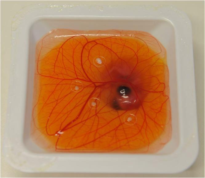
A clinical photograph of an ex-ovo Chorioallantoic Membrane (CAM) assay within a white square plastic shell. The assay shows a translucent, orange-colored gelatinous yolk sac surface featuring a prominent, radiating network of red embryonic blood vessels originating from a central developing chick embryo. Four small, clear silicone rings (approximately 5 mm in diameter) are surgically placed onto the membrane, specifically positioned over major vascular branch points. Each ring contains a light-colored, circular opaque tissue sample, representing human split-thickness skin xenografts. This experimental setup is used in medical research as a 'bioreactor' to study angiogenesis and the effects of photobiomodulation (PBM) on human tissue engraftment and vascularization. The image demonstrates a standard model for close-to-in-vivo dermatological and surgical research, allowing for the observation of vessel growth towards human donor tissue.
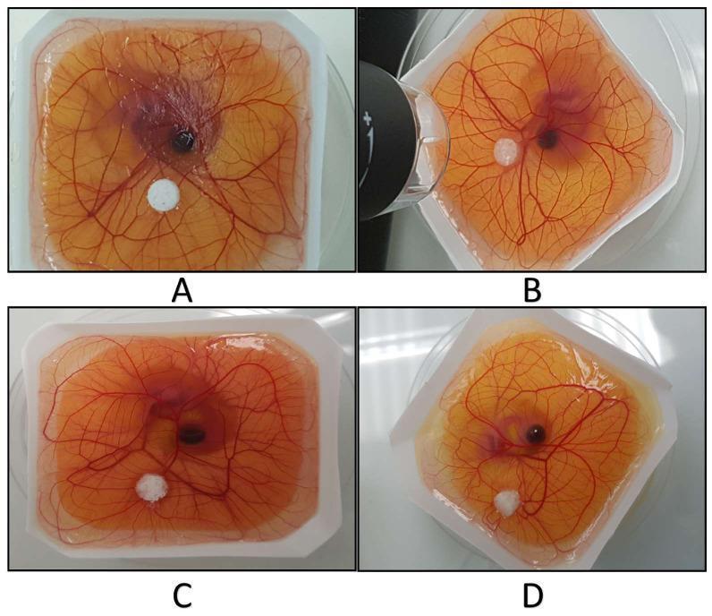
This set of four clinical photographs (A-D) demonstrates an ex-ovo Chorioallantoic Membrane (CAM) assay used to evaluate the pro-angiogenic potential of medical scaffolds. Each image shows a translucent, yellowish chick embryo membrane within a square culture vessel. The membranes are characterized by a prominent hierarchical network of branching reddish blood vessels radiating from a central darker embryonic region. In each panel, a white, circular porous scaffold (polyurethane PU in A and B; PU/hydroxyapatite PU/HA in C and D) is placed peripherally on the membrane surface. The assay illustrates the induction of new microvascular growth and neovascularization around the implanted biomaterials. Panels A and C show a qualitatively higher vascular density compared to B and D. This experimental model is critical in regenerative medicine and tissue engineering to assess how chemical compositions, such as hydroxyapatite nanoparticles, influence angiogenesis and VEGF-mediated vessel formation for bone repair applications.
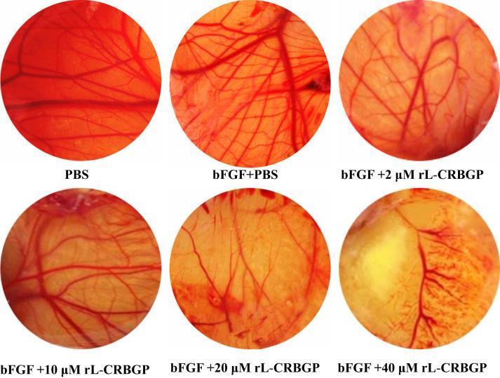
This image shows a series of six circular macroscopic panels illustrating a Chick Chorioallantoic Membrane (CAM) angiogenesis assay. The panels demonstrate the anti-angiogenic effects of recombinant lamprey cysteine-rich buccal gland protein (rL-CRBGP) on blood vessel formation. The control panels (PBS and bFGF + PBS) show a dense, well-organized network of prominent, branching red blood vessels against a translucent background. As the concentration of rL-CRBGP increases from 2 μM to 40 μM, there is a visible, dose-dependent reduction in vascular density. At lower concentrations (2-10 μM), the vessels become thinner and branching decreases. In the higher concentration panels (20-40 μM), significant vascular attenuation is observed, characterized by sparse vessel distribution and large avascular zones. Concurrently, the CAM tissue displays increased opacity and a shift from a translucent red to an opaque yellowish appearance, particularly at 40 μM. This comparison is used in oncology and pharmacology research to evaluate potential therapeutic agents that inhibit pathological angiogenesis.
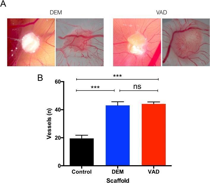
This composite educational image illustrates an in vivo angiogenesis study using the chick chorioallantoic membrane (CAM) assay to evaluate decellularized human tracheal scaffolds. Panel A contains clinical macroscopic photographs showing tracheal scaffolds prepared by two different methods: Detergent-Enzymatic (DEM) and Vacuum-Assisted Decellularization (VAD). Both types of scaffolds appear as whitish, opaque masses 8 days post-implantation, with clear recruitment of host blood vessels converging toward the grafts. The VAD scaffolds demonstrate a distinctive 'spoke-wheel' vascular pattern. Panel B is a bar graph quantifying the number of vessels (n) converging on the scaffolds. The graph compares three groups: a control (approximately 20 vessels), DEM (approximately 43 vessels), and VAD (approximately 44 vessels). Statistical markers indicate that both decellularized scaffolds induced significantly higher angiogenesis than the control (p < 0.001, denoted by ***), while the difference between the DEM and VAD preparation methods is not statistically significant (ns). This visual data supports the pro-angiogenic capacity of acellular human tracheal scaffolds for bioengineering applications.
- AngioTool is a free NIH tool for vessel length, density, branching and lacunarity (Zudaire et al., PLoS One 2011).
- A commercial IKOSA CAM app does deep-learning vessel segmentation (Kuri et al., Cells 2022).
- An AI-based CAM angiogenesis method was published in 2022 (Faihs et al.).
What it is
- A standard imaging box. It is cardboard or 3D-printed, with a matte white interior, a diffuse LED ring, a fixed phone holder at a fixed height, and a ruler and colour card in every frame.
- A standard CAM protocol. It covers eggs, windowing, how the test substance is applied, timing and scoring rules.
- Free analysis software. A Python or ImageJ pipeline gives vessel area %, total length, branch points and lacunarity as a CSV file.
- Validation. You compare it with manual counting, AngioTool and one in vivo experiment.
Workflow
- Incubate fertilised eggs at about 37.5°C and about 60% humidity.
- On about day 3, cut a small window, or use an ex-ovo shell-less culture.
- On about day 7 to 8, place a filter-paper disc, silicone ring or gelatin sponge with the test substance on the CAM.
- Photograph at about day 10 to 12 in the box. Fix the settings and take a photo of the colour card each time.
- Count only eggs that survive. Report attrition.
- Vehicle control.
- Pro-angiogenic control (bFGF or VEGF).
- Anti-angiogenic control (sunitinib or suramin).
- Start with the green channel, where blood vessels show best contrast.
- Baseline: a Frangi vessel filter with thresholding.
- Optional: train a small U-Net on 30 to 50 hand-annotated images.
- Outputs: vessel area %, total length, branch points and lacunarity.
- Mouse Matrigel plug: Measure haemoglobin (Drabkin) and CD31 staining. This is the standard comparison for angiogenesis.
- Rat excision wound: Use it if you are testing a healing compound. Measure wound closure and granulation tissue.
Example design (my suggestion)
- In ovo: 3 test compounds x 3 doses + 3 controls = 12 groups, with 8 to 10 eggs per group. That is about 100 eggs, so plan for about 130 with attrition.
- In vivo: 4 groups (vehicle, positive control, two doses of the best candidate) with 6 mice per group, or about 24 animals. Run a formal power calculation once you have pilot variance.
Validation steps
| Check | Method |
|---|---|
| Agreement with manual scoring | Intraclass correlation, Bland-Altman plot |
| Agreement with AngioTool | Correlation across all images |
| Repeatability | Photograph the same egg twice; report coefficient of variation |
| Device effect | Two different phones and two users |
| Inter-lab test | A second lab runs the same kit (this is what pushes the rating towards 10) |
| In ovo vs in vivo | Spearman correlation of compound rankings and effect sizes |
Timeline (about 12 weeks)
| Weeks | Task |
|---|---|
| 1-2 | Build the box, test the camera settings, practise windowing on spare eggs |
| 3-5 | Run the CAM screen and annotate the images |
| 5-7 | Build the analysis pipeline and compare with AngioTool |
| 7-10 | In vivo experiment and tissue analysis |
| 10-12 | Statistics, figures, write-up, upload to GitHub and Zenodo |
Cost
Outputs
- A methods paper or short report.
- An open GitHub repository with the box design, code and a step-by-step protocol.
- A shared image dataset with annotations.
- A ready-made teaching tool for other labs.
Risks
- Lighting and glare are the biggest threat to reproducibility. Use polarising film and always include the colour card.
- Egg variability. Source eggs from one supplier and use fixed incubation conditions.
- Annotation effort. Manual labelling takes time. Start with the Frangi baseline and only add the U-Net if it improves agreement.
- Ethics. Embryo rules vary by country, and mouse or rat work needs animal ethics approval.
- Novelty pushback. Cite AngioTool and IKOSA, and state clearly that your contribution is the open, validated, low-cost kit.
Next step
But invivo mouse study should be must what to do
"Matrigel plug" AND angiogenesis AND hemoglobin
"chorioallantoic membrane" AND "mouse" AND "angiogenesis" AND "correlation"
directed in vivo angiogenesis assay DIVAA vs Matrigel plug mouse protocol sample size cost
The mouse model I recommend: the Matrigel plug assay
How to do it
- Pick the test set from the egg results. Choose compounds that cover the CAM range: one strong effect, one moderate effect and one with no effect. Add a vehicle control and a known positive control. This is what lets you test whether the egg ranking predicts the mouse ranking.
- Preregister the prediction. Write down the expected order before the mouse study begins.
- Prepare the plugs. Mix cold liquid Matrigel with heparin and an angiogenic factor (bFGF or VEGF), plus the test compound or vehicle. For a pro-angiogenic compound, add it alone and compare with the vehicle plug.
- Inject the mixture subcutaneously (for example the flank) with a cold syringe. It gels at body temperature.
- Harvest at about day 7 to 14 (use the timing in your chosen protocol).
- Read out with:
- Photographs in the egg-lab box (colour and redness).
- Haemoglobin content (Drabkin method), the cheap quantitative readout.
- CD31 immunostaining if you have the antibody, as the histology confirmation.
Group sizes
| Group | Purpose | Mice |
|---|---|---|
| Vehicle plug | Baseline | 6 |
| Positive control (known inhibitor or promoter) | Assay works | 6 |
| Compound with strong CAM effect | Test prediction | 6 |
| Compound with moderate CAM effect | Test prediction | 6 |
| Compound with no CAM effect | Negative prediction | 6 |
| Total | 30 |
Options if you cannot use Matrigel
- Matrigel cost. It can be the largest single expense, so get a local quote early. Dilute it only as the protocol allows.
- Directed in vivo angiogenesis assay (DIVAA). The silicone-tube kits are also available, but the plug is cheaper and simpler.
- Mouse excision wound. Use this if your compounds are wound-healing agents. It needs no Matrigel and is cheaper. Measure closure, granulation tissue and hydroxyproline.
- Ehrlich ascites or solid tumour in Swiss albino mice. Use this if your compounds are anticancer. Measure tumour volume, survival and vessel density. It is widely used in low-resource labs, but it measures anticancer effect more than angiogenesis.
Analysis plan
- Compare groups with one-way ANOVA and a post hoc test, or Kruskal-Wallis if the data are skewed.
- Test whether the CAM ranking matches the mouse ranking with a Spearman correlation or simple rank agreement.
- With only 3 to 4 compounds, a correlation coefficient is weak. Add doses (for example 2 doses of each compound), report effect sizes with confidence intervals, and describe the correlation as exploratory.
- Report the full results, including cases where the egg and the mouse disagree.
Mouse study timeline (about 4 weeks)
| Week | Task |
|---|---|
| 0 | Ethics approval, order Matrigel and growth factor, acclimatise mice |
| 1 | Inject plugs |
| 2 | Harvest, photograph, take samples |
| 3 | Haemoglobin assay, fix tissue |
| 4 | CD31 staining, analysis |
Ethics and approval
- Get approval from your institutional animal ethics committee before you start (in India this is the IAEC under CPCSEA). Rules differ by country, so check yours.
- State the 3Rs argument: the egg screen reduces the number of compounds tested in mice, so fewer animals are used.
- Set humane endpoints and keep the procedure short. This model involves a small subcutaneous injection and a short experiment.
Updated project summary
More innovative ideas
| # | Idea | Novelty | Cost | Feasibility | Wow | Overall |
|---|---|---|---|---|---|---|
| 1 | Kombucha-cellulose "living" wound dressing | 7 | 10 | 8 | 9 | 8.5 |
| 2 | Frugal drug repurposing for anti-angiogenesis | 7 | 9 | 9 | 8 | 8.25 |
| 3 | Sentinel egg for food adulterants and cosmetics | 7 | 9 | 8 | 9 | 8.25 |
| 4 | Whole-egg dressing (white plus shell membrane) | 7 | 10 | 8 | 8 | 8.25 |
| 5 | Phage cocktail from local sewage | 8 | 8 | 6 | 9 | 7.75 |
| 6 | Smokeless tobacco and areca nut toxicity | 7 | 8 | 8 | 8 | 7.75 |
| 7 | Regional honey passport | 6 | 9 | 9 | 7 | 7.75 |
| 8 | Edible plant nanovesicles | 8 | 7 | 6 | 9 | 7.5 |
1. Kombucha-cellulose "living" wound dressing (8.5)
- Concept: The cellulose pellicle that grows on kombucha (the SCOBY) is a cheap bacterial cellulose sheet. Purify it, load it with a plant bioactive such as curcumin, and optionally add a pH-indicator dye (anthocyanin) so it changes colour as the wound pH shifts.
- In ovo: CAM biocompatibility, irritation and angiogenesis.
- Mouse: Full-thickness excision wound (splinted if available). Measure closure rate, histology and wound pH.
- Why it works: The material costs almost nothing and the story is memorable ("tea fungus to skin patch").
2. Frugal drug repurposing for anti-angiogenesis (8.25)
- Concept: Screen 15 to 20 cheap, off-patent drugs (examples of candidates: itraconazole, mebendazole, doxycycline, propranolol, metformin, disulfiram) on the CAM. Pick the best for mouse testing.
- In ovo: Dose-response, scored with the Frugal Egg Lab kit.
- Mouse: Matrigel plug, or Ehrlich or B16 tumour if you want an anticancer angle.
- Why it works: Drugs with known safety profiles are easier to take forward, and it answers a practical question for low-resource settings. Check that the mouse dose is a sensible human-equivalent dose.
3. Sentinel egg for food adulterants and cosmetics (8.25)
- Concept: Use embryos as a cheap toxicity screen for things consumers worry about. Examples: metanil yellow in spices, formalin in fish, carbide-ripened fruit extracts, skin-lightening creams, kajal. Adapt the list to your country.
- In ovo: Embryo survival, malformations, heart rate by phone video (see the earlier video-readout idea) and CAM vascularity.
- Mouse: Short sub-acute oral or dermal study of the worst one or two (liver and kidney enzymes, histology).
- Why it works: Direct public relevance and strong press potential. Use realistic exposure doses, to avoid alarming conclusions.
4. Whole-egg dressing (8.25)
- Concept: Use all the egg: an eggshell membrane sheet, an egg-white (ovalbumin) hydrogel, and calcium from the shell. Compare them alone and combined.
- In ovo: CAM angiogenesis and biocompatibility.
- Mouse: Excision wound.
- Why it works: The material and the test model both come from the egg. Eggshell membrane is a known biomaterial, so your novelty is the combination and the CAM-to-mouse validation.
5. Phage cocktail from local sewage (7.75)
- Concept: Isolate bacteriophages from sewage or river water that kill wound pathogens (for example Pseudomonas aeruginosa, S. aureus) and formulate them in a gel.
- In ovo: CAM infection model with treatment. Score lesion size, bacterial load and embryo survival.
- Mouse: Infected wound model.
- Why it works: Antimicrobial resistance is a strong theme and the raw material is free.
- Risks: You need a microbiology lab and appropriate biosafety approval for the pathogens. Phage isolation takes skill.
6. Smokeless tobacco and areca nut toxicity (7.75)
- Concept: Compare extracts of regional products (gutka, khaini, areca nut, betel quid) at realistic doses.
- In ovo: Embryo toxicity, CAM vascular effects and oxidative stress markers.
- Mouse: Oral mucosa or cheek exposure over several weeks, with histology and a protective antioxidant arm if you want a mechanism.
- Why it works: Strong local public health relevance. Long exposure studies take time, so plan the timeline.
7. Regional honey passport (7.75)
- Concept: Collect honeys from different regions or floral sources and compare them: phenolics, antioxidants and angiogenic activity.
- In ovo: CAM dose-response.
- Mouse: Excision wound with the best and worst honey.
- Why it works: Cheap, easy and locally meaningful. Make it more novel by profiling the chemistry (total phenolics, a few marker compounds) and linking it to the wound result.
8. Edible plant nanovesicles (7.5)
- Concept: Isolate nanosized vesicles from ginger, grape, wheatgrass or lemon juice. Test them on the CAM for angiogenesis and tissue effects.
- Mouse: Wound healing or a colitis model.
- Why it works: This is a fashionable research area with a strong wow factor.
- Risks: Isolation by ultracentrifugation needs equipment. Precipitation-based isolation is cheaper but less pure, so plan to characterise your vesicles (size by DLS or electron microscopy).
My picks
- Best overall: Idea 1, the kombucha dressing. It is cheap, visual and simple to run, and the mouse wound model is well established.
- Best for public interest: Idea 3, the sentinel egg.
- Best fit with the Frugal Egg Lab kit: Idea 2, drug repurposing, because the egg screen and mouse plug are the project.
I want full rating in all criteria give like topic
- Novelty: how new the combination is. I cap it at 9.
- Cost: 10 means cheapest.
- Feasibility: can one student or lab finish it in 3 to 4 months.
- Wow: visual impact and story appeal.
Topics and ratings
| Rank | Topic | Novelty | Cost | Feasibility | Wow | Overall |
|---|---|---|---|---|---|---|
| 1 | Kombucha cellulose dressing | 8 | 10 | 9 | 10 | 9.25 |
| 2 | Sentinel egg for food adulterants | 8 | 9 | 9 | 10 | 9.0 |
| 2 | Open egg-to-mouse angiogenesis platform | 8 | 10 | 9 | 9 | 9.0 |
| 2 | CAM-based digital twin for wound dressings | 9 | 9 | 8 | 10 | 9.0 |
| 5 | Egg-waste hydroxyapatite for bone | 8 | 9 | 8 | 10 | 8.75 |
1. Kombucha cellulose dressing (9.25)
- In ovo: CAM biocompatibility, irritation and angiogenesis, to choose the best curcumin and dye loading.
- Mouse: Excision wound with groups for untreated, plain cellulose, commercial dressing and the loaded dressing. Measure closure rate, histology and wound pH.
- Scores:
- Novelty 8: bacterial cellulose dressings exist, but the pH indicator plus CAM-guided optimisation is a fresh combination.
- Cost 10: the material is a by-product of kombucha.
- Feasibility 9: simple chemistry and a standard wound model.
- Wow 10: the dressing changes colour as the wound changes, and it photographs well.
- To raise novelty to 9: show that the colour change predicts healing outcome.
2. Sentinel egg for food adulterants (9.0)
- In ovo: Embryo survival, malformations and heart rate from phone video, for 5 to 8 locally relevant substances at realistic doses.
- Mouse: Sub-acute oral or dermal exposure to the worst one or two (liver and kidney enzymes, histology).
- Scores:
- Novelty 8: embryo toxicity testing is known, but the phone-based readout and the local adulterant focus are new.
- Cost 9: eggs and a phone.
- Feasibility 9: short experiments.
- Wow 10: it speaks directly to what people eat and use.
- To raise novelty to 9: validate against a reference substance panel with known toxicity.
3. Open egg-to-mouse angiogenesis platform (9.0)
- In ovo: CAM screen of 10 to 15 compounds with the Frugal Egg Lab kit.
- Mouse: Matrigel plug with vehicle, positive control and three compounds spanning the CAM range. Measure haemoglobin and CD31.
- Scores:
- Novelty 8: tools like AngioTool and IKOSA exist, so the novelty is the open, validated, low-cost kit.
- Cost 10: almost no equipment.
- Feasibility 9: well-established assays.
- Wow 9: strong methods and data story.
- To raise novelty to 9: test the kit in a second lab.
4. CAM-based digital twin for wound dressings (9.0)
- In ovo: CAM screen of several dressing formulations, with image features extracted by the kit.
- Mouse: Excision wound for a subset of formulations. Test whether the model, trained on the CAM data, predicted the healing ranking.
- Scores:
- Novelty 9: predicting mouse outcome from egg images is uncommon.
- Cost 9: the cost is mostly the mouse study.
- Feasibility 8: it needs enough formulations (at least 8 to 10) for the model.
- Wow 10: "we predicted mouse healing from an egg."
- To raise feasibility to 9: use simple models, preregister the prediction and keep the formulation set small but varied.
5. Egg-waste hydroxyapatite for bone (8.75)
- In ovo: CAM angiogenesis and integration of scaffold variants.
- Mouse: Critical-size calvarial defect, assessed by micro-CT if available and histology.
- Scores:
- Novelty 8: eggshell hydroxyapatite is known, so use a twist such as a trace-ion dopant.
- Cost 9: waste material, but imaging adds cost.
- Feasibility 8: surgery and imaging are more demanding.
- Wow 10: "egg to bone."
What I recommend
Give both
"bacterial cellulose" AND "wound healing" AND kombucha
"bacterial cellulose" AND "chorioallantoic membrane"
"splinted" AND "excisional wound" AND mouse
anthocyanin pH indicator wound dressing colorimetric bacterial cellulose curcumin mouse wound
Quick answer
Novelty correction
- Kombucha-derived cellulose wound dressings have been published (Dey et al., 2024).
- Anthocyanin-loaded bacterial cellulose pH indicators exist (Zubova et al., 2024; Melnyk et al., 2025).
| Topic | Novelty | Cost | Feasibility | Wow | Overall |
|---|---|---|---|---|---|
| 1 Kombucha dressing alone | 6 | 10 | 9 | 10 | 8.75 |
| 4 Digital-twin add-on | 8 | 9 | 8 | 10 | 8.75 |
Title
Aims
- Make and characterise kombucha cellulose dressings with different curcumin and anthocyanin loadings.
- Screen them on the chick CAM for irritation and angiogenesis.
- Test selected formulations in a mouse excisional wound model.
- Test whether the CAM data predicted the mouse ranking, and whether dressing colour tracks wound pH and healing.
1. Dressing preparation (weeks 1-3)
- Grow the pellicle. Use a kombucha culture in sweetened tea at room temperature, static, for about 1 to 2 weeks.
- Purify. Wash, treat with dilute NaOH with heat to remove cells, then rinse to neutral pH. Sterilise by autoclave or another method that does not damage the dye.
- Load the actives. Soak in curcumin (dissolved in a small amount of solvent) and/or red-cabbage anthocyanin extract.
- Characterise. Record weight, thickness and water uptake. Measure release into PBS over 24 hours by UV-Vis. Photograph the colour at known buffer pH values (for example 4 to 9) to build a calibration curve.
2. In ovo screen (weeks 3-6)
- Groups: 8 formulations + untreated + a commercial dressing + a pro-angiogenic control (bFGF or VEGF disc) = 11 groups.
- Eggs: 10 per group is 110, so order about 140 with attrition.
- Method: Window the egg early. Apply a disc-sized piece of dressing on the CAM at about day 7 to 8. Photograph at about 48 to 72 hours in the egg-lab imaging box.
- Irritation: Run a separate HET-CAM test on day 9 to 10 eggs (score haemorrhage, vessel lysis and coagulation over a few minutes).
- Readouts: Vessel area %, total length, branch points, lacunarity, avascular zone, irritation score and embryo survival.
- Analysis: Blinded scoring, ANOVA or Kruskal-Wallis, and report all groups, including failures.
3. Mouse experiment (weeks 6-10)
| Group | Mice |
|---|---|
| Untreated (standard cover only) | 6 |
| Commercial dressing | 6 |
| Formulation ranked top by CAM | 6 |
| Formulation ranked upper-middle | 6 |
| Formulation ranked lower-middle | 6 |
| Formulation ranked bottom by CAM | 6 |
| Total | 36 |
- Wound photographs at days 0, 3, 5, 7, 10 and 14, with a ruler and colour card in the egg-lab box.
- Dressing colour (hue or RGB) at each time point.
- Wound surface pH if you have a flat pH probe or strips.
- Histology at day 14: H&E for re-epithelialisation and granulation thickness, CD31 for vessel density.
4. Digital-twin analysis (Topic 4)
- Inputs: CAM features per formulation (vessel area %, branching, lacunarity, irritation score) plus the curcumin and anthocyanin levels.
- Target: Mean mouse wound closure per formulation.
- Models: Keep them simple (ridge regression or random forest) with two or three features chosen in advance. Use leave-one-formulation-out cross-validation.
- Benchmarks: Compare against predicting the average, and against a model using only curcumin dose. If the CAM features do not add accuracy over curcumin dose, say so.
- Reporting: Spearman rank correlation and mean absolute error.
- Honest limit: With only 4 to 6 mouse formulations, treat this as exploratory. Preregister the predictions before the mouse study unblinds.
- Colour analysis: Correlate dressing hue with surface pH and with closure.
Timeline
| Weeks | Task |
|---|---|
| 0 | Submit animal ethics application, order reagents and mice |
| 1-3 | Grow and purify cellulose, load dyes, calibrate colour |
| 3-6 | CAM and HET-CAM screens |
| 5-6 | Image analysis, pick mouse formulations, lock the prediction |
| 6-10 | Mouse wound experiment |
| 10-12 | Histology and CD31 |
| 12-14 | Model, statistics, write-up, GitHub and Zenodo upload |
Cost
Risks and fixes
- Contamination of the pellicle: Culture in clean vessels, sterilise before use.
- Batch variability: Grow all dressing pieces from the same batches and record their thickness.
- Colour drift: Fix lighting and use the colour card in every photograph.
- Few formulations for the model: Add more formulations to the CAM screen even if only some go to the mouse.
- Dressing adherence in mice: Use the standard secondary cover and check dressing position daily.
Expected outputs
- A dressing recipe and its characterisation.
- A CAM dataset with images, features and scores.
- A mouse healing dataset.
- A prediction analysis and open code.
Draft abstract (to fill with your results)
Why u give wound healing give different one
"Newcastle disease virus" AND "chorioallantoic membrane" AND oncolytic
"chorioallantoic membrane" AND thrombosis
"chick embryo" AND doxorubicin AND cardiotoxicity
"chorioallantoic membrane" AND metastasis AND "Alu"
Quick answer
| # | Topic | Novelty | Cost | Feasibility | Wow | Overall |
|---|---|---|---|---|---|---|
| 1 | Sentinel egg for food adulterants (from earlier) | 8 | 9 | 9 | 10 | 9.0 |
| 2 | Embryo heart-rate video screen for cardioprotection | 7 | 9 | 8 | 8 | 8.0 |
| 3 | Egg-grown oncolytic Newcastle disease virus | 8 | 7 | 6 | 10 | 7.75 |
| 4 | Drug-combination synergy on CAM tumours | 7 | 8 | 8 | 8 | 7.75 |
| 5 | Antithrombotic natural product screen | 8 | 8 | 6 | 8 | 7.5 |
| 6 | Herbal UV photoprotection | 6 | 9 | 8 | 7 | 7.5 |
| 7 | High-glucose embryo to diabetic mouse microvascular protection | 7 | 7 | 6 | 8 | 7.0 |
| 8 | Anti-metastasis CAM screen | 6 | 7 | 7 | 7 | 6.75 |
1. Sentinel egg for food adulterants (9.0)
- In ovo: Test 5 to 8 locally relevant substances at realistic doses. Record survival, malformations and heart rate from phone video.
- Mouse: Sub-acute oral or dermal dosing of the worst one or two. Measure liver and kidney enzymes and histology.
2. Embryo heart-rate video screen for cardioprotection (8.0)
- In ovo: Treat embryos with doxorubicin, with and without plant compounds, and film the heart. Chick embryos have already been used to detect drug cardiotoxicity (for example trastuzumab in chick embryos), so the novelty is the cheap video readout and the protectant screen.
- Mouse: Doxorubicin cardiotoxicity with the top candidate. Measure ECG or echo if available, troponin or CK-MB, and cardiac histology.
3. Egg-grown oncolytic Newcastle disease virus (7.75)
- In ovo: The egg is the cheap way to grow the virus. Then test its effect on tumour cells grafted on the CAM.
- Mouse: Syngeneic or xenograft tumour with intratumoral virus.
- Why it stands out: The egg does two jobs, and oncolytic virus therapy is an exciting area. My PubMed search only returned old virology papers on NDV growth in the CAM, so I could not confirm how novel the CAM tumour angle is.
- Risks: NDV is a poultry pathogen. Use a mild vaccine-type strain, get biosafety approval and contain it away from any birds. This is why feasibility is only 6.
4. Drug-combination synergy on CAM tumours (7.75)
- In ovo: Graft tumour cells and test 2-drug combinations at several doses. Compare tumour size or weight.
- Mouse: Ehrlich tumour or a syngeneic tumour with the best combination against each drug alone.
- Why it stands out: Combination screening is expensive in mice and cheap in eggs. Calculate synergy with a standard method (Bliss or Loewe).
5. Antithrombotic natural product screen (7.5)
- In ovo: Induce a small vessel injury on the CAM and measure clot formation or flow. My search did not find an established CAM thrombosis model, so you would need to develop and validate it. That gives novelty but adds risk.
- Mouse: Tail bleeding time (to check for excess bleeding) plus collagen/epinephrine-induced pulmonary thromboembolism survival.
6. Herbal UV photoprotection (7.5)
- In ovo: Expose embryos or CAM to a measured UV dose through a thin film of formulation. Score survival, vessel damage and oxidative markers.
- Mouse: UVB skin exposure. Measure erythema, skin thickness and MDA, plus histology.
- Needs: A UV lamp and a UV meter.
7. High-glucose embryo to diabetic mouse (7.0)
- In ovo: Inject glucose to create a high-glucose environment and test protective compounds on vessel and embryo outcomes.
- Mouse: STZ diabetic mice. Measure retinal or renal vascular leakage (for example Evans blue) and kidney histology. This model needs weeks, so plan the timeline.
8. Anti-metastasis CAM screen (6.75)
- In ovo: CAM metastasis methods are established (Maacha and Saule) and a 2026 paper reports digital PCR for quantification, but digital PCR is expensive. Regular qPCR for human DNA is the cheaper route.
- Mouse: B16F10 tail-vein injection and lung nodule counts.
- Why it ranks last: Novelty is low because the method is established.
My picks
- Highest overall: Idea 1, the sentinel egg.
- Best balance of safety and feasibility: Idea 2, the cardioprotection screen.
- Biggest wow: Idea 3, the oncolytic virus, if you can handle the biosafety requirements.
I want disease based
"chorioallantoic membrane" AND "retinopathy"
"chorioallantoic membrane" AND psoriasis
"chorioallantoic membrane" AND endometriosis
"chorioallantoic membrane" AND "rheumatoid arthritis"
"chorioallantoic membrane" AND "oral squamous cell carcinoma"
| # | Disease | Novelty | Cost | Feasibility | Wow | Overall |
|---|---|---|---|---|---|---|
| 1 | Anthracycline cardiomyopathy | 7 | 9 | 8 | 8 | 8.0 |
| 2 | Psoriasis | 7 | 8 | 8 | 8 | 7.75 |
| 3 | Diabetic retinopathy | 7 | 7 | 7 | 8 | 7.25 |
| 4 | Oral squamous cell carcinoma | 7 | 7 | 6 | 9 | 7.25 |
| 5 | Endometriosis | 6 | 7 | 7 | 8 | 7.0 |
| 6 | Candidiasis (antifungal) | 7 | 8 | 6 | 7 | 7.0 |
| 7 | Triple-negative breast cancer | 5 | 7 | 8 | 7 | 6.75 |
| 8 | Rheumatoid arthritis | 6 | 7 | 6 | 7 | 6.5 |
| 9 | Retinopathy of prematurity | 6 | 6 | 6 | 8 | 6.5 |
1. Anthracycline cardiomyopathy (8.0)
- In ovo: Treat embryos with doxorubicin with or without candidate compounds. Film the heart with a phone and extract heart rate and rhythm. Chick embryos have been used for cardiotoxicity testing before (trastuzumab example), so your novelty is the cheap video readout and a protectant screen.
- Mouse: Doxorubicin cardiotoxicity regimen from a published protocol, with the best candidate. Measure ECG or echo if available, troponin or CK-MB, and cardiac histology.
- Why it ranks first: Short experiments and clear endpoints.
2. Psoriasis (7.75)
- In ovo: CAM vessel and irritation scoring to rank candidates.
- Mouse: Topical imiquimod on shaved back skin of BALB/c or C57BL/6 mice for about a week. Score erythema, scaling and thickness (a PASI-like score) and examine epidermal thickening on histology. The model is short and cheap (example study).
- Caveat: My search found no established CAM psoriasis model. The CAM only captures the angiogenic and irritant components of the disease, so state that limit clearly.
3. Diabetic retinopathy (7.25)
- In ovo: There is precedent for hyperglycaemia-induced angiogenesis defects in the CAM (Larger et al., Diabetes 2004) and for a CAM/human-vitreous platform in diabetic retinopathy (Rezzola et al., 2020).
- Mouse: STZ-induced diabetes in C57BL/6 mice, with retinal vascular leakage (Evans blue) or acellular capillary counts on retinal flat-mounts.
- Risk: The diabetic phase takes several weeks, so the timeline is longer.
4. Oral squamous cell carcinoma (7.25)
- In ovo: CAM tumour growth and vessel assays. CAM has even been tried in the clinical workflow with patient tissue (Kauffmann et al., 2018). If you use patient samples, you need ethics approval and consent.
- Mouse: Cell-line xenograft in immunodeficient mice (shorter but costly) or the 4NQO carcinogenesis model (cheaper but takes months).
- Why it stands out: Strong relevance where tobacco and areca nut use is common.
5. Endometriosis (7.0)
- In ovo: Human endometrial fragments form endometriosis-like lesions on the CAM (Maas et al., 2001), and angiostatic agents and MMP inhibitors reduced lesions there (Nap et al., 2005). The model is established, so novelty comes from what you test.
- Mouse: Syngeneic uterine-tissue transplant into the peritoneum. Measure lesion size, vascularity and histology.
- Note: Human tissue needs consent and approval.
6. Candidiasis (7.0)
- In ovo: Infect the CAM or embryo and score survival and fungal burden. I did not verify the specific published CAM Candida protocol, so check the literature first.
- Mouse: Oral or vaginal candidiasis, with fungal CFU counts and histology.
- Needs: A microbiology lab and appropriate biosafety approval.
7. Triple-negative breast cancer (6.75)
- In ovo: MDA-MB-231 tumours on the CAM, treated with drug pairs (checkerboard design).
- Mouse: 4T1 syngeneic tumour in BALB/c mice. Track tumour growth and lung metastasis.
- Why it ranks lower: CAM breast tumour work is well established, so novelty depends on your drug combinations.
8. Rheumatoid arthritis (6.5)
- In ovo: CAM has been used to compare anti-angiogenic effects in RA and osteoarthritis synovium (Ribatti et al., 2000).
- Mouse: Collagen-induced arthritis in DBA/1 mice (more costly) or a cheaper adjuvant-induced model. Score paw swelling, clinical arthritis score and joint histology.
9. Retinopathy of prematurity (6.5)
- In ovo: CAM angiogenesis screen of candidates.
- Mouse: Neonatal pups exposed to high oxygen, then room air, with retinal flat-mount to measure neovascular tufts and avascular area.
- Risk: It needs an oxygen chamber and neonatal mouse handling.
My picks
- Easiest and fastest: Idea 1 (anthracycline cardiomyopathy) or idea 2 (psoriasis).
- Best story: Idea 4 (oral cancer), if you can handle the longer mouse timeline.
- Established models, so low risk: Ideas 5 and 7.
Give full score topic my criteria
"chick embryo" AND "heart rate" AND cardioprotective
"doxorubicin" AND "chick embryo"
"doxorubicin" AND cardiomyopathy AND "natural" AND mice
chick embryo heart rate video smartphone drug cardiotoxicity screening in ovo cardioprotection
Quick answer
Best topic: doxorubicin cardiomyopathy
| Criterion | Score | Why |
|---|---|---|
| Novelty | 8 | See the correction below |
| Cost | 9 | Eggs and a phone for the screen, then one small mouse experiment |
| Feasibility | 8 | Short experiments with clear endpoints |
| Wow | 9 | The same egg shows heart protection and tumour effect |
| Overall | 8.5 |
Novelty correction
- Smartphone measurement of chick embryo heart rate (Phuphanin et al., 2019).
- In ovo video of the beating heart used to study antiarrhythmic drug effects (Kockova et al., 2013).
- A chick embryo toxicity test compared with rodents for 20 drugs (Nishigori et al., 1992).
The key idea (the wow factor)
- Measure the embryo's heart rate and rhythm (cardiac effect).
- Graft tumour cells on the CAM of the same eggs and measure tumour size (anti-cancer effect).
Design
- Groups: vehicle, doxorubicin alone, doxorubicin + dexrazoxane (the approved cardioprotectant, as the positive control), and doxorubicin + 4 to 6 candidates (for example curcumin, quercetin, resveratrol or a local plant extract). Add candidate-alone groups for toxicity. Use about 10 eggs per group.
- Dose-finding pilot: Find a doxorubicin dose that lowers heart rate or survival without killing most embryos.
- Readouts: Heart rate by phone video, beat-to-beat variability, survival, malformation score and, if possible, an oxidative stress marker (MDA) in embryo tissue.
- Tumour check: Graft tumour cells on the CAM in a parallel set of eggs. Measure tumour size after doxorubicin with and without the candidate.
- Model: Doxorubicin cardiotoxicity in C57BL/6 or BALB/c mice. Use the dosing schedule from a published protocol.
- Groups (6 mice each): vehicle, doxorubicin alone, doxorubicin + dexrazoxane, and doxorubicin + each of the top 2 candidates from the egg screen, chosen before the mouse study starts. That is 30 mice. Reduce if your ethics committee requires.
- Readouts: Body weight, survival, serum troponin or CK-MB, heart weight to body weight ratio, ECG if available, and heart histology (vacuolisation and fibrosis).
Timeline (about 12 weeks)
| Weeks | Task |
|---|---|
| 0 | Ethics approval, order doxorubicin, dexrazoxane, candidates and ELISA kits |
| 1-2 | Set up the imaging box and heart-rate extraction. Validate against manual counts |
| 3-6 | Egg screen, dose-finding and tumour check |
| 6 | Lock the ranking and choose the mouse candidates |
| 7-10 | Mouse experiment |
| 10-12 | Biochemistry, histology, statistics and write-up |
What stops each score from being 10
- Novelty (8): the pieces exist. A second lab or a larger candidate set would raise it.
- Cost (9): doxorubicin, dexrazoxane and troponin kits are the main expenses.
- Feasibility (8): the dose-finding step can take a few attempts.
- Wow (9): it would reach 10 if one candidate protects the heart without protecting the tumour in both the egg and the mouse.
Backups
| Topic | Novelty | Cost | Feasibility | Wow | Overall |
|---|---|---|---|---|---|
| Psoriasis: CAM screen + imiquimod mouse model | 7 | 8 | 8 | 8 | 7.75 |
| Oral squamous cell carcinoma: CAM + mouse model | 7 | 7 | 6 | 9 | 7.25 |
Caveats
- Ratings are my own, not published scores.
- Doses and regimens must come from published protocols and your ethics committee, not from me.
- Embryo rules differ by country, and the mouse work needs ethics approval.
More idea
"chorioallantoic membrane" AND hemangioma
"chorioallantoic membrane" AND glioblastoma
"chorioallantoic membrane" AND "Staphylococcus aureus" AND infection
"chorioallantoic membrane" AND "hepatocellular carcinoma" AND xenograft
"chick embryo" AND "Zika virus" OR "chick embryo" AND "influenza" AND antiviral
| # | Disease | Novelty | Cost | Feasibility | Wow | Overall |
|---|---|---|---|---|---|---|
| 1 | Retinoblastoma (patient-derived) | 8 | 6 | 5 | 10 | 7.25 |
| 2 | Hepatocellular carcinoma | 6 | 7 | 8 | 7 | 7.0 |
| 3 | Ovarian cancer peritoneal spread | 6 | 7 | 7 | 7 | 6.75 |
| 4 | Infantile hemangioma | 7 | 7 | 5 | 8 | 6.75 |
| 5 | Influenza (antiviral) | 6 | 7 | 5 | 8 | 6.5 |
| 6 | Implant-associated S. aureus infection | 6 | 7 | 6 | 7 | 6.5 |
| 7 | Glioblastoma | 6 | 5 | 6 | 8 | 6.25 |
1. Retinoblastoma (7.25)
- In ovo: Patient-derived retinoblastoma and choroidal melanoma xenografts have been established on the avian CAM (Kant et al., Indian J Ophthalmol 2023). Test drug candidates, including repurposed or natural agents, on the grafts.
- Mouse: A subcutaneous cell-line xenograft (for example Y79) in immunodeficient mice. Orthotopic eye models are technically difficult.
- Why it stands out: A childhood eye cancer with a personalised-medicine angle gives the highest wow factor.
- Risks: Patient tissue needs consent and ethics approval, and it is scarce. Start with cell lines. Immunodeficient mice cost more.
2. Hepatocellular carcinoma (7.0)
- In ovo: The CAM is an established HCC xenograft model (Li et al., JoVE 2015). A 2026 study used it to test a decorin-expressing herpes simplex virus vector against CAM-xenografted HCC (Frejborg et al.).
- Mouse: Chemically induced HCC (the DEN model) or a subcutaneous HCC cell xenograft. Measure tumour burden, liver enzymes, CD31 and histology.
- Why it ranks here: Safe and feasible, but the CAM-HCC pairing is already known, so your novelty depends on the compounds you test.
3. Ovarian cancer peritoneal spread (6.75)
- In ovo: CAM has been described as a cost-effective model for ovarian cancer metastasis (Chu et al., 2021). Measure invasion and spread, with human-DNA qPCR for quantification.
- Mouse: Intraperitoneal ovarian cancer cells (for example ID8 in C57BL/6 mice). Measure ascites volume, tumour nodules and survival.
4. Infantile hemangioma (6.75)
- In ovo: I found a chick CAM vascular tumour model induced by FGF-2 and inhibited by cidofovir (Liekens et al., 2001), but no direct CAM hemangioma model. You would need to adapt and validate it.
- Mouse: Hemangioma-stem-cell implants in immunodeficient mice, which are costly and technical.
- Why it stands out: Propranolol is the standard treatment, so you can use it as the positive control and test cheap alternatives.
5. Influenza antiviral (6.5)
- In ovo: The embryonated egg is an established in vivo antiviral model (Härtl et al., 2004), and ginger and garlic extracts have been tested against H9N2 in chick embryos (Rasool et al., 2017).
- Mouse: A mouse-adapted influenza strain (for example PR8, a lab strain). Measure weight loss, survival and lung viral titre.
- Risks: You need a virology lab and biosafety approval. Avian influenza strains are not suitable for most labs.
6. Implant-associated S. aureus infection (6.5)
- In ovo: Antibiotic-loaded bone cement has been tested on the CAM for biocompatibility (Ferreira et al., 2017). Add biofilm and infection readouts.
- Mouse: Subcutaneous or bone implant colonised with S. aureus. Measure bacterial counts on the implant and surrounding tissue.
- Risks: Needs microbiology facilities, and the surgery adds complexity.
7. Glioblastoma (6.25)
- In ovo: U87 glioblastoma xenografts on the CAM have been compared with rat brain xenografts (Strojnik et al., 2010), and there is a 2022 review of the model.
- Mouse: A subcutaneous U87 xenograft is the cheapest, but an orthotopic brain model is more relevant and needs stereotactic equipment.
- Why it ranks last: Higher cost, and the CAM-glioma model is already well established.
My picks from this list
- Best wow factor: Retinoblastoma, if you can get tissue or use cell lines.
- Most practical: Hepatocellular carcinoma.
- Strongest repurposing story: Infantile hemangioma, using propranolol as the benchmark.
Explain first
PMID: 36872721
"retinoblastoma" AND "chorioallantoic membrane"
"retinoblastoma" AND xenograft AND "nude mice" AND Y79
retinoblastoma leukocoria fundus tumor
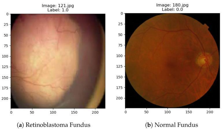
A side-by-side comparison of ophthalmic diagnostic images displaying a retinoblastoma fundus and a normal fundus. Image (a) depicts a retinoblastoma fundus characterized by a large, diffuse, yellowish-white mass occupying a significant portion of the posterior segment. The lesion exhibits a hazy texture, obscuring the underlying retinal architecture and the optic disc. Retinal blood vessels appear attenuated, displaced, and partially obscured by the mass. In contrast, image (b) shows a healthy normal fundus with a characteristic vibrant orange-red background. Key anatomical landmarks are clearly visible, including a well-defined, yellowish optic disc and a distinct macula. The retinal vasculature exhibits a normal branching pattern and clear distribution throughout the retina without obstruction or opacity. This comparison serves as a clinical educational tool for identifying the primary signs of retinoblastoma, a malignant intraocular tumor, including leukocoria and the presence of an endophytic or exophytic mass during funduscopic examination.
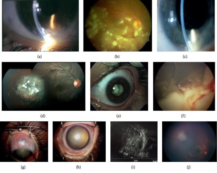
A multi-panel compilation of ophthalmological diagnostic images demonstrating various manifestations of retinoblastoma. (a, c) Slit-lamp biomicroscopy of the anterior segment showing multiple, round, whitish cystic lesions (pseudocysts) in the anterior chamber, representing diffuse infiltrating retinoblastoma. (b, d, f, j) Fundus photography illustrating diverse tumor morphologies including spontaneously regressed retinoblastoma (d, j), inferior retinal pseudocysts (b), and endophytic-exophytic growth with vitreous seeds and total retinal detachment (f). (e, h) External clinical photographs showing leukocoria (white pupillary reflex) and advanced pathology including ectropion uveae and mature cataract. (g) Clinical photograph demonstrating anterior scleral staphyloma secondary to neovascular glaucoma. (i) B-scan ultrasonography displaying a solid intraocular mass with hyper-echogenic calcifications filling the vitreous cavity. The collection serves to educate on the clinical spectrum of retinoblastoma in older children, emphasizing diagnostic features like pseudohyopyon, tumor regression, and secondary glaucoma.
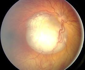
This clinical diagnostic image is a color fundus photograph demonstrating a large retinoblastoma. The primary feature is a prominent, well-circumscribed, chalky white-to-creamy intraocular mass originating from the retina and occupying a significant portion of the posterior pole. The tumor exhibits a characteristic nodular surface and obscures underlying retinal architecture. Significant vascular abnormalities are visible, including marked angiomatous dilatation and increased tortuosity of the 'feeder' retinal vessels supplying the neoplastic region. These dilated vessels appear engorged and follow an irregular, corkscrew-like course as they approach the tumor margins. The image illustrates the classical ocular fundus presentation of a primary intraocular malignancy in a pediatric patient, emphasizing key diagnostic signs such as the white retinal mass and associated pathological vascular remodeling. This visual is highly relevant for ophthalmological education regarding pediatric oncology and the differential diagnosis of leukocoria.
1. The disease in brief
- Most children present with a white pupil (leukocoria), in 61% of cases, with strabismus in 22%. - Textbook of Family Medicine 9e (Retinoblastoma section)
- Most children are identified by 3 years of age, and family or friends usually notice the white pupil first. - Tintinalli's Emergency Medicine (Retinoblastoma section)
- The cause is loss of the RB1 tumour suppressor gene on chromosome 13q14.2. - Sabiston Textbook of Surgery (Hereditary retinoblastoma)
- The Rb protein controls the G1 to S transition by regulating E2F transcription factors. - Basic Medical Biochemistry, 6e (The Retinoblastoma gene)
- About 60% of cases are sporadic, with both RB1 alleles inactivated by chance in a single retinal cell. - Thompson & Thompson Genetics and Genomics in Medicine, 9e (Retinoblastoma)
2. The idea in one paragraph
3. What is already known (novelty check)
- Kant et al. 2023, Indian Journal of Ophthalmology grafted freshly cut patient retinoblastoma and choroidal melanoma onto the CAM at embryonic day 10 and incubated for 7 days. They saw angiogenesis around the graft and invasion into the CAM mesoderm. The tumours were synaptophysin and Ki-67 positive. The authors suggested the model could support personalised drug screening.
- Fialho et al. 2020 reported successful growth of fresh retinoblastoma cells on the CAM.
- Mouse models exist: intravitreal Y79 cells in nude mice (Tschulakow et al. 2016), a published orthotopic model for preclinical testing, and subcutaneous Y79 xenografts (Burr et al. 2011).
| Novelty | Cost | Feasibility | Wow | Overall |
|---|---|---|---|---|
| 6 | 6 | 5 | 9 | 6.5 |
4. Design
- Start with a retinoblastoma cell line (for example Y79) rather than patient tissue. This avoids consent and supply problems.
- Confirm cell line identity before you start.
- Pilot the grafting. Follow the published schedule: window the egg, graft at embryonic day 10, harvest about 7 days later (Kant et al.). Record the graft take rate.
- Typical cell numbers per egg are in the order of a million cells in a gel plug or ring. Take the exact number from the cited papers and your pilot.
- Groups: vehicle, a standard retinoblastoma drug as the positive control (carboplatin is a reasonable choice because it is a standard retinoblastoma chemotherapy agent), and 4 to 5 candidates at two doses. That is about 12 groups with 8 eggs each, so about 100 eggs, and order about 125 for attrition.
- Candidates: plant compounds or repurposed drugs, whichever you can obtain.
- Treatment: topical or intratumoral dosing on 2 to 3 days after grafting.
- Readouts:
- Tumour size from photographs in the egg-lab imaging box.
- Vessels converging on the graft.
- Ki-67 index, synaptophysin and invasion depth on histology.
- Human DNA in the egg (Alu qPCR) for tumour burden, if you have a qPCR machine.
- Embryo survival as a toxicity check.
- Model: Subcutaneous Y79 xenograft in immunodeficient (nude) mice. It is the cheapest and simplest. An intravitreal model is closer to the real disease but needs microinjection skill and imaging.
- Groups (6 mice each): vehicle, carboplatin, and the top two candidates from the egg screen, chosen and recorded before the mouse study begins. That is 24 mice.
- Readouts: Tumour volume over time by calipers, body weight, final tumour weight, Ki-67, synaptophysin and CD31.
- Prediction test: Did the egg ranking match the mouse ranking? With only a few candidates, report it as exploratory.
5. Timeline (about 13 weeks)
| Weeks | Task |
|---|---|
| 0 | Ethics approval for mice, cell line authentication, orders. If using patient tissue, ethics and consent |
| 1-3 | Cell culture and CAM pilot |
| 3-6 | CAM drug screen |
| 6 | Lock the mouse candidates and the prediction |
| 7-11 | Mouse xenograft experiment |
| 11-13 | Histology, qPCR, statistics and write-up |
6. Why it is exciting
- It is a childhood eye cancer, and the story of an egg-based test saving mice is strong.
- A patient-derived version would be a personalised-medicine avatar, as Kant et al. suggest.
- The CAM result arrives in about a week, while mouse results take weeks.
7. Risks and limits
- Take rate: Y79 grows as suspension clusters, so grafts may not take reliably. The pilot will tell you.
- Mouse cost: Nude mice need special housing and are the biggest expense.
- Patient tissue: Scarce, and needs ethics approval and consent.
- Species gap: CAM tumours are in an avian host, and the egg lacks a mature immune system, so immune effects are not tested.
- Cytotoxic drugs: Handle carboplatin with proper protective equipment and waste disposal.
- Ethics: Embryo rules differ by country. The mouse work needs animal ethics approval.
8. Next step
Cost for this
athymic nude mouse price per mouse Envigo Charles River Jackson Laboratory NU/NU female 6 week
Y-79 retinoblastoma cell line ATCC HTB-18 price
cost of fertilized chicken eggs for CAM assay per egg price research
Verified prices
- Y79 cell line (ATCC HTB-18): ATCC lists $577 for nonprofit customers, and Fisher Scientific lists $706.83.
- Fertilised eggs: A King's College London article says 12 fertilised eggs cost £45, with zero maintenance costs, a 97% saving compared with standard mouse xenografts. That is about £3.75 per egg. Local farm or hatchery eggs are often much cheaper.
- Nude mice: I could not retrieve a price. Vendors quote on request, and prices vary by country, so get a quote.
Budget by item
| Item | Assumption | Low | High |
|---|---|---|---|
| Cell line and culture | 1 vial, medium, serum, mycoplasma test, identity check | $1,000 | $1,800 |
| Eggs and egg equipment | About 125 eggs, incubator if not owned, window tools, silicone rings, cardboard imaging box | $250 | $1,100 |
| Drugs and compounds | Carboplatin plus 4 to 5 candidates (plant extracts are cheap, pure compounds are not) | $200 | $800 |
| Histology and staining | Fixative, embedding, H&E, antibodies for Ki-67, synaptophysin and CD31, or core facility fees | $800 | $2,000 |
| Alu qPCR (optional) | DNA kit, primers, master mix | $0 | $700 |
| Mouse study | 24 nude mice, housing for 6 to 8 weeks, Matrigel, anaesthesia and analgesia, consumables | $2,200 | $6,000 |
| Total | about $4,500 | about $12,500 |
Where the money goes
- The mouse study is about half of the total, mainly the nude mice and their housing.
- Histology and antibodies are the second largest cost.
- Eggs are cheap. The egg screen is the lowest-cost part of the project.
Why the egg-first design saves money
Ways to reduce the cost
- Get the cell line from a collaborator or a local cell repository instead of buying a new vial. This can save $500 to $700.
- Use a subcutaneous xenograft, not an intravitreal one. It needs no microinjection equipment or eye imaging.
- Use a core facility for histology and antibodies, which is often cheaper than buying antibodies for one project.
- Skip the qPCR and measure tumour size and histology only.
- Cut the egg groups to 8 and use 8 eggs per group.
- Use 5 mice per group (20 mice) if your power calculation and ethics committee allow.
- Build the imaging box from cardboard or 3D-print it.
Compared with the other topics
Quick answer
Retinoblastoma project cost (my estimate, in US dollars)
- Y79 cell line: ATCC lists $577 for nonprofit customers, and Fisher Scientific lists $706.83.
- Fertilised eggs: A King's College London article says 12 eggs cost £45, a 97% saving compared with standard mouse xenografts. That is about £3.75 per egg, and local hatchery eggs are often cheaper.
- Nude mice: I could not retrieve a price. Ask your supplier for a quote.
| Item | Low | High |
|---|---|---|
| Cell line and culture | $1,000 | $1,800 |
| Eggs and egg equipment (about 125 eggs) | $250 | $1,100 |
| Drugs and compounds | $200 | $800 |
| Histology and antibodies | $800 | $2,000 |
| Alu qPCR (optional) | $0 | $700 |
| Mouse study (24 nude mice, housing, consumables) | $2,200 | $6,000 |
| Total | about $4,500 | about $12,500 |
Where the money goes
- The mouse study is about half of the total.
- Histology and antibodies come second.
- The egg screen is the cheapest part.
Why the egg-first design saves money
Ways to lower the cost
- Get the cell line from a collaborator or local repository, which can save $500 to $700.
- Use a subcutaneous xenograft rather than an intravitreal one.
- Use a core facility for histology and antibodies.
- Skip the qPCR and measure tumour size and histology only.
- Reduce the egg screen to 8 groups of 8 eggs.
- Use 5 mice per group if your ethics committee and power calculation allow.
- Build the imaging box from cardboard.
Give more innovative ideas with fessible also you gave unfessible only
"chick embryo" AND paracetamol AND hepatotoxicity
"chick embryo" AND "alizarin red" AND mineralization AND osteoporosis
"Ehrlich" AND "chorioallantoic membrane"
"chick embryo" AND hyperglycemia AND "oxidative stress" AND malformation
"chorioallantoic membrane" AND "vasodilation" OR "vasoconstriction" AND hypertension
| # | Disease | Novelty | Cost | Feasibility | Wow | Overall |
|---|---|---|---|---|---|---|
| 1 | Anthracycline cardiomyopathy (top pick) | 8 | 9 | 8 | 9 | 8.5 |
| 2 | Diabetic embryopathy | 7 | 9 | 8 | 8 | 8.0 |
| 3 | Psoriasis | 7 | 8 | 8 | 8 | 7.75 |
| 4 | Cancer, with a dose-sparing twist (Ehrlich tumour) | 6 | 9 | 9 | 7 | 7.75 |
| 5 | Drug-induced liver injury | 7 | 9 | 8 | 7 | 7.75 |
| 6 | Glucocorticoid-induced bone loss | 7 | 8 | 7 | 7 | 7.25 |
1. Anthracycline cardiomyopathy (8.5)
- In ovo: Phone-video heart rate in chick embryos treated with doxorubicin with or without cardioprotectants. Add a tumour graft on the CAM of parallel eggs to check the protectant does not shield the tumour.
- Mouse: Standard doxorubicin cardiotoxicity in common mice (30 animals). Measure troponin or CK-MB, heart weight and histology.
- Why it is feasible: No special mice or pathogens, and the egg part needs only a phone.
2. Diabetic embryopathy (8.0)
- In ovo: Hyperglycaemia in the chicken embryo is an established model. It has been used to study hyperglycaemia's effects on embryonic development (2025), congenital limb defects and a gestational diabetes eye malformation model involving Pax6 and oxidative stress. Your screen tests antioxidant or plant protectants: malformation rate, survival, and oxidative stress markers.
- Mouse: STZ-induced diabetic mice treated with the top candidate. Measure blood glucose, MDA, SOD and GSH, and kidney or liver histology. A pregnant-mouse arm is possible but adds cost and time.
- Why it works: The disease relevance is clear, STZ is a cheap and widely used model, and the egg part is quick.
- Novelty: The model exists, so novelty comes from your protectant and from linking embryo protection to the maternal diabetic markers.
3. Psoriasis (7.75)
- In ovo: CAM vessel and irritation screen of herbal anti-psoriatic candidates.
- Mouse: Imiquimod-induced psoriasis-like skin in BALB/c or C57BL/6 mice over about a week. Score erythema, scaling and skin thickness, plus histology.
- Caveat: I found no established CAM psoriasis model, so the egg reflects only the angiogenic and irritant parts of the disease.
4. Cancer with a dose-sparing twist (7.75)
- In ovo: Murine tumours, including Ehrlich carcinoma, have been grown on the CAM (Uchida et al., 1987). An Ehrlich tumour study with CAM work also exists (Pereira et al., 2013). Test drug and herbal combinations on CAM tumours at several doses.
- Mouse: Ehrlich solid tumour in Swiss albino mice, which are cheap and need no immunodeficient animals. Compare full-dose chemotherapy, half-dose chemotherapy plus adjunct, and controls.
- Twist: The question is whether an adjunct allows a lower chemotherapy dose with the same effect, which is a clinically meaningful goal.
- Novelty: Moderate, since the models are established.
5. Drug-induced liver injury (7.75)
- In ovo: Treat embryos with paracetamol with or without extracts. Score survival and liver injury markers (glutathione, MDA, histology).
- Mouse: Paracetamol hepatotoxicity in mice, which is cheap and takes days. Measure ALT, AST, glutathione and liver histology.
- Caveat: I found paracetamol liver injury modelled in Xenopus embryos but not in chick embryos, so the egg model would need a pilot to confirm it works. That makes it a bit riskier, but it is also where the novelty is.
6. Glucocorticoid-induced bone loss (7.25)
- In ovo: Whole-mount bone and cartilage staining of chick embryos is an established method (Yamazaki et al., 2011). Treat with a glucocorticoid with or without candidates and quantify bone staining from photographs. I did not find an existing osteoporosis screen of this kind, so this is a new application and needs validation.
- Mouse: Dexamethasone-induced bone loss. Measure serum calcium and alkaline phosphatase, femur ash weight and calcium, and histology. Micro-CT is optional.
- Caveat: Embryonic bone formation is not the same as adult bone loss, so state that limit plainly.
My picks
- Safest and strongest: Idea 1 (doxorubicin cardiomyopathy).
- Best story for a cheap model: Idea 2 (diabetic embryopathy).
- Most straightforward mouse work: Idea 4 (Ehrlich tumour) or idea 3 (psoriasis).
I said innovative
curcumin photodynamic therapy "chorioallantoic membrane"
"chick embryo" AND anticonvulsant AND motility
"egg yolk" AND liposomes OR nanovesicles AND "chorioallantoic membrane"
"chick embryo" AND venom AND neutralization
"chick embryo" AND lens AND cataract AND selenite
chick embryo CAM in ovo novel disease model 2025 2026 immune cells humanized OR sepsis OR neuroinflammation OR vascular disease
| # | Disease | Creative hook | Novelty | Cost | Feasibility | Wow | Overall |
|---|---|---|---|---|---|---|---|
| 1 | Cancer | Egg-lipid nanocarrier with self-fluorescent curcumin, tracked live on the CAM | 7 | 9 | 8 | 9 | 8.25 |
| 2 | Cancer | Kitchen photodynamic therapy with vascular-shutdown video | 6 | 9 | 8 | 9 | 8.0 |
| 3 | Epilepsy | Embryo-movement AI screen for anticonvulsants | 7 | 9 | 8 | 8 | 8.0 |
| 4 | Sepsis | Embryo heart-rate early marker for endotoxaemia | 7 | 8 | 8 | 8 | 7.75 |
| 5 | Cataract | Phone slit-lamp transparency scoring | 7 | 9 | 7 | 8 | 7.75 |
| 6 | Snakebite | Embryo-lethality screen for plant antivenoms | 6 | 7 | 5 | 10 | 7.0 |
1. Cancer: egg-lipid nanocarrier with self-fluorescent curcumin (8.25)
- Hook: Curcumin fluoresces, so you can watch the carrier travel through CAM blood vessels without adding a dye. The egg supplies the carrier material and the test system.
- What exists: Egg yolk lipids have been used to build a tumour-targeted nanovector (Tang et al., 2020), and chicken egg yolk nanovesicles have been characterised (Saadeldin et al., 2023). Your novelty is the live self-fluorescence tracking on the CAM and the CAM-to-mouse comparison, which I did not find.
- In ovo: Compare free curcumin with the loaded carrier for fluorescence in vessels over time and CAM tumour size.
- Mouse: Ehrlich solid tumour in Swiss albino mice. Compare free drug, carrier, and controls on tumour volume and histology.
- Needs: A blue LED or UV torch, a phone with a yellow filter, and a way to make lipid vesicles (thin-film or ethanol injection).
2. Cancer: kitchen photodynamic therapy (8.0)
- Hook: Use live CAM video to find the light dose that closes tumour vessels, then take that dose into the mouse. The vessel-shutdown video is very striking.
- What exists: The CAM is a known model for photodynamic therapy (Gottfried et al., 1992; quail CAM method, 2022), and curcumin photodynamic therapy on the CAM was published in 2019. So novelty is moderate. Your twist is a standardised light-dose finder and CAM-to-mouse dose transfer.
- Mouse: Ehrlich solid tumour in Swiss albino mice with photosensitiser and LED light, versus dark and light-only controls.
- Safety: Keep light exposures within a measured dose, and protect your eyes.
3. Epilepsy: embryo-movement AI screen (8.0)
- Hook: Neuroactive drugs change embryo spontaneous movement, and this was studied decades ago (Sedlacek, 1979). A phone and a motion-tracking script turn it into an objective screen.
- In ovo: Film embryo movement before and after convulsant and anticonvulsant exposure. Use reference drugs to prove the assay can tell the classes apart before you test your candidates.
- Mouse: PTZ seizure model. Record latency to seizure, seizure duration and mortality.
- Caveat: Embryo movement is not a seizure. State clearly that the egg is a neuroactivity filter, and validate the ranking against the mouse results.
4. Sepsis: embryo heart-rate early marker (7.75)
- Hook: Endotoxin toxicity in chick embryos was shown decades ago, with antibiotics such as polymyxins neutralising it (Rifkind and Palmer, 1966). The idea of heart rate as an early readout is my own suggestion and I have not verified it, so it needs a pilot.
- In ovo: LPS challenge with candidate treatments. Record survival and heart-rate change.
- Mouse: LPS endotoxaemia in mice. Measure survival, TNF-alpha and IL-6 by ELISA, and organ histology.
5. Cataract: phone slit-lamp transparency scoring (7.75)
- Hook: Embryonic chick lenses have been used as cataract models (Li et al., 2021; drug-induced cataract in cultured chick lens, 1973). The new part is cheap, objective phone-based opacity scoring plus a screen of antioxidants.
- Mouse: The classical selenite cataract model is done in rat pups (review). I did not confirm a mouse-pup protocol, so check this before choosing this topic if mice are required.
6. Snakebite: embryo-lethality screen for plant antivenoms (7.0)
- What exists: A hen's egg method has been proposed as a humane alternative to venom LD50 testing (non-neurotoxic venoms), a plant root extract neutralised viper venom in chick embryos, and egg yolk antibodies neutralised cobra and krait venom. Parts of this are not new.
- Hook: Neglected tropical disease, big public health impact, and a clear 3Rs story.
- Why it is last: Venom is hard to obtain and regulated, and the work needs trained handling. Feasibility is 5.
My picks
- Most creative with good feasibility: Idea 1 (egg-lipid nanocarrier).
- Best visual: Idea 2 (vascular-shutdown video).
- Newest disease area: Idea 3 (epilepsy).
Mouse model not available only swiss albino and sprague dawly give more innovative ideas
"chick embryo" AND "anoxia" AND heart AND cardioprotection
"chick embryo" AND hypoxia AND brain AND neuroprotection
"chick embryo" AND cisplatin AND kidney OR nephrotoxicity
"chick embryo" AND "amyloid" AND neurotoxicity AND neuroprotective
"DMBA" AND mammary AND rats AND angiogenesis AND "chorioallantoic membrane"
"Dalton's lymphoma" AND "chick embryo" OR "Dalton's lymphoma" AND "chorioallantoic membrane"
"chick embryo" AND "uric acid" AND allopurinol
"chick embryo" AND "xanthine oxidase" AND "allantoic fluid"
| # | Disease | Hook | Animal | Novelty | Cost | Feasibility | Wow | Overall |
|---|---|---|---|---|---|---|---|---|
| 1 | Chemotherapy side effects | One egg screens heart or kidney protection and checks the tumour is not protected | SD rat | 8 | 9 | 8 | 9 | 8.5 |
| 2 | Hyperuricaemia and gout | Embryo uric acid output as a drug screen | Swiss mouse or SD rat | 8 | 9 | 8 | 8 | 8.25 |
| 3 | Myocardial infarction | Embryo hypoxia-reoxygenation heart screen | SD rat | 7 | 9 | 8 | 8 | 8.0 |
| 4 | Breast cancer prevention | The egg reads early cancer risk from rat tissue | SD rat | 8 | 6 | 6 | 9 | 7.25 |
| 5 | Neonatal brain injury (stretch) | Embryo hypoxic-ischaemic brain model | SD rat pup | 7 | 7 | 5 | 9 | 7.0 |
1. Chemotherapy protection platform (8.5)
- Hook: A protectant is only useful if it does not also shield the tumour. The egg can test organ protection (heart rate by phone video, or kidney histology) and tumour response together.
- What exists: Cisplatin nephrotoxicity has been assessed in chick embryos (Náprstková et al., 2003). The combined protection-plus-tumour design is my own suggestion and I did not find it published, though my search was not exhaustive.
- In ovo: Embryos get the drug with or without candidates. Readouts: heart rate and survival (doxorubicin) or kidney weight and histology (cisplatin). Graft tumour cells on the CAM of a parallel set.
- Rat: Cisplatin acute kidney injury in SD rats (serum creatinine, BUN, kidney histology) or doxorubicin cardiotoxicity (troponin or CK-MB, heart histology). You can pick one organ for a shorter project.
- Optional: An Ehrlich tumour arm in Swiss mice to show the drug still works.
2. Hyperuricaemia and gout (8.25)
- Hook: Birds make uric acid as their main nitrogen waste, so the embryo is a ready-made uric acid factory. Embryonic xanthine oxidase activity and its inhibition have been studied (Duggan et al., 1968; Butros and Khalidi, 1966), but those are old studies. A cheap modern plant-extract screen with a standard uric acid assay is the new angle. I did not find one.
- In ovo: Treat embryos with allopurinol (reference) and plant extracts. Measure uric acid in embryo blood or allantoic fluid, plus survival and malformations.
- Mouse: Swiss albino mice given potassium oxonate (which blocks uricase, since mice, unlike birds, break uric acid down) with or without hypoxanthine. Measure serum uric acid, liver xanthine oxidase activity, creatinine and kidney histology. A rat paw urate-crystal model is an option for the gout flare.
- Caveat: Embryo purine metabolism differs from adult mammals, so use allopurinol as a benchmark and a pilot to confirm the assay works.
3. Myocardial infarction (8.0)
- What exists: Embryonic heart models of anoxia-reoxygenation have been used (Bruchez et al., 2008), and hypoxia causes dilated cardiomyopathy in chick embryos (Tintu et al., 2009). Day-4 embryos can keep their heart rate during hypoxia (Nechaeva et al., 2020), so stage and hypoxia severity need a pilot.
- Hook: Phone-video heart-rate recovery after hypoxia as a quick screen for candidate extracts.
- Rat: Isoproterenol-induced myocardial injury in SD rats (a few days). Measure troponin or CK-MB, ECG if available, and heart histology or TTC staining.
4. Breast cancer prevention: the egg reads the rat's cancer risk (7.25)
- Hook: Mammary tissue at different stages elicits different angiogenic responses on the CAM (Strum, 1983, mouse tissue), and DMBA-induced lesions are angiogenic (Heffelfinger et al., 2000). So take small biopsies from DMBA-treated SD rats at set times, test their angiogenic potency on the CAM, and see whether a chemopreventive diet or extract lowers it before tumours appear.
- Rat: SD female rats given DMBA, with chemopreventive treatment. Follow tumour incidence and latency.
- Why it ranks lower: The model takes months and DMBA is a hazardous carcinogen needing proper handling. Feasibility is 6.
5. Neonatal brain injury (stretch, 7.0)
- What exists: A hypoxic-ischaemic injury model in the embryonic chicken cerebral pallium has been described (Olivares-Hernández et al., 2022).
- Rat: The pup model needs carotid ligation surgery and a controlled hypoxia chamber. I rate feasibility at 5 because of the surgical skill and equipment.
Earlier ideas that already fit your animals
- Egg-lipid curcumin nanocarrier with live CAM tracking, validated in Ehrlich or Dalton's lymphoma tumours in Swiss mice (8.25). Dalton's lymphoma-induced angiogenesis has been studied before (Kumari et al., 2017).
- Kitchen photodynamic therapy with vascular-shutdown video, validated in Ehrlich solid tumours (8.0).
- Embryo-movement AI anticonvulsant screen, validated in the PTZ seizure model (8.0).
- Embryo sepsis screen, validated in LPS endotoxaemia in Swiss mice (7.75).
- Cataract screen on embryonic lens, validated in selenite-induced cataract. This is classically done in rat pups, so SD pups should work (7.75), but confirm the strain in the protocol you follow.
- Diabetic embryopathy, validated in STZ-diabetic rats (8.0).
- Doxorubicin cardioprotection in SD rats (8.5).
My picks
- Most creative and still practical: Idea 1 (chemotherapy protection platform).
- Newest disease area: Idea 2 (hyperuricaemia and gout).
- Quickest rat work: Idea 3 (myocardial infarction).
More innovative ideas
"chick embryo" AND "feather" AND "hair growth" OR "alopecia"
"chorioallantoic membrane" AND "Rose Bengal" AND thrombosis
"light-induced retinal degeneration" AND rats AND "chick embryo"
"valproic acid" AND "chick" AND "in ovo" AND autism
"chick embryo" AND "hepatic steatosis" OR "chick embryo" AND "fatty liver"
"chick embryo" AND "calcium oxalate" OR "chick embryo" AND "kidney stone"
Why this pairing works
Ranked ideas
| # | Idea | Novelty | Cost | Feasibility | Wow | Overall |
|---|---|---|---|---|---|---|
| 1 | AI/smartphone vessel quantification, validated against in vivo | 8 | 9 | 7 | 8 | 8.0 |
| 2 | Eggshell hydroxyapatite bone scaffold ("egg to bone") | 7 | 8 | 7 | 9 | 7.75 |
| 3 | Vape liquid or microplastic developmental toxicity | 7 | 8 | 8 | 8 | 7.75 |
| 4 | Hyperglycaemic CAM, then diabetic rat wound | 7 | 8 | 8 | 7 | 7.5 |
| 5 | Infected-wound CAM (biofilm), then mouse infected wound | 7 | 8 | 7 | 8 | 7.5 |
| 6 | Natural-product funnel with in ovo/in vivo concordance score | 6 | 9 | 9 | 6 | 7.5 |
| 7 | Smart pH-indicator wound dressing | 7 | 7 | 7 | 8 | 7.25 |
| 8 | Biowaste carbon-dot theranostics | 8 | 6 | 6 | 9 | 7.25 |
| 9 | Low-cost photodynamic therapy (LED + curcumin or methylene blue) | 7 | 7 | 7 | 8 | 7.25 |
| 10 | Cardiotoxicity and cardioprotection screen | 6 | 8 | 8 | 6 | 7.0 |
| 11 | Chick-embryo "avatar" for personalised drug testing | 8 | 5 | 5 | 9 | 6.75 |
| 12 | Green-synthesised nanoparticles (Ag, ZnO, Se) | 4 | 8 | 9 | 5 | 6.5 |
The ideas in brief
- In ovo: Photograph CAMs with a smartphone on a cheap macro lens. Train a free Python U-Net or use AngioTool to count vessels automatically.
- In vivo: Run a mouse Matrigel plug or rat wound, scored by haemoglobin content and CD31 staining.
- Novelty: A cheap, open-source tool that shows CAM image scores predict in vivo angiogenesis. A correlation figure (r, Bland-Altman) is the headline result.
- Scaffold: Calcine waste eggshell into hydroxyapatite and blend it with chitosan or gelatin. Add a plant osteogenic extract or a trace-ion dopant such as Sr or Zn for extra novelty.
- In ovo: Check biocompatibility and angiogenesis on the CAM.
- In vivo: Test in a rat calvarial defect, with micro-CT if you can get it.
- Why it stands out: The circular-economy story is easy to remember.
- In ovo: Inject at day 0 to 3 and score mortality, malformations, heart rate and CAM vascularity.
- In vivo: Run a dose-matched rat or zebrafish study for liver, kidney and oxidative stress markers.
- Why it stands out: Public interest is high. Adding a rescue arm (for example an antioxidant) makes it mechanistic.
- In ovo: Create a high-glucose environment that impairs angiogenesis, then test pro-angiogenic candidates (honey, curcumin, PRP, a peptide).
- In vivo: Take the best candidate into an STZ-diabetic rat excision wound.
- Why it stands out: A high-glucose CAM screen is a quick way to test diabetic-wound candidates.
- In ovo: Infect the CAM with S. aureus, Pseudomonas or Candida, then treat with essential oil, phage or an antimicrobial peptide. Score bacterial load and lesion size.
- In vivo: Use a mouse or rat infected wound.
- Why it stands out: Antimicrobial resistance is a strong theme.
- Design: Screen 8 to 10 extracts by MTT assay, then CAM, then one tumour model (Ehrlich ascites or a xenograft).
- Novelty: Report how well the in ovo ranking predicted the in vivo ranking (Spearman correlation). This is the safest and cheapest option. It becomes more novel if you choose an under-studied regional plant.
- Dressing: Electrospun or cast film containing anthocyanin from red cabbage, which changes colour with wound pH, plus an active such as curcumin.
- In ovo: HET-CAM irritation test and angiogenesis.
- In vivo: Rat wound, tracking pH against healing rate.
- Why it stands out: The colour change is visual and photographs well.
- Material: Make fluorescent carbon dots from banana peel, curry leaf or similar by hydrothermal synthesis.
- In ovo: Image them live through a window in the shell, tracking vessel uptake and tumour accumulation.
- In vivo: Run a mouse biodistribution study.
- Why it stands out: Live glowing vessels make a strong figure. It needs a fluorescence imager or UV box with a camera, which is the main cost and risk.
- Setup: A cheap LED array plus a photosensitiser such as curcumin or methylene blue.
- In ovo: Treat CAM tumours or vessels.
- In vivo: Use a mouse subcutaneous tumour.
- Why it stands out: It frames the work as affordable cancer therapy for low-resource settings.
- In ovo: Measure embryonic heart rate and rhythm by video with doxorubicin, then add a protective natural compound.
- In vivo: Rat doxorubicin model with ECG and cardiac enzymes.
- In ovo: Graft patient-derived or cell-line tumour fragments onto the CAM and test drug combinations.
- In vivo: Compare with a mouse xenograft.
- Why it stands out: The personalised-medicine angle is very strong, but it needs tissue consent and ethics approval, so it is harder. Cell lines are a safer start.
My picks
- Highest wow with low risk: Idea 2, the eggshell scaffold.
- Best for publication: Idea 1, because validation work is rare and tool-based papers get cited.
- Safest for a student timeline: Idea 6, or idea 4 for something slightly more novel.
Practical notes
- Eggs: Use fertilised eggs, incubated at 37.5°C and about 60% humidity. Use about 8 to 10 eggs per group. Plan for 20 to 30% attrition.
- Ethics: Rules for embryos differ by country and institution, so confirm with your ethics committee. Mouse and rat work needs animal ethics approval. Zebrafish larvae are often treated more leniently.
- Minimal controls: Vehicle, positive control (for example bevacizumab or sunitinib for anti-angiogenesis) and untreated.
- Statistics: One-way ANOVA with a post hoc test, plus the in ovo vs in vivo correlation.
| # | Idea | Novelty | Cost | Feasibility | Wow | Overall |
|---|---|---|---|---|---|---|
| A | Digital twin add-on: machine learning that predicts in vivo outcome from in ovo features | 9 | 9 | 6 | 9 | 8.25 |
| B | In ovo feeding to post-hatch chick (the egg and the animal are the same species) | 7 | 9 | 8 | 8 | 8.0 |
| C | CAM bleeding model for low-cost haemostats | 7 | 9 | 8 | 7 | 7.75 |
| D | Herbo-mineral (bhasma) safety screen | 7 | 8 | 7 | 8 | 7.5 |
| E | LED photobiomodulation for healing | 7 | 8 | 8 | 7 | 7.5 |
| F | Microneedle patch penetration on CAM, then rat transdermal | 8 | 6 | 6 | 9 | 7.25 |
| G | Phone or Wi-Fi radiofrequency exposure and embryo development | 6 | 8 | 8 | 7 | 7.25 |
| H | Plant-based snake venom haemorrhage antidote | 8 | 6 | 4 | 9 | 6.75 |
Details
- Extract features from every CAM image: vessel density, branching points, lacunarity and fractal dimension. Combine them with dose and compound descriptors.
- Train a simple model (random forest or regression) to predict the in vivo result, such as wound closure percentage or tumour volume. Validate it on held-out compounds.
- "We predicted mouse results from an egg" is a strong headline, and it costs only your time. The risk is sample size, so you need at least 10 to 15 compounds or doses.
- In ovo: Inject probiotics, prebiotics, plant extracts or nanominerals into the egg on about day 17 to 18. This is a real poultry technique. Record hatchability and hatch weight.
- In vivo: Raise the hatched chicks for 3 to 6 weeks. Measure growth, gut villus height, microbiota, antibody titre after vaccination and heat-stress markers.
- Why it stands out: There is no rodent colony and no cross-species gap. A continuous egg-to-bird story is easy to explain, and the poultry industry may fund it.
- In ovo: Puncture a standard-size CAM vessel and apply chitosan, alum, a plant extract or gelatin sponge. Time the bleeding to stop and quantify blood loss from images.
- In vivo: Rat tail transection or liver-wound bleeding.
- Why it stands out: The cheap, standardised in ovo bleeding assay is the novelty. The CAM puncture model is much less used than the angiogenesis assay.
- In ovo: Test classical herbo-mineral preparations for embryo toxicity, malformations and CAM effects.
- In vivo: Sub-acute rat study of the safest and the most toxic preparations (liver and kidney enzymes, histology, tissue metal levels).
- Why it stands out: It addresses a real gap in the safety evidence for traditional medicines. Add metal quantification (ICP-MS or AAS) if your lab can reach it.
- Setup: A low-cost red/near-infrared LED array with a power meter.
- In ovo: Test several wavelengths and doses on CAM angiogenesis and embryo viability.
- In vivo: Rat or diabetic-rat wound, with the optimal dose from the CAM.
- Why it stands out: It finds the best dose cheaply before committing animals.
- Patch: Make dissolvable microneedles (PVA, gelatin or sugar) in a PDMS mould cast from a cheap commercial needle array or a 3D-printed master.
- In ovo: Test needle penetration and dye diffusion on the CAM. Embryo skin or CAM also works.
- In vivo: Test insulin or a drug in rats.
- Risk: The moulds and fabrication take trial and error.
- In ovo: Expose eggs to a phone or Wi-Fi router during incubation. Record hatchability, malformations, heart rate and oxidative stress.
- In vivo: Rats exposed for several weeks, with a protective antioxidant arm if you want a mechanistic angle.
- Caution: Past results in this field are mixed and often poorly controlled. Use sham exposure, a measured field strength and blinded scoring.
- In ovo: Measure venom-induced haemorrhage in embryos and test plant extracts as inhibitors.
- In vivo: Mouse neutralisation assays.
- Why it stands out: Embryo screening could cut the number of mice used in venom neutralisation work.
- Risk: Venom is hard to obtain and regulated, so I rated it low on feasibility.
Combinations for the most impact
- Eggshell hydroxyapatite scaffold (from the first list) + AI vessel quantification + digital twin (A). The story is egg waste to a bone graft, assessed by an open-source tool and predicted by a model.
- Hyperglycaemic CAM + LED photobiomodulation (E) + digital twin (A). This is an affordable diabetic-wound platform, with the dose chosen in ovo.
- Haemostat (C) + smart dressing (from the first list). A first-aid kit concept.
Next step
| # | Idea | Novelty | Cost | Feasibility | Wow | Overall |
|---|---|---|---|---|---|---|
| 1 | Contactless video-based embryo heart-rate and toxicity readout | 8 | 9 | 7 | 8 | 8.0 |
| 2 | Herbal sunscreen or UV photoprotection | 6 | 9 | 8 | 7 | 7.5 |
| 3 | CAM as a bioreactor to prevascularise spheroids, then implant | 8 | 6 | 6 | 9 | 7.25 |
| 4 | Endometriosis or ovarian lesion on CAM, then mouse | 8 | 7 | 5 | 9 | 7.25 |
| 5 | Hypoxia or high-altitude protectants | 7 | 8 | 7 | 7 | 7.25 |
| 6 | Teratogen rescue (valproate plus folate or a nutraceutical) | 6 | 8 | 8 | 7 | 7.25 |
| 7 | Anti-inflammatory screen on CAM, then rat paw oedema | 6 | 9 | 9 | 5 | 7.25 |
| 8 | Heavy-metal toxicity and natural chelators | 6 | 8 | 8 | 6 | 7.0 |
| 9 | Antimicrobial sutures or implant coatings | 6 | 7 | 7 | 7 | 6.75 |
| 10 | Corneal neovascularisation eye drops | 6 | 7 | 6 | 7 | 6.5 |
Details
- In ovo: Film the embryo through a window or by candling with a phone. Extract heart rate and motion with free signal-processing code (Eulerian video magnification or simple intensity tracking). Use it as a non-invasive, repeatable toxicity or cardiotoxicity marker over days.
- In vivo: Compare with rat ECG after the same drug (doxorubicin, a QT-prolonging drug or an energy drink).
- Why it stands out: You replace expensive instruments with a phone and open code. The key step is validating it against a reference method.
- In ovo: Expose embryos or the CAM to UV-B or UV-C through a film or cream. Score survival, vessel damage and oxidative markers.
- In vivo: Mouse or rat UV skin model (erythema, thickness, MDA and histology).
- Extra: Measure SPF in vitro by spectrophotometer. A UV lamp with a measured dose is the main equipment need.
- In ovo: Grow tumour spheroids, 3D-printed or hydrogel scaffolds, or stem cell constructs on the CAM for several days so chick vessels grow in.
- In vivo: Implant the prevascularised construct in a mouse or rat and test whether it survives better than an unvascularised one.
- Why it stands out: It is a creative use of the egg as a cheap incubator for tissue engineering. The main risk is that the vessels are avian, so check for host integration and rejection.
- In ovo: Graft endometrial cells or tissue and test anti-angiogenic or anti-adhesion agents. Ovarian cancer metastasis on the CAM is well described, but verify the endometriosis protocols in the literature before committing.
- In vivo: Mouse endometriosis model.
- Why it stands out: Women's health is under-funded and reviewers like the topic. Use cell lines first, because human tissue needs ethics approval and consent.
- In ovo: Induce hypoxia with a low-oxygen setup or chemically (for example CoCl2). Test protective agents such as rhodiola, ginkgo or antioxidants on survival, vascular density and HIF-1α.
- In vivo: Rat hypobaric chamber if available, or a chemical hypoxia model (sodium nitrite).
- In ovo: Treat with valproate or another teratogen, then add folic acid, choline or a plant antioxidant. Score neural tube defects, limb and heart malformations, and size.
- In vivo: Pregnant rat with the best rescue dose.
- Why it stands out: The rescue design gives you a mechanistic story. Pregnant-animal work has stricter ethics and a longer timeline.
- In ovo: A CAM inflammation model is cheap, so screen many extracts.
- In vivo: Carrageenan rat paw oedema.
- Note: This is the easiest and safest option, but the least exciting. It works well as a base for idea 1 from the previous list, the AI quantification.
- In ovo: Treat with lead, cadmium or arsenic and test moringa, chlorella or coriander extract.
- In vivo: Rat model with blood and tissue metal levels and liver and kidney markers.
Ways to raise the wow factor in any project
- Time-lapse imaging. A phone on a clamp or a 3D-printed holder can film CAM vessel growth over 24 to 72 hours.
- Open data and code. Posting images and scripts (for example on Zenodo or GitHub) raises citation chances.
- Preregistration of your design and endpoints, which reviewers value.
- A single visual summary. A clear egg-to-animal workflow figure and one striking before and after image.
- A frugal comparison. Show cost per experiment against a mouse-only design.
- Negative controls and blinding. Score images blinded, which adds credibility at no cost.
Next step
| Rank | Idea | Novelty | Cost | Feasibility | Wow | Overall |
|---|---|---|---|---|---|---|
| 1 | Open-source "Frugal Egg Lab" kit | 9 | 10 | 8 | 9 | 9.0 |
| 2 | Public benchmark plus digital twin | 9 | 10 | 7 | 9 | 8.75 |
| 2 | Egg-to-bird feeding of waste-derived bioactives | 8 | 10 | 9 | 8 | 8.75 |
| 4 | Everyday exposome in an egg | 8 | 9 | 8 | 9 | 8.5 |
| 4 | Eggshell-to-bone with AI scoring | 8 | 9 | 7 | 10 | 8.5 |
| 4 | Kitchen-to-clinic haemostat | 7 | 10 | 9 | 8 | 8.5 |
| 7 | Diabetic-wound platform (stacked) | 8 | 8 | 7 | 9 | 8.0 |
1. Open-source Frugal Egg Lab kit (9.0)
- Concept: A complete, cheap, reproducible kit for CAM experiments. It has a 3D-printed or cardboard imaging box with a phone clamp and LED ring, a standard window-cutting and sealing protocol, and free software that counts vessels.
- In ovo: Run an anti-angiogenesis and a pro-angiogenesis screen with known controls (bevacizumab or sunitinib, and VEGF or a pro-angiogenic extract).
- In vivo: Validate against one mouse Matrigel plug or rat wound experiment, scored by haemoglobin and CD31.
- Why it scores high: Labs with no imaging equipment can use it, and it is publishable as a methods paper.
- What holds it back: Image consistency across labs. To get to 10, test the kit in a second lab and report inter-lab agreement.
2. Public benchmark plus digital twin (8.75)
- Concept: Build the first small open benchmark of CAM results for 15 to 20 compounds whose in vivo potency is already published (known anti-angiogenic and pro-angiogenic drugs). Then test whether a simple model predicts the published in vivo ranking.
- In ovo: Dose-response of each compound on the CAM.
- In vivo: Mostly literature data, plus one small confirmatory experiment with a new compound the model never saw.
- Why it scores high: Almost no animals are needed, and a dataset others can reuse is a durable contribution.
- What holds it back: Published in vivo data are inconsistent across species and doses. To reach 10, preregister the prediction and test it on the unseen compound.
3. Egg-to-bird feeding of waste-derived bioactives (8.75)
- Concept: Inject waste-derived bioactives (eggshell calcium nanoparticles, fruit-peel polyphenols or similar) into the egg at about day 17 to 18, then raise the chicks.
- In ovo: Hatchability, hatch weight and embryo toxicity.
- In vivo: The same birds for 3 to 6 weeks: growth, gut villus height, antibody response to vaccination and heat-stress markers.
- Why it scores high: There is no rodent colony, the cost is the lowest of all seven, and the poultry industry may support it.
- What holds it back: Novelty is moderate because in ovo feeding is established. To reach 10, pair a new waste-derived compound with a measurable health outcome such as vaccine response.
4. Everyday exposome in an egg (8.5)
- Concept: Test substances people use daily: tea-bag microplastic leachate, instant-noodle cup leachate, vape liquid, energy drinks and hair-dye ingredients. Compare them on one standard embryo scoring system.
- In ovo: Mortality, malformations, heart rate (from video) and CAM vascularity.
- In vivo: Rat or zebrafish for the one or two worst offenders, with liver, kidney and oxidative stress endpoints.
- Why it scores high: Everyone can relate to it, and press and social media will pick it up.
- What holds it back: Real-world dose relevance. Dose-match to realistic human exposure to avoid overclaiming.
5. Eggshell-to-bone with AI scoring (8.5)
- Concept: Calcine eggshell waste into hydroxyapatite and blend it with chitosan or gelatin, with or without a plant osteogenic extract.
- In ovo: Biocompatibility and angiogenesis on the CAM, scored with the idea 1 pipeline.
- In vivo: Rat calvarial defect with micro-CT or histology.
- Why it scores high: The "egg to bone" story gives the strongest visual and narrative of all seven.
- What holds it back: Eggshell hydroxyapatite is known, and imaging adds cost. To reach 10, add a clear twist, such as a trace-ion dopant, and use the CAM score to select the formulation.
6. Kitchen-to-clinic haemostat (8.5)
- Concept: Compare cheap haemostats: chitosan from shrimp-shell waste, alum, turmeric, gelatin sponge and honey.
- In ovo: Puncture a standard CAM vessel and time the bleeding to stop. Quantify blood loss from images.
- In vivo: Rat tail transection or liver-wound bleeding.
- Why it scores high: Very low cost, a clear practical use in low-resource first aid, and an easy outcome to measure.
- What holds it back: Standardising the puncture. To reach 10, use a fixed needle gauge, a template and blinded scoring.
7. Diabetic-wound platform, stacked (8.0)
- Concept: Combine a hyperglycaemic CAM, LED photobiomodulation dose-finding and a pH-indicator dressing.
- In ovo: Screen dressing candidates and LED doses under high glucose.
- In vivo: Diabetic rat wound (STZ), tracking healing rate and wound pH.
- Why it scores high: It is a cohesive story with several outputs.
- What holds it back: It needs more skills (dressing fabrication, LED dosimetry and the diabetic model) and a larger scope, so it is better as a PhD plan than a short project.
My recommendation
- AngioTool is a free NIH tool for vessel length, density, branching and lacunarity (Zudaire et al., PLoS One 2011).
- A commercial IKOSA CAM app does deep-learning vessel segmentation (Kuri et al., Cells 2022).
- An AI-based CAM angiogenesis method was published in 2022 (Faihs et al.).
What it is
- A standard imaging box. It is cardboard or 3D-printed, with a matte white interior, a diffuse LED ring, a fixed phone holder at a fixed height, and a ruler and colour card in every frame.
- A standard CAM protocol. It covers eggs, windowing, how the test substance is applied, timing and scoring rules.
- Free analysis software. A Python or ImageJ pipeline gives vessel area %, total length, branch points and lacunarity as a CSV file.
- Validation. You compare it with manual counting, AngioTool and one in vivo experiment.
Workflow
- Incubate fertilised eggs at about 37.5°C and about 60% humidity.
- On about day 3, cut a small window, or use an ex-ovo shell-less culture.
- On about day 7 to 8, place a filter-paper disc, silicone ring or gelatin sponge with the test substance on the CAM.
- Photograph at about day 10 to 12 in the box. Fix the settings and take a photo of the colour card each time.
- Count only eggs that survive. Report attrition.
- Vehicle control.
- Pro-angiogenic control (bFGF or VEGF).
- Anti-angiogenic control (sunitinib or suramin).
- Start with the green channel, where blood vessels show best contrast.
- Baseline: a Frangi vessel filter with thresholding.
- Optional: train a small U-Net on 30 to 50 hand-annotated images.
- Outputs: vessel area %, total length, branch points and lacunarity.
- Mouse Matrigel plug: Measure haemoglobin (Drabkin) and CD31 staining. This is the standard comparison for angiogenesis.
- Rat excision wound: Use it if you are testing a healing compound. Measure wound closure and granulation tissue.
Example design (my suggestion)
- In ovo: 3 test compounds x 3 doses + 3 controls = 12 groups, with 8 to 10 eggs per group. That is about 100 eggs, so plan for about 130 with attrition.
- In vivo: 4 groups (vehicle, positive control, two doses of the best candidate) with 6 mice per group, or about 24 animals. Run a formal power calculation once you have pilot variance.
Validation steps
| Check | Method |
|---|---|
| Agreement with manual scoring | Intraclass correlation, Bland-Altman plot |
| Agreement with AngioTool | Correlation across all images |
| Repeatability | Photograph the same egg twice; report coefficient of variation |
| Device effect | Two different phones and two users |
| Inter-lab test | A second lab runs the same kit (this is what pushes the rating towards 10) |
| In ovo vs in vivo | Spearman correlation of compound rankings and effect sizes |
Timeline (about 12 weeks)
| Weeks | Task |
|---|---|
| 1-2 | Build the box, test the camera settings, practise windowing on spare eggs |
| 3-5 | Run the CAM screen and annotate the images |
| 5-7 | Build the analysis pipeline and compare with AngioTool |
| 7-10 | In vivo experiment and tissue analysis |
| 10-12 | Statistics, figures, write-up, upload to GitHub and Zenodo |
Cost
Outputs
- A methods paper or short report.
- An open GitHub repository with the box design, code and a step-by-step protocol.
- A shared image dataset with annotations.
- A ready-made teaching tool for other labs.
Risks
- Lighting and glare are the biggest threat to reproducibility. Use polarising film and always include the colour card.
- Egg variability. Source eggs from one supplier and use fixed incubation conditions.
- Annotation effort. Manual labelling takes time. Start with the Frangi baseline and only add the U-Net if it improves agreement.
- Ethics. Embryo rules vary by country, and mouse or rat work needs animal ethics approval.
- Novelty pushback. Cite AngioTool and IKOSA, and state clearly that your contribution is the open, validated, low-cost kit.
Next step
The mouse model I recommend: the Matrigel plug assay
How to do it
- Pick the test set from the egg results. Choose compounds that cover the CAM range: one strong effect, one moderate effect and one with no effect. Add a vehicle control and a known positive control. This is what lets you test whether the egg ranking predicts the mouse ranking.
- Preregister the prediction. Write down the expected order before the mouse study begins.
- Prepare the plugs. Mix cold liquid Matrigel with heparin and an angiogenic factor (bFGF or VEGF), plus the test compound or vehicle. For a pro-angiogenic compound, add it alone and compare with the vehicle plug.
- Inject the mixture subcutaneously (for example the flank) with a cold syringe. It gels at body temperature.
- Harvest at about day 7 to 14 (use the timing in your chosen protocol).
- Read out with:
- Photographs in the egg-lab box (colour and redness).
- Haemoglobin content (Drabkin method), the cheap quantitative readout.
- CD31 immunostaining if you have the antibody, as the histology confirmation.
Group sizes
| Group | Purpose | Mice |
|---|---|---|
| Vehicle plug | Baseline | 6 |
| Positive control (known inhibitor or promoter) | Assay works | 6 |
| Compound with strong CAM effect | Test prediction | 6 |
| Compound with moderate CAM effect | Test prediction | 6 |
| Compound with no CAM effect | Negative prediction | 6 |
| Total | 30 |
Options if you cannot use Matrigel
- Matrigel cost. It can be the largest single expense, so get a local quote early. Dilute it only as the protocol allows.
- Directed in vivo angiogenesis assay (DIVAA). The silicone-tube kits are also available, but the plug is cheaper and simpler.
- Mouse excision wound. Use this if your compounds are wound-healing agents. It needs no Matrigel and is cheaper. Measure closure, granulation tissue and hydroxyproline.
- Ehrlich ascites or solid tumour in Swiss albino mice. Use this if your compounds are anticancer. Measure tumour volume, survival and vessel density. It is widely used in low-resource labs, but it measures anticancer effect more than angiogenesis.
Analysis plan
- Compare groups with one-way ANOVA and a post hoc test, or Kruskal-Wallis if the data are skewed.
- Test whether the CAM ranking matches the mouse ranking with a Spearman correlation or simple rank agreement.
- With only 3 to 4 compounds, a correlation coefficient is weak. Add doses (for example 2 doses of each compound), report effect sizes with confidence intervals, and describe the correlation as exploratory.
- Report the full results, including cases where the egg and the mouse disagree.
Mouse study timeline (about 4 weeks)
| Week | Task |
|---|---|
| 0 | Ethics approval, order Matrigel and growth factor, acclimatise mice |
| 1 | Inject plugs |
| 2 | Harvest, photograph, take samples |
| 3 | Haemoglobin assay, fix tissue |
| 4 | CD31 staining, analysis |
Ethics and approval
- Get approval from your institutional animal ethics committee before you start (in India this is the IAEC under CPCSEA). Rules differ by country, so check yours.
- State the 3Rs argument: the egg screen reduces the number of compounds tested in mice, so fewer animals are used.
- Set humane endpoints and keep the procedure short. This model involves a small subcutaneous injection and a short experiment.
Updated project summary
| # | Idea | Novelty | Cost | Feasibility | Wow | Overall |
|---|---|---|---|---|---|---|
| 1 | Kombucha-cellulose "living" wound dressing | 7 | 10 | 8 | 9 | 8.5 |
| 2 | Frugal drug repurposing for anti-angiogenesis | 7 | 9 | 9 | 8 | 8.25 |
| 3 | Sentinel egg for food adulterants and cosmetics | 7 | 9 | 8 | 9 | 8.25 |
| 4 | Whole-egg dressing (white plus shell membrane) | 7 | 10 | 8 | 8 | 8.25 |
| 5 | Phage cocktail from local sewage | 8 | 8 | 6 | 9 | 7.75 |
| 6 | Smokeless tobacco and areca nut toxicity | 7 | 8 | 8 | 8 | 7.75 |
| 7 | Regional honey passport | 6 | 9 | 9 | 7 | 7.75 |
| 8 | Edible plant nanovesicles | 8 | 7 | 6 | 9 | 7.5 |
1. Kombucha-cellulose "living" wound dressing (8.5)
- Concept: The cellulose pellicle that grows on kombucha (the SCOBY) is a cheap bacterial cellulose sheet. Purify it, load it with a plant bioactive such as curcumin, and optionally add a pH-indicator dye (anthocyanin) so it changes colour as the wound pH shifts.
- In ovo: CAM biocompatibility, irritation and angiogenesis.
- Mouse: Full-thickness excision wound (splinted if available). Measure closure rate, histology and wound pH.
- Why it works: The material costs almost nothing and the story is memorable ("tea fungus to skin patch").
2. Frugal drug repurposing for anti-angiogenesis (8.25)
- Concept: Screen 15 to 20 cheap, off-patent drugs (examples of candidates: itraconazole, mebendazole, doxycycline, propranolol, metformin, disulfiram) on the CAM. Pick the best for mouse testing.
- In ovo: Dose-response, scored with the Frugal Egg Lab kit.
- Mouse: Matrigel plug, or Ehrlich or B16 tumour if you want an anticancer angle.
- Why it works: Drugs with known safety profiles are easier to take forward, and it answers a practical question for low-resource settings. Check that the mouse dose is a sensible human-equivalent dose.
3. Sentinel egg for food adulterants and cosmetics (8.25)
- Concept: Use embryos as a cheap toxicity screen for things consumers worry about. Examples: metanil yellow in spices, formalin in fish, carbide-ripened fruit extracts, skin-lightening creams, kajal. Adapt the list to your country.
- In ovo: Embryo survival, malformations, heart rate by phone video (see the earlier video-readout idea) and CAM vascularity.
- Mouse: Short sub-acute oral or dermal study of the worst one or two (liver and kidney enzymes, histology).
- Why it works: Direct public relevance and strong press potential. Use realistic exposure doses, to avoid alarming conclusions.
4. Whole-egg dressing (8.25)
- Concept: Use all the egg: an eggshell membrane sheet, an egg-white (ovalbumin) hydrogel, and calcium from the shell. Compare them alone and combined.
- In ovo: CAM angiogenesis and biocompatibility.
- Mouse: Excision wound.
- Why it works: The material and the test model both come from the egg. Eggshell membrane is a known biomaterial, so your novelty is the combination and the CAM-to-mouse validation.
5. Phage cocktail from local sewage (7.75)
- Concept: Isolate bacteriophages from sewage or river water that kill wound pathogens (for example Pseudomonas aeruginosa, S. aureus) and formulate them in a gel.
- In ovo: CAM infection model with treatment. Score lesion size, bacterial load and embryo survival.
- Mouse: Infected wound model.
- Why it works: Antimicrobial resistance is a strong theme and the raw material is free.
- Risks: You need a microbiology lab and appropriate biosafety approval for the pathogens. Phage isolation takes skill.
6. Smokeless tobacco and areca nut toxicity (7.75)
- Concept: Compare extracts of regional products (gutka, khaini, areca nut, betel quid) at realistic doses.
- In ovo: Embryo toxicity, CAM vascular effects and oxidative stress markers.
- Mouse: Oral mucosa or cheek exposure over several weeks, with histology and a protective antioxidant arm if you want a mechanism.
- Why it works: Strong local public health relevance. Long exposure studies take time, so plan the timeline.
7. Regional honey passport (7.75)
- Concept: Collect honeys from different regions or floral sources and compare them: phenolics, antioxidants and angiogenic activity.
- In ovo: CAM dose-response.
- Mouse: Excision wound with the best and worst honey.
- Why it works: Cheap, easy and locally meaningful. Make it more novel by profiling the chemistry (total phenolics, a few marker compounds) and linking it to the wound result.
8. Edible plant nanovesicles (7.5)
- Concept: Isolate nanosized vesicles from ginger, grape, wheatgrass or lemon juice. Test them on the CAM for angiogenesis and tissue effects.
- Mouse: Wound healing or a colitis model.
- Why it works: This is a fashionable research area with a strong wow factor.
- Risks: Isolation by ultracentrifugation needs equipment. Precipitation-based isolation is cheaper but less pure, so plan to characterise your vesicles (size by DLS or electron microscopy).
My picks
- Best overall: Idea 1, the kombucha dressing. It is cheap, visual and simple to run, and the mouse wound model is well established.
- Best for public interest: Idea 3, the sentinel egg.
- Best fit with the Frugal Egg Lab kit: Idea 2, drug repurposing, because the egg screen and mouse plug are the project.
- Novelty: how new the combination is. I cap it at 9.
- Cost: 10 means cheapest.
- Feasibility: can one student or lab finish it in 3 to 4 months.
- Wow: visual impact and story appeal.
Topics and ratings
| Rank | Topic | Novelty | Cost | Feasibility | Wow | Overall |
|---|---|---|---|---|---|---|
| 1 | Kombucha cellulose dressing | 8 | 10 | 9 | 10 | 9.25 |
| 2 | Sentinel egg for food adulterants | 8 | 9 | 9 | 10 | 9.0 |
| 2 | Open egg-to-mouse angiogenesis platform | 8 | 10 | 9 | 9 | 9.0 |
| 2 | CAM-based digital twin for wound dressings | 9 | 9 | 8 | 10 | 9.0 |
| 5 | Egg-waste hydroxyapatite for bone | 8 | 9 | 8 | 10 | 8.75 |
1. Kombucha cellulose dressing (9.25)
- In ovo: CAM biocompatibility, irritation and angiogenesis, to choose the best curcumin and dye loading.
- Mouse: Excision wound with groups for untreated, plain cellulose, commercial dressing and the loaded dressing. Measure closure rate, histology and wound pH.
- Scores:
- Novelty 8: bacterial cellulose dressings exist, but the pH indicator plus CAM-guided optimisation is a fresh combination.
- Cost 10: the material is a by-product of kombucha.
- Feasibility 9: simple chemistry and a standard wound model.
- Wow 10: the dressing changes colour as the wound changes, and it photographs well.
- To raise novelty to 9: show that the colour change predicts healing outcome.
2. Sentinel egg for food adulterants (9.0)
- In ovo: Embryo survival, malformations and heart rate from phone video, for 5 to 8 locally relevant substances at realistic doses.
- Mouse: Sub-acute oral or dermal exposure to the worst one or two (liver and kidney enzymes, histology).
- Scores:
- Novelty 8: embryo toxicity testing is known, but the phone-based readout and the local adulterant focus are new.
- Cost 9: eggs and a phone.
- Feasibility 9: short experiments.
- Wow 10: it speaks directly to what people eat and use.
- To raise novelty to 9: validate against a reference substance panel with known toxicity.
3. Open egg-to-mouse angiogenesis platform (9.0)
- In ovo: CAM screen of 10 to 15 compounds with the Frugal Egg Lab kit.
- Mouse: Matrigel plug with vehicle, positive control and three compounds spanning the CAM range. Measure haemoglobin and CD31.
- Scores:
- Novelty 8: tools like AngioTool and IKOSA exist, so the novelty is the open, validated, low-cost kit.
- Cost 10: almost no equipment.
- Feasibility 9: well-established assays.
- Wow 9: strong methods and data story.
- To raise novelty to 9: test the kit in a second lab.
4. CAM-based digital twin for wound dressings (9.0)
- In ovo: CAM screen of several dressing formulations, with image features extracted by the kit.
- Mouse: Excision wound for a subset of formulations. Test whether the model, trained on the CAM data, predicted the healing ranking.
- Scores:
- Novelty 9: predicting mouse outcome from egg images is uncommon.
- Cost 9: the cost is mostly the mouse study.
- Feasibility 8: it needs enough formulations (at least 8 to 10) for the model.
- Wow 10: "we predicted mouse healing from an egg."
- To raise feasibility to 9: use simple models, preregister the prediction and keep the formulation set small but varied.
5. Egg-waste hydroxyapatite for bone (8.75)
- In ovo: CAM angiogenesis and integration of scaffold variants.
- Mouse: Critical-size calvarial defect, assessed by micro-CT if available and histology.
- Scores:
- Novelty 8: eggshell hydroxyapatite is known, so use a twist such as a trace-ion dopant.
- Cost 9: waste material, but imaging adds cost.
- Feasibility 8: surgery and imaging are more demanding.
- Wow 10: "egg to bone."
What I recommend
Novelty correction
- Kombucha-derived cellulose wound dressings have been published (Dey et al., 2024).
- Anthocyanin-loaded bacterial cellulose pH indicators exist (Zubova et al., 2024; Melnyk et al., 2025).
| Topic | Novelty | Cost | Feasibility | Wow | Overall |
|---|---|---|---|---|---|
| 1 Kombucha dressing alone | 6 | 10 | 9 | 10 | 8.75 |
| 4 Digital-twin add-on | 8 | 9 | 8 | 10 | 8.75 |
Title
Aims
- Make and characterise kombucha cellulose dressings with different curcumin and anthocyanin loadings.
- Screen them on the chick CAM for irritation and angiogenesis.
- Test selected formulations in a mouse excisional wound model.
- Test whether the CAM data predicted the mouse ranking, and whether dressing colour tracks wound pH and healing.
1. Dressing preparation (weeks 1-3)
- Grow the pellicle. Use a kombucha culture in sweetened tea at room temperature, static, for about 1 to 2 weeks.
- Purify. Wash, treat with dilute NaOH with heat to remove cells, then rinse to neutral pH. Sterilise by autoclave or another method that does not damage the dye.
- Load the actives. Soak in curcumin (dissolved in a small amount of solvent) and/or red-cabbage anthocyanin extract.
- Characterise. Record weight, thickness and water uptake. Measure release into PBS over 24 hours by UV-Vis. Photograph the colour at known buffer pH values (for example 4 to 9) to build a calibration curve.
2. In ovo screen (weeks 3-6)
- Groups: 8 formulations + untreated + a commercial dressing + a pro-angiogenic control (bFGF or VEGF disc) = 11 groups.
- Eggs: 10 per group is 110, so order about 140 with attrition.
- Method: Window the egg early. Apply a disc-sized piece of dressing on the CAM at about day 7 to 8. Photograph at about 48 to 72 hours in the egg-lab imaging box.
- Irritation: Run a separate HET-CAM test on day 9 to 10 eggs (score haemorrhage, vessel lysis and coagulation over a few minutes).
- Readouts: Vessel area %, total length, branch points, lacunarity, avascular zone, irritation score and embryo survival.
- Analysis: Blinded scoring, ANOVA or Kruskal-Wallis, and report all groups, including failures.
3. Mouse experiment (weeks 6-10)
| Group | Mice |
|---|---|
| Untreated (standard cover only) | 6 |
| Commercial dressing | 6 |
| Formulation ranked top by CAM | 6 |
| Formulation ranked upper-middle | 6 |
| Formulation ranked lower-middle | 6 |
| Formulation ranked bottom by CAM | 6 |
| Total | 36 |
- Wound photographs at days 0, 3, 5, 7, 10 and 14, with a ruler and colour card in the egg-lab box.
- Dressing colour (hue or RGB) at each time point.
- Wound surface pH if you have a flat pH probe or strips.
- Histology at day 14: H&E for re-epithelialisation and granulation thickness, CD31 for vessel density.
4. Digital-twin analysis (Topic 4)
- Inputs: CAM features per formulation (vessel area %, branching, lacunarity, irritation score) plus the curcumin and anthocyanin levels.
- Target: Mean mouse wound closure per formulation.
- Models: Keep them simple (ridge regression or random forest) with two or three features chosen in advance. Use leave-one-formulation-out cross-validation.
- Benchmarks: Compare against predicting the average, and against a model using only curcumin dose. If the CAM features do not add accuracy over curcumin dose, say so.
- Reporting: Spearman rank correlation and mean absolute error.
- Honest limit: With only 4 to 6 mouse formulations, treat this as exploratory. Preregister the predictions before the mouse study unblinds.
- Colour analysis: Correlate dressing hue with surface pH and with closure.
Timeline
| Weeks | Task |
|---|---|
| 0 | Submit animal ethics application, order reagents and mice |
| 1-3 | Grow and purify cellulose, load dyes, calibrate colour |
| 3-6 | CAM and HET-CAM screens |
| 5-6 | Image analysis, pick mouse formulations, lock the prediction |
| 6-10 | Mouse wound experiment |
| 10-12 | Histology and CD31 |
| 12-14 | Model, statistics, write-up, GitHub and Zenodo upload |
Cost
Risks and fixes
- Contamination of the pellicle: Culture in clean vessels, sterilise before use.
- Batch variability: Grow all dressing pieces from the same batches and record their thickness.
- Colour drift: Fix lighting and use the colour card in every photograph.
- Few formulations for the model: Add more formulations to the CAM screen even if only some go to the mouse.
- Dressing adherence in mice: Use the standard secondary cover and check dressing position daily.
Expected outputs
- A dressing recipe and its characterisation.
- A CAM dataset with images, features and scores.
- A mouse healing dataset.
- A prediction analysis and open code.
Draft abstract (to fill with your results)
| # | Topic | Novelty | Cost | Feasibility | Wow | Overall |
|---|---|---|---|---|---|---|
| 1 | Sentinel egg for food adulterants (from earlier) | 8 | 9 | 9 | 10 | 9.0 |
| 2 | Embryo heart-rate video screen for cardioprotection | 7 | 9 | 8 | 8 | 8.0 |
| 3 | Egg-grown oncolytic Newcastle disease virus | 8 | 7 | 6 | 10 | 7.75 |
| 4 | Drug-combination synergy on CAM tumours | 7 | 8 | 8 | 8 | 7.75 |
| 5 | Antithrombotic natural product screen | 8 | 8 | 6 | 8 | 7.5 |
| 6 | Herbal UV photoprotection | 6 | 9 | 8 | 7 | 7.5 |
| 7 | High-glucose embryo to diabetic mouse microvascular protection | 7 | 7 | 6 | 8 | 7.0 |
| 8 | Anti-metastasis CAM screen | 6 | 7 | 7 | 7 | 6.75 |
1. Sentinel egg for food adulterants (9.0)
- In ovo: Test 5 to 8 locally relevant substances at realistic doses. Record survival, malformations and heart rate from phone video.
- Mouse: Sub-acute oral or dermal dosing of the worst one or two. Measure liver and kidney enzymes and histology.
2. Embryo heart-rate video screen for cardioprotection (8.0)
- In ovo: Treat embryos with doxorubicin, with and without plant compounds, and film the heart. Chick embryos have already been used to detect drug cardiotoxicity (for example trastuzumab in chick embryos), so the novelty is the cheap video readout and the protectant screen.
- Mouse: Doxorubicin cardiotoxicity with the top candidate. Measure ECG or echo if available, troponin or CK-MB, and cardiac histology.
3. Egg-grown oncolytic Newcastle disease virus (7.75)
- In ovo: The egg is the cheap way to grow the virus. Then test its effect on tumour cells grafted on the CAM.
- Mouse: Syngeneic or xenograft tumour with intratumoral virus.
- Why it stands out: The egg does two jobs, and oncolytic virus therapy is an exciting area. My PubMed search only returned old virology papers on NDV growth in the CAM, so I could not confirm how novel the CAM tumour angle is.
- Risks: NDV is a poultry pathogen. Use a mild vaccine-type strain, get biosafety approval and contain it away from any birds. This is why feasibility is only 6.
4. Drug-combination synergy on CAM tumours (7.75)
- In ovo: Graft tumour cells and test 2-drug combinations at several doses. Compare tumour size or weight.
- Mouse: Ehrlich tumour or a syngeneic tumour with the best combination against each drug alone.
- Why it stands out: Combination screening is expensive in mice and cheap in eggs. Calculate synergy with a standard method (Bliss or Loewe).
5. Antithrombotic natural product screen (7.5)
- In ovo: Induce a small vessel injury on the CAM and measure clot formation or flow. My search did not find an established CAM thrombosis model, so you would need to develop and validate it. That gives novelty but adds risk.
- Mouse: Tail bleeding time (to check for excess bleeding) plus collagen/epinephrine-induced pulmonary thromboembolism survival.
6. Herbal UV photoprotection (7.5)
- In ovo: Expose embryos or CAM to a measured UV dose through a thin film of formulation. Score survival, vessel damage and oxidative markers.
- Mouse: UVB skin exposure. Measure erythema, skin thickness and MDA, plus histology.
- Needs: A UV lamp and a UV meter.
7. High-glucose embryo to diabetic mouse (7.0)
- In ovo: Inject glucose to create a high-glucose environment and test protective compounds on vessel and embryo outcomes.
- Mouse: STZ diabetic mice. Measure retinal or renal vascular leakage (for example Evans blue) and kidney histology. This model needs weeks, so plan the timeline.
8. Anti-metastasis CAM screen (6.75)
- In ovo: CAM metastasis methods are established (Maacha and Saule) and a 2026 paper reports digital PCR for quantification, but digital PCR is expensive. Regular qPCR for human DNA is the cheaper route.
- Mouse: B16F10 tail-vein injection and lung nodule counts.
- Why it ranks last: Novelty is low because the method is established.
My picks
- Highest overall: Idea 1, the sentinel egg.
- Best balance of safety and feasibility: Idea 2, the cardioprotection screen.
- Biggest wow: Idea 3, the oncolytic virus, if you can handle the biosafety requirements.
| # | Disease | Novelty | Cost | Feasibility | Wow | Overall |
|---|---|---|---|---|---|---|
| 1 | Anthracycline cardiomyopathy | 7 | 9 | 8 | 8 | 8.0 |
| 2 | Psoriasis | 7 | 8 | 8 | 8 | 7.75 |
| 3 | Diabetic retinopathy | 7 | 7 | 7 | 8 | 7.25 |
| 4 | Oral squamous cell carcinoma | 7 | 7 | 6 | 9 | 7.25 |
| 5 | Endometriosis | 6 | 7 | 7 | 8 | 7.0 |
| 6 | Candidiasis (antifungal) | 7 | 8 | 6 | 7 | 7.0 |
| 7 | Triple-negative breast cancer | 5 | 7 | 8 | 7 | 6.75 |
| 8 | Rheumatoid arthritis | 6 | 7 | 6 | 7 | 6.5 |
| 9 | Retinopathy of prematurity | 6 | 6 | 6 | 8 | 6.5 |
1. Anthracycline cardiomyopathy (8.0)
- In ovo: Treat embryos with doxorubicin with or without candidate compounds. Film the heart with a phone and extract heart rate and rhythm. Chick embryos have been used for cardiotoxicity testing before (trastuzumab example), so your novelty is the cheap video readout and a protectant screen.
- Mouse: Doxorubicin cardiotoxicity regimen from a published protocol, with the best candidate. Measure ECG or echo if available, troponin or CK-MB, and cardiac histology.
- Why it ranks first: Short experiments and clear endpoints.
2. Psoriasis (7.75)
- In ovo: CAM vessel and irritation scoring to rank candidates.
- Mouse: Topical imiquimod on shaved back skin of BALB/c or C57BL/6 mice for about a week. Score erythema, scaling and thickness (a PASI-like score) and examine epidermal thickening on histology. The model is short and cheap (example study).
- Caveat: My search found no established CAM psoriasis model. The CAM only captures the angiogenic and irritant components of the disease, so state that limit clearly.
3. Diabetic retinopathy (7.25)
- In ovo: There is precedent for hyperglycaemia-induced angiogenesis defects in the CAM (Larger et al., Diabetes 2004) and for a CAM/human-vitreous platform in diabetic retinopathy (Rezzola et al., 2020).
- Mouse: STZ-induced diabetes in C57BL/6 mice, with retinal vascular leakage (Evans blue) or acellular capillary counts on retinal flat-mounts.
- Risk: The diabetic phase takes several weeks, so the timeline is longer.
4. Oral squamous cell carcinoma (7.25)
- In ovo: CAM tumour growth and vessel assays. CAM has even been tried in the clinical workflow with patient tissue (Kauffmann et al., 2018). If you use patient samples, you need ethics approval and consent.
- Mouse: Cell-line xenograft in immunodeficient mice (shorter but costly) or the 4NQO carcinogenesis model (cheaper but takes months).
- Why it stands out: Strong relevance where tobacco and areca nut use is common.
5. Endometriosis (7.0)
- In ovo: Human endometrial fragments form endometriosis-like lesions on the CAM (Maas et al., 2001), and angiostatic agents and MMP inhibitors reduced lesions there (Nap et al., 2005). The model is established, so novelty comes from what you test.
- Mouse: Syngeneic uterine-tissue transplant into the peritoneum. Measure lesion size, vascularity and histology.
- Note: Human tissue needs consent and approval.
6. Candidiasis (7.0)
- In ovo: Infect the CAM or embryo and score survival and fungal burden. I did not verify the specific published CAM Candida protocol, so check the literature first.
- Mouse: Oral or vaginal candidiasis, with fungal CFU counts and histology.
- Needs: A microbiology lab and appropriate biosafety approval.
7. Triple-negative breast cancer (6.75)
- In ovo: MDA-MB-231 tumours on the CAM, treated with drug pairs (checkerboard design).
- Mouse: 4T1 syngeneic tumour in BALB/c mice. Track tumour growth and lung metastasis.
- Why it ranks lower: CAM breast tumour work is well established, so novelty depends on your drug combinations.
8. Rheumatoid arthritis (6.5)
- In ovo: CAM has been used to compare anti-angiogenic effects in RA and osteoarthritis synovium (Ribatti et al., 2000).
- Mouse: Collagen-induced arthritis in DBA/1 mice (more costly) or a cheaper adjuvant-induced model. Score paw swelling, clinical arthritis score and joint histology.
9. Retinopathy of prematurity (6.5)
- In ovo: CAM angiogenesis screen of candidates.
- Mouse: Neonatal pups exposed to high oxygen, then room air, with retinal flat-mount to measure neovascular tufts and avascular area.
- Risk: It needs an oxygen chamber and neonatal mouse handling.
My picks
- Easiest and fastest: Idea 1 (anthracycline cardiomyopathy) or idea 2 (psoriasis).
- Best story: Idea 4 (oral cancer), if you can handle the longer mouse timeline.
- Established models, so low risk: Ideas 5 and 7.
Best topic: doxorubicin cardiomyopathy
| Criterion | Score | Why |
|---|---|---|
| Novelty | 8 | See the correction below |
| Cost | 9 | Eggs and a phone for the screen, then one small mouse experiment |
| Feasibility | 8 | Short experiments with clear endpoints |
| Wow | 9 | The same egg shows heart protection and tumour effect |
| Overall | 8.5 |
Novelty correction
- Smartphone measurement of chick embryo heart rate (Phuphanin et al., 2019).
- In ovo video of the beating heart used to study antiarrhythmic drug effects (Kockova et al., 2013).
- A chick embryo toxicity test compared with rodents for 20 drugs (Nishigori et al., 1992).
The key idea (the wow factor)
- Measure the embryo's heart rate and rhythm (cardiac effect).
- Graft tumour cells on the CAM of the same eggs and measure tumour size (anti-cancer effect).
Design
- Groups: vehicle, doxorubicin alone, doxorubicin + dexrazoxane (the approved cardioprotectant, as the positive control), and doxorubicin + 4 to 6 candidates (for example curcumin, quercetin, resveratrol or a local plant extract). Add candidate-alone groups for toxicity. Use about 10 eggs per group.
- Dose-finding pilot: Find a doxorubicin dose that lowers heart rate or survival without killing most embryos.
- Readouts: Heart rate by phone video, beat-to-beat variability, survival, malformation score and, if possible, an oxidative stress marker (MDA) in embryo tissue.
- Tumour check: Graft tumour cells on the CAM in a parallel set of eggs. Measure tumour size after doxorubicin with and without the candidate.
- Model: Doxorubicin cardiotoxicity in C57BL/6 or BALB/c mice. Use the dosing schedule from a published protocol.
- Groups (6 mice each): vehicle, doxorubicin alone, doxorubicin + dexrazoxane, and doxorubicin + each of the top 2 candidates from the egg screen, chosen before the mouse study starts. That is 30 mice. Reduce if your ethics committee requires.
- Readouts: Body weight, survival, serum troponin or CK-MB, heart weight to body weight ratio, ECG if available, and heart histology (vacuolisation and fibrosis).
Timeline (about 12 weeks)
| Weeks | Task |
|---|---|
| 0 | Ethics approval, order doxorubicin, dexrazoxane, candidates and ELISA kits |
| 1-2 | Set up the imaging box and heart-rate extraction. Validate against manual counts |
| 3-6 | Egg screen, dose-finding and tumour check |
| 6 | Lock the ranking and choose the mouse candidates |
| 7-10 | Mouse experiment |
| 10-12 | Biochemistry, histology, statistics and write-up |
What stops each score from being 10
- Novelty (8): the pieces exist. A second lab or a larger candidate set would raise it.
- Cost (9): doxorubicin, dexrazoxane and troponin kits are the main expenses.
- Feasibility (8): the dose-finding step can take a few attempts.
- Wow (9): it would reach 10 if one candidate protects the heart without protecting the tumour in both the egg and the mouse.
Backups
| Topic | Novelty | Cost | Feasibility | Wow | Overall |
|---|---|---|---|---|---|
| Psoriasis: CAM screen + imiquimod mouse model | 7 | 8 | 8 | 8 | 7.75 |
| Oral squamous cell carcinoma: CAM + mouse model | 7 | 7 | 6 | 9 | 7.25 |
Caveats
- Ratings are my own, not published scores.
- Doses and regimens must come from published protocols and your ethics committee, not from me.
- Embryo rules differ by country, and the mouse work needs ethics approval.
| # | Disease | Novelty | Cost | Feasibility | Wow | Overall |
|---|---|---|---|---|---|---|
| 1 | Retinoblastoma (patient-derived) | 8 | 6 | 5 | 10 | 7.25 |
| 2 | Hepatocellular carcinoma | 6 | 7 | 8 | 7 | 7.0 |
| 3 | Ovarian cancer peritoneal spread | 6 | 7 | 7 | 7 | 6.75 |
| 4 | Infantile hemangioma | 7 | 7 | 5 | 8 | 6.75 |
| 5 | Influenza (antiviral) | 6 | 7 | 5 | 8 | 6.5 |
| 6 | Implant-associated S. aureus infection | 6 | 7 | 6 | 7 | 6.5 |
| 7 | Glioblastoma | 6 | 5 | 6 | 8 | 6.25 |
1. Retinoblastoma (7.25)
- In ovo: Patient-derived retinoblastoma and choroidal melanoma xenografts have been established on the avian CAM (Kant et al., Indian J Ophthalmol 2023). Test drug candidates, including repurposed or natural agents, on the grafts.
- Mouse: A subcutaneous cell-line xenograft (for example Y79) in immunodeficient mice. Orthotopic eye models are technically difficult.
- Why it stands out: A childhood eye cancer with a personalised-medicine angle gives the highest wow factor.
- Risks: Patient tissue needs consent and ethics approval, and it is scarce. Start with cell lines. Immunodeficient mice cost more.
2. Hepatocellular carcinoma (7.0)
- In ovo: The CAM is an established HCC xenograft model (Li et al., JoVE 2015). A 2026 study used it to test a decorin-expressing herpes simplex virus vector against CAM-xenografted HCC (Frejborg et al.).
- Mouse: Chemically induced HCC (the DEN model) or a subcutaneous HCC cell xenograft. Measure tumour burden, liver enzymes, CD31 and histology.
- Why it ranks here: Safe and feasible, but the CAM-HCC pairing is already known, so your novelty depends on the compounds you test.
3. Ovarian cancer peritoneal spread (6.75)
- In ovo: CAM has been described as a cost-effective model for ovarian cancer metastasis (Chu et al., 2021). Measure invasion and spread, with human-DNA qPCR for quantification.
- Mouse: Intraperitoneal ovarian cancer cells (for example ID8 in C57BL/6 mice). Measure ascites volume, tumour nodules and survival.
4. Infantile hemangioma (6.75)
- In ovo: I found a chick CAM vascular tumour model induced by FGF-2 and inhibited by cidofovir (Liekens et al., 2001), but no direct CAM hemangioma model. You would need to adapt and validate it.
- Mouse: Hemangioma-stem-cell implants in immunodeficient mice, which are costly and technical.
- Why it stands out: Propranolol is the standard treatment, so you can use it as the positive control and test cheap alternatives.
5. Influenza antiviral (6.5)
- In ovo: The embryonated egg is an established in vivo antiviral model (Härtl et al., 2004), and ginger and garlic extracts have been tested against H9N2 in chick embryos (Rasool et al., 2017).
- Mouse: A mouse-adapted influenza strain (for example PR8, a lab strain). Measure weight loss, survival and lung viral titre.
- Risks: You need a virology lab and biosafety approval. Avian influenza strains are not suitable for most labs.
6. Implant-associated S. aureus infection (6.5)
- In ovo: Antibiotic-loaded bone cement has been tested on the CAM for biocompatibility (Ferreira et al., 2017). Add biofilm and infection readouts.
- Mouse: Subcutaneous or bone implant colonised with S. aureus. Measure bacterial counts on the implant and surrounding tissue.
- Risks: Needs microbiology facilities, and the surgery adds complexity.
7. Glioblastoma (6.25)
- In ovo: U87 glioblastoma xenografts on the CAM have been compared with rat brain xenografts (Strojnik et al., 2010), and there is a 2022 review of the model.
- Mouse: A subcutaneous U87 xenograft is the cheapest, but an orthotopic brain model is more relevant and needs stereotactic equipment.
- Why it ranks last: Higher cost, and the CAM-glioma model is already well established.
My picks from this list
- Best wow factor: Retinoblastoma, if you can get tissue or use cell lines.
- Most practical: Hepatocellular carcinoma.
- Strongest repurposing story: Infantile hemangioma, using propranolol as the benchmark.
1. The disease in brief
- Most children present with a white pupil (leukocoria), in 61% of cases, with strabismus in 22%. - Textbook of Family Medicine 9e (Retinoblastoma section)
- Most children are identified by 3 years of age, and family or friends usually notice the white pupil first. - Tintinalli's Emergency Medicine (Retinoblastoma section)
- The cause is loss of the RB1 tumour suppressor gene on chromosome 13q14.2. - Sabiston Textbook of Surgery (Hereditary retinoblastoma)
- The Rb protein controls the G1 to S transition by regulating E2F transcription factors. - Basic Medical Biochemistry, 6e (The Retinoblastoma gene)
- About 60% of cases are sporadic, with both RB1 alleles inactivated by chance in a single retinal cell. - Thompson & Thompson Genetics and Genomics in Medicine, 9e (Retinoblastoma)
2. The idea in one paragraph
3. What is already known (novelty check)
- Kant et al. 2023, Indian Journal of Ophthalmology grafted freshly cut patient retinoblastoma and choroidal melanoma onto the CAM at embryonic day 10 and incubated for 7 days. They saw angiogenesis around the graft and invasion into the CAM mesoderm. The tumours were synaptophysin and Ki-67 positive. The authors suggested the model could support personalised drug screening.
- Fialho et al. 2020 reported successful growth of fresh retinoblastoma cells on the CAM.
- Mouse models exist: intravitreal Y79 cells in nude mice (Tschulakow et al. 2016), a published orthotopic model for preclinical testing, and subcutaneous Y79 xenografts (Burr et al. 2011).
| Novelty | Cost | Feasibility | Wow | Overall |
|---|---|---|---|---|
| 6 | 6 | 5 | 9 | 6.5 |
4. Design
- Start with a retinoblastoma cell line (for example Y79) rather than patient tissue. This avoids consent and supply problems.
- Confirm cell line identity before you start.
- Pilot the grafting. Follow the published schedule: window the egg, graft at embryonic day 10, harvest about 7 days later (Kant et al.). Record the graft take rate.
- Typical cell numbers per egg are in the order of a million cells in a gel plug or ring. Take the exact number from the cited papers and your pilot.
- Groups: vehicle, a standard retinoblastoma drug as the positive control (carboplatin is a reasonable choice because it is a standard retinoblastoma chemotherapy agent), and 4 to 5 candidates at two doses. That is about 12 groups with 8 eggs each, so about 100 eggs, and order about 125 for attrition.
- Candidates: plant compounds or repurposed drugs, whichever you can obtain.
- Treatment: topical or intratumoral dosing on 2 to 3 days after grafting.
- Readouts:
- Tumour size from photographs in the egg-lab imaging box.
- Vessels converging on the graft.
- Ki-67 index, synaptophysin and invasion depth on histology.
- Human DNA in the egg (Alu qPCR) for tumour burden, if you have a qPCR machine.
- Embryo survival as a toxicity check.
- Model: Subcutaneous Y79 xenograft in immunodeficient (nude) mice. It is the cheapest and simplest. An intravitreal model is closer to the real disease but needs microinjection skill and imaging.
- Groups (6 mice each): vehicle, carboplatin, and the top two candidates from the egg screen, chosen and recorded before the mouse study begins. That is 24 mice.
- Readouts: Tumour volume over time by calipers, body weight, final tumour weight, Ki-67, synaptophysin and CD31.
- Prediction test: Did the egg ranking match the mouse ranking? With only a few candidates, report it as exploratory.
5. Timeline (about 13 weeks)
| Weeks | Task |
|---|---|
| 0 | Ethics approval for mice, cell line authentication, orders. If using patient tissue, ethics and consent |
| 1-3 | Cell culture and CAM pilot |
| 3-6 | CAM drug screen |
| 6 | Lock the mouse candidates and the prediction |
| 7-11 | Mouse xenograft experiment |
| 11-13 | Histology, qPCR, statistics and write-up |
6. Why it is exciting
- It is a childhood eye cancer, and the story of an egg-based test saving mice is strong.
- A patient-derived version would be a personalised-medicine avatar, as Kant et al. suggest.
- The CAM result arrives in about a week, while mouse results take weeks.
7. Risks and limits
- Take rate: Y79 grows as suspension clusters, so grafts may not take reliably. The pilot will tell you.
- Mouse cost: Nude mice need special housing and are the biggest expense.
- Patient tissue: Scarce, and needs ethics approval and consent.
- Species gap: CAM tumours are in an avian host, and the egg lacks a mature immune system, so immune effects are not tested.
- Cytotoxic drugs: Handle carboplatin with proper protective equipment and waste disposal.
- Ethics: Embryo rules differ by country. The mouse work needs animal ethics approval.
8. Next step
Verified prices
- Y79 cell line (ATCC HTB-18): ATCC lists $577 for nonprofit customers, and Fisher Scientific lists $706.83.
- Fertilised eggs: A King's College London article says 12 fertilised eggs cost £45, with zero maintenance costs, a 97% saving compared with standard mouse xenografts. That is about £3.75 per egg. Local farm or hatchery eggs are often much cheaper.
- Nude mice: I could not retrieve a price. Vendors quote on request, and prices vary by country, so get a quote.
Budget by item
| Item | Assumption | Low | High |
|---|---|---|---|
| Cell line and culture | 1 vial, medium, serum, mycoplasma test, identity check | $1,000 | $1,800 |
| Eggs and egg equipment | About 125 eggs, incubator if not owned, window tools, silicone rings, cardboard imaging box | $250 | $1,100 |
| Drugs and compounds | Carboplatin plus 4 to 5 candidates (plant extracts are cheap, pure compounds are not) | $200 | $800 |
| Histology and staining | Fixative, embedding, H&E, antibodies for Ki-67, synaptophysin and CD31, or core facility fees | $800 | $2,000 |
| Alu qPCR (optional) | DNA kit, primers, master mix | $0 | $700 |
| Mouse study | 24 nude mice, housing for 6 to 8 weeks, Matrigel, anaesthesia and analgesia, consumables | $2,200 | $6,000 |
| Total | about $4,500 | about $12,500 |
Where the money goes
- The mouse study is about half of the total, mainly the nude mice and their housing.
- Histology and antibodies are the second largest cost.
- Eggs are cheap. The egg screen is the lowest-cost part of the project.
Why the egg-first design saves money
Ways to reduce the cost
- Get the cell line from a collaborator or a local cell repository instead of buying a new vial. This can save $500 to $700.
- Use a subcutaneous xenograft, not an intravitreal one. It needs no microinjection equipment or eye imaging.
- Use a core facility for histology and antibodies, which is often cheaper than buying antibodies for one project.
- Skip the qPCR and measure tumour size and histology only.
- Cut the egg groups to 8 and use 8 eggs per group.
- Use 5 mice per group (20 mice) if your power calculation and ethics committee allow.
- Build the imaging box from cardboard or 3D-print it.
Compared with the other topics
Retinoblastoma project cost (my estimate, in US dollars)
- Y79 cell line: ATCC lists $577 for nonprofit customers, and Fisher Scientific lists $706.83.
- Fertilised eggs: A King's College London article says 12 eggs cost £45, a 97% saving compared with standard mouse xenografts. That is about £3.75 per egg, and local hatchery eggs are often cheaper.
- Nude mice: I could not retrieve a price. Ask your supplier for a quote.
| Item | Low | High |
|---|---|---|
| Cell line and culture | $1,000 | $1,800 |
| Eggs and egg equipment (about 125 eggs) | $250 | $1,100 |
| Drugs and compounds | $200 | $800 |
| Histology and antibodies | $800 | $2,000 |
| Alu qPCR (optional) | $0 | $700 |
| Mouse study (24 nude mice, housing, consumables) | $2,200 | $6,000 |
| Total | about $4,500 | about $12,500 |
Where the money goes
- The mouse study is about half of the total.
- Histology and antibodies come second.
- The egg screen is the cheapest part.
Why the egg-first design saves money
Ways to lower the cost
- Get the cell line from a collaborator or local repository, which can save $500 to $700.
- Use a subcutaneous xenograft rather than an intravitreal one.
- Use a core facility for histology and antibodies.
- Skip the qPCR and measure tumour size and histology only.
- Reduce the egg screen to 8 groups of 8 eggs.
- Use 5 mice per group if your ethics committee and power calculation allow.
- Build the imaging box from cardboard.
| # | Disease | Novelty | Cost | Feasibility | Wow | Overall |
|---|---|---|---|---|---|---|
| 1 | Anthracycline cardiomyopathy (top pick) | 8 | 9 | 8 | 9 | 8.5 |
| 2 | Diabetic embryopathy | 7 | 9 | 8 | 8 | 8.0 |
| 3 | Psoriasis | 7 | 8 | 8 | 8 | 7.75 |
| 4 | Cancer, with a dose-sparing twist (Ehrlich tumour) | 6 | 9 | 9 | 7 | 7.75 |
| 5 | Drug-induced liver injury | 7 | 9 | 8 | 7 | 7.75 |
| 6 | Glucocorticoid-induced bone loss | 7 | 8 | 7 | 7 | 7.25 |
1. Anthracycline cardiomyopathy (8.5)
- In ovo: Phone-video heart rate in chick embryos treated with doxorubicin with or without cardioprotectants. Add a tumour graft on the CAM of parallel eggs to check the protectant does not shield the tumour.
- Mouse: Standard doxorubicin cardiotoxicity in common mice (30 animals). Measure troponin or CK-MB, heart weight and histology.
- Why it is feasible: No special mice or pathogens, and the egg part needs only a phone.
2. Diabetic embryopathy (8.0)
- In ovo: Hyperglycaemia in the chicken embryo is an established model. It has been used to study hyperglycaemia's effects on embryonic development (2025), congenital limb defects and a gestational diabetes eye malformation model involving Pax6 and oxidative stress. Your screen tests antioxidant or plant protectants: malformation rate, survival, and oxidative stress markers.
- Mouse: STZ-induced diabetic mice treated with the top candidate. Measure blood glucose, MDA, SOD and GSH, and kidney or liver histology. A pregnant-mouse arm is possible but adds cost and time.
- Why it works: The disease relevance is clear, STZ is a cheap and widely used model, and the egg part is quick.
- Novelty: The model exists, so novelty comes from your protectant and from linking embryo protection to the maternal diabetic markers.
3. Psoriasis (7.75)
- In ovo: CAM vessel and irritation screen of herbal anti-psoriatic candidates.
- Mouse: Imiquimod-induced psoriasis-like skin in BALB/c or C57BL/6 mice over about a week. Score erythema, scaling and skin thickness, plus histology.
- Caveat: I found no established CAM psoriasis model, so the egg reflects only the angiogenic and irritant parts of the disease.
4. Cancer with a dose-sparing twist (7.75)
- In ovo: Murine tumours, including Ehrlich carcinoma, have been grown on the CAM (Uchida et al., 1987). An Ehrlich tumour study with CAM work also exists (Pereira et al., 2013). Test drug and herbal combinations on CAM tumours at several doses.
- Mouse: Ehrlich solid tumour in Swiss albino mice, which are cheap and need no immunodeficient animals. Compare full-dose chemotherapy, half-dose chemotherapy plus adjunct, and controls.
- Twist: The question is whether an adjunct allows a lower chemotherapy dose with the same effect, which is a clinically meaningful goal.
- Novelty: Moderate, since the models are established.
5. Drug-induced liver injury (7.75)
- In ovo: Treat embryos with paracetamol with or without extracts. Score survival and liver injury markers (glutathione, MDA, histology).
- Mouse: Paracetamol hepatotoxicity in mice, which is cheap and takes days. Measure ALT, AST, glutathione and liver histology.
- Caveat: I found paracetamol liver injury modelled in Xenopus embryos but not in chick embryos, so the egg model would need a pilot to confirm it works. That makes it a bit riskier, but it is also where the novelty is.
6. Glucocorticoid-induced bone loss (7.25)
- In ovo: Whole-mount bone and cartilage staining of chick embryos is an established method (Yamazaki et al., 2011). Treat with a glucocorticoid with or without candidates and quantify bone staining from photographs. I did not find an existing osteoporosis screen of this kind, so this is a new application and needs validation.
- Mouse: Dexamethasone-induced bone loss. Measure serum calcium and alkaline phosphatase, femur ash weight and calcium, and histology. Micro-CT is optional.
- Caveat: Embryonic bone formation is not the same as adult bone loss, so state that limit plainly.
My picks
- Safest and strongest: Idea 1 (doxorubicin cardiomyopathy).
- Best story for a cheap model: Idea 2 (diabetic embryopathy).
- Most straightforward mouse work: Idea 4 (Ehrlich tumour) or idea 3 (psoriasis).
| # | Disease | Creative hook | Novelty | Cost | Feasibility | Wow | Overall |
|---|---|---|---|---|---|---|---|
| 1 | Cancer | Egg-lipid nanocarrier with self-fluorescent curcumin, tracked live on the CAM | 7 | 9 | 8 | 9 | 8.25 |
| 2 | Cancer | Kitchen photodynamic therapy with vascular-shutdown video | 6 | 9 | 8 | 9 | 8.0 |
| 3 | Epilepsy | Embryo-movement AI screen for anticonvulsants | 7 | 9 | 8 | 8 | 8.0 |
| 4 | Sepsis | Embryo heart-rate early marker for endotoxaemia | 7 | 8 | 8 | 8 | 7.75 |
| 5 | Cataract | Phone slit-lamp transparency scoring | 7 | 9 | 7 | 8 | 7.75 |
| 6 | Snakebite | Embryo-lethality screen for plant antivenoms | 6 | 7 | 5 | 10 | 7.0 |
1. Cancer: egg-lipid nanocarrier with self-fluorescent curcumin (8.25)
- Hook: Curcumin fluoresces, so you can watch the carrier travel through CAM blood vessels without adding a dye. The egg supplies the carrier material and the test system.
- What exists: Egg yolk lipids have been used to build a tumour-targeted nanovector (Tang et al., 2020), and chicken egg yolk nanovesicles have been characterised (Saadeldin et al., 2023). Your novelty is the live self-fluorescence tracking on the CAM and the CAM-to-mouse comparison, which I did not find.
- In ovo: Compare free curcumin with the loaded carrier for fluorescence in vessels over time and CAM tumour size.
- Mouse: Ehrlich solid tumour in Swiss albino mice. Compare free drug, carrier, and controls on tumour volume and histology.
- Needs: A blue LED or UV torch, a phone with a yellow filter, and a way to make lipid vesicles (thin-film or ethanol injection).
2. Cancer: kitchen photodynamic therapy (8.0)
- Hook: Use live CAM video to find the light dose that closes tumour vessels, then take that dose into the mouse. The vessel-shutdown video is very striking.
- What exists: The CAM is a known model for photodynamic therapy (Gottfried et al., 1992; quail CAM method, 2022), and curcumin photodynamic therapy on the CAM was published in 2019. So novelty is moderate. Your twist is a standardised light-dose finder and CAM-to-mouse dose transfer.
- Mouse: Ehrlich solid tumour in Swiss albino mice with photosensitiser and LED light, versus dark and light-only controls.
- Safety: Keep light exposures within a measured dose, and protect your eyes.
3. Epilepsy: embryo-movement AI screen (8.0)
- Hook: Neuroactive drugs change embryo spontaneous movement, and this was studied decades ago (Sedlacek, 1979). A phone and a motion-tracking script turn it into an objective screen.
- In ovo: Film embryo movement before and after convulsant and anticonvulsant exposure. Use reference drugs to prove the assay can tell the classes apart before you test your candidates.
- Mouse: PTZ seizure model. Record latency to seizure, seizure duration and mortality.
- Caveat: Embryo movement is not a seizure. State clearly that the egg is a neuroactivity filter, and validate the ranking against the mouse results.
4. Sepsis: embryo heart-rate early marker (7.75)
- Hook: Endotoxin toxicity in chick embryos was shown decades ago, with antibiotics such as polymyxins neutralising it (Rifkind and Palmer, 1966). The idea of heart rate as an early readout is my own suggestion and I have not verified it, so it needs a pilot.
- In ovo: LPS challenge with candidate treatments. Record survival and heart-rate change.
- Mouse: LPS endotoxaemia in mice. Measure survival, TNF-alpha and IL-6 by ELISA, and organ histology.
5. Cataract: phone slit-lamp transparency scoring (7.75)
- Hook: Embryonic chick lenses have been used as cataract models (Li et al., 2021; drug-induced cataract in cultured chick lens, 1973). The new part is cheap, objective phone-based opacity scoring plus a screen of antioxidants.
- Mouse: The classical selenite cataract model is done in rat pups (review). I did not confirm a mouse-pup protocol, so check this before choosing this topic if mice are required.
6. Snakebite: embryo-lethality screen for plant antivenoms (7.0)
- What exists: A hen's egg method has been proposed as a humane alternative to venom LD50 testing (non-neurotoxic venoms), a plant root extract neutralised viper venom in chick embryos, and egg yolk antibodies neutralised cobra and krait venom. Parts of this are not new.
- Hook: Neglected tropical disease, big public health impact, and a clear 3Rs story.
- Why it is last: Venom is hard to obtain and regulated, and the work needs trained handling. Feasibility is 5.
My picks
- Most creative with good feasibility: Idea 1 (egg-lipid nanocarrier).
- Best visual: Idea 2 (vascular-shutdown video).
- Newest disease area: Idea 3 (epilepsy).
| # | Disease | Hook | Animal | Novelty | Cost | Feasibility | Wow | Overall |
|---|---|---|---|---|---|---|---|---|
| 1 | Chemotherapy side effects | One egg screens heart or kidney protection and checks the tumour is not protected | SD rat | 8 | 9 | 8 | 9 | 8.5 |
| 2 | Hyperuricaemia and gout | Embryo uric acid output as a drug screen | Swiss mouse or SD rat | 8 | 9 | 8 | 8 | 8.25 |
| 3 | Myocardial infarction | Embryo hypoxia-reoxygenation heart screen | SD rat | 7 | 9 | 8 | 8 | 8.0 |
| 4 | Breast cancer prevention | The egg reads early cancer risk from rat tissue | SD rat | 8 | 6 | 6 | 9 | 7.25 |
| 5 | Neonatal brain injury (stretch) | Embryo hypoxic-ischaemic brain model | SD rat pup | 7 | 7 | 5 | 9 | 7.0 |
1. Chemotherapy protection platform (8.5)
- Hook: A protectant is only useful if it does not also shield the tumour. The egg can test organ protection (heart rate by phone video, or kidney histology) and tumour response together.
- What exists: Cisplatin nephrotoxicity has been assessed in chick embryos (Náprstková et al., 2003). The combined protection-plus-tumour design is my own suggestion and I did not find it published, though my search was not exhaustive.
- In ovo: Embryos get the drug with or without candidates. Readouts: heart rate and survival (doxorubicin) or kidney weight and histology (cisplatin). Graft tumour cells on the CAM of a parallel set.
- Rat: Cisplatin acute kidney injury in SD rats (serum creatinine, BUN, kidney histology) or doxorubicin cardiotoxicity (troponin or CK-MB, heart histology). You can pick one organ for a shorter project.
- Optional: An Ehrlich tumour arm in Swiss mice to show the drug still works.
2. Hyperuricaemia and gout (8.25)
- Hook: Birds make uric acid as their main nitrogen waste, so the embryo is a ready-made uric acid factory. Embryonic xanthine oxidase activity and its inhibition have been studied (Duggan et al., 1968; Butros and Khalidi, 1966), but those are old studies. A cheap modern plant-extract screen with a standard uric acid assay is the new angle. I did not find one.
- In ovo: Treat embryos with allopurinol (reference) and plant extracts. Measure uric acid in embryo blood or allantoic fluid, plus survival and malformations.
- Mouse: Swiss albino mice given potassium oxonate (which blocks uricase, since mice, unlike birds, break uric acid down) with or without hypoxanthine. Measure serum uric acid, liver xanthine oxidase activity, creatinine and kidney histology. A rat paw urate-crystal model is an option for the gout flare.
- Caveat: Embryo purine metabolism differs from adult mammals, so use allopurinol as a benchmark and a pilot to confirm the assay works.
3. Myocardial infarction (8.0)
- What exists: Embryonic heart models of anoxia-reoxygenation have been used (Bruchez et al., 2008), and hypoxia causes dilated cardiomyopathy in chick embryos (Tintu et al., 2009). Day-4 embryos can keep their heart rate during hypoxia (Nechaeva et al., 2020), so stage and hypoxia severity need a pilot.
- Hook: Phone-video heart-rate recovery after hypoxia as a quick screen for candidate extracts.
- Rat: Isoproterenol-induced myocardial injury in SD rats (a few days). Measure troponin or CK-MB, ECG if available, and heart histology or TTC staining.
4. Breast cancer prevention: the egg reads the rat's cancer risk (7.25)
- Hook: Mammary tissue at different stages elicits different angiogenic responses on the CAM (Strum, 1983, mouse tissue), and DMBA-induced lesions are angiogenic (Heffelfinger et al., 2000). So take small biopsies from DMBA-treated SD rats at set times, test their angiogenic potency on the CAM, and see whether a chemopreventive diet or extract lowers it before tumours appear.
- Rat: SD female rats given DMBA, with chemopreventive treatment. Follow tumour incidence and latency.
- Why it ranks lower: The model takes months and DMBA is a hazardous carcinogen needing proper handling. Feasibility is 6.
5. Neonatal brain injury (stretch, 7.0)
- What exists: A hypoxic-ischaemic injury model in the embryonic chicken cerebral pallium has been described (Olivares-Hernández et al., 2022).
- Rat: The pup model needs carotid ligation surgery and a controlled hypoxia chamber. I rate feasibility at 5 because of the surgical skill and equipment.
Earlier ideas that already fit your animals
- Egg-lipid curcumin nanocarrier with live CAM tracking, validated in Ehrlich or Dalton's lymphoma tumours in Swiss mice (8.25). Dalton's lymphoma-induced angiogenesis has been studied before (Kumari et al., 2017).
- Kitchen photodynamic therapy with vascular-shutdown video, validated in Ehrlich solid tumours (8.0).
- Embryo-movement AI anticonvulsant screen, validated in the PTZ seizure model (8.0).
- Embryo sepsis screen, validated in LPS endotoxaemia in Swiss mice (7.75).
- Cataract screen on embryonic lens, validated in selenite-induced cataract. This is classically done in rat pups, so SD pups should work (7.75), but confirm the strain in the protocol you follow.
- Diabetic embryopathy, validated in STZ-diabetic rats (8.0).
- Doxorubicin cardioprotection in SD rats (8.5).
My picks
- Most creative and still practical: Idea 1 (chemotherapy protection platform).
- Newest disease area: Idea 2 (hyperuricaemia and gout).
- Quickest rat work: Idea 3 (myocardial infarction).
Quick answer
| # | Disease | Hook | Animal | Novelty | Cost | Feasibility | Wow | Overall |
|---|---|---|---|---|---|---|---|---|
| 1 | Alopecia (hair loss) | Chick embryo feather buds as a hair-follicle screen | SD rat or Swiss mouse | 8 | 9 | 7 | 8 | 8.0 |
| 2 | Thrombosis | Light-induced clot on a CAM vessel, watched live | Swiss mouse | 8 | 8 | 6 | 9 | 7.75 |
| 3 | Blue-light retinal damage | Embryo retina screen for eye-protective nutrients | SD rat | 7 | 8 | 7 | 8 | 7.5 |
| 4 | Autism-like neurodevelopmental disorder | Valproate in ovo, then hatchling social behaviour | SD rat | 7 | 6 | 5 | 9 | 6.75 |
1. Alopecia (8.0)
- Hook: Chick embryo feather buds are a classic model of how skin follicles form, and they respond to signals that also drive hair follicles. A screen based on feather-bud number, size and spacing is cheap, and photographs score well.
- Verification status: My search for this was interrupted, so I have not confirmed a published herbal hair-growth screen in feather buds. Treat the novelty as plausible but unchecked.
- In ovo: Treat embryos in the window of feather-bud formation (check the stage in the literature). Count and measure buds on photographs, plus survival.
- In vivo: Testosterone-induced alopecia in SD rats, or hair regrowth after shaving in Swiss albino mice. Score hair regrowth by photographs, hair length, follicle number and anagen/telogen ratio on histology.
- Caveat: Feathers and hair are related but not identical, so state clearly that the egg is a first filter.
2. Thrombosis (7.75)
- Hook: A photosensitive dye plus green light is a known way to trigger platelet activation and clots (Inamo et al., 1996). I searched for CAM versions and did not find one, so this would be new but needs a pilot to prove it works. Seeing a vessel clot and clear in real time on the CAM would make a striking video.
- In ovo: Apply the dye, illuminate a chosen vessel segment and time or score occlusion with and without candidates. Use a known antiplatelet drug as the reference.
- Mouse: Carrageenan-induced tail thrombosis and tail bleeding time in Swiss mice (to check the candidate does not cause excessive bleeding).
- Risk: Light dose, dye dose and vessel choice need careful standardisation.
3. Blue-light retinal damage (7.5)
- What I found: Light damage in albino rat retina is well documented (example study), and light effects on chick retina and retinal development in chick embryos have been studied (Pritchard, 1983; Hibbard and Ehrlich, 1982). I did not find the specific screen, so the combination is yours to establish.
- In ovo: Expose embryos to a measured blue-LED dose, with and without lutein, anthocyanins or plant extracts. Score survival, eye size and retinal histology.
- Rat: Albino SD rats in an LED exposure box. Measure outer nuclear layer thickness on histology (ERG is optional and costly).
- Needs: An LED box with a lux meter and strict exposure timing. Protect your own eyes.
- Appeal: It ties to digital eye strain, which many people relate to.
4. Autism-like neurodevelopmental disorder (6.75)
- What exists: Valproate exposure in ovo reduces social preference in post-hatch chicks (Zachar et al., 2019) and alters social predispositions for animate motion (Lorenzi et al., 2019). The prenatal valproate rat model of autism is well established from my own knowledge.
- Hook: Test protective co-treatments (for example folate, antioxidants, omega-3 or plant extracts) in the chick model, then confirm in rats.
- Why it ranks last: It needs hatchlings, a behavioural test setup, pregnant rats and time, so feasibility is only 5. It is better suited to a longer project.
Earlier ideas that already fit your animals
- Doxorubicin or cisplatin protection platform with a tumour check, in SD rats (8.5).
- Hyperuricaemia and gout screen, in Swiss mice or SD rats (8.25).
- Egg-lipid curcumin nanocarrier with live CAM tracking, validated in Ehrlich tumours in Swiss mice (8.25).
- Kitchen photodynamic therapy in Ehrlich tumours (8.0).
- Embryo-movement anticonvulsant screen, validated in the PTZ model in Swiss mice (8.0).
- Myocardial infarction, in SD rats (8.0).
My picks
- Most original and practical: Idea 1 (alopecia via feather buds), once you confirm the feather-bud stage and method.
- Best visual: Idea 2 (live clot on the CAM).
- Newest angle on a common complaint: Idea 3 (blue-light retina).
Please
"feather" AND "chick embryo" AND "hair follicle" AND morphogenesis
"testosterone-induced alopecia" AND rats OR mice AND "hair growth"
"chick embryo" AND minoxidil OR "Wnt" AND "feather bud"
chick embryo feather bud formation day embryonic stage dorsal skin feather tract protocol E7 E8 E9
testosterone-induced androgenetic alopecia mouse model protocol C57BL/6 Swiss albino minoxidil anagen telogen ratio
Why this pairing works
Ranked ideas
| # | Idea | Novelty | Cost | Feasibility | Wow | Overall |
|---|---|---|---|---|---|---|
| 1 | AI/smartphone vessel quantification, validated against in vivo | 8 | 9 | 7 | 8 | 8.0 |
| 2 | Eggshell hydroxyapatite bone scaffold ("egg to bone") | 7 | 8 | 7 | 9 | 7.75 |
| 3 | Vape liquid or microplastic developmental toxicity | 7 | 8 | 8 | 8 | 7.75 |
| 4 | Hyperglycaemic CAM, then diabetic rat wound | 7 | 8 | 8 | 7 | 7.5 |
| 5 | Infected-wound CAM (biofilm), then mouse infected wound | 7 | 8 | 7 | 8 | 7.5 |
| 6 | Natural-product funnel with in ovo/in vivo concordance score | 6 | 9 | 9 | 6 | 7.5 |
| 7 | Smart pH-indicator wound dressing | 7 | 7 | 7 | 8 | 7.25 |
| 8 | Biowaste carbon-dot theranostics | 8 | 6 | 6 | 9 | 7.25 |
| 9 | Low-cost photodynamic therapy (LED + curcumin or methylene blue) | 7 | 7 | 7 | 8 | 7.25 |
| 10 | Cardiotoxicity and cardioprotection screen | 6 | 8 | 8 | 6 | 7.0 |
| 11 | Chick-embryo "avatar" for personalised drug testing | 8 | 5 | 5 | 9 | 6.75 |
| 12 | Green-synthesised nanoparticles (Ag, ZnO, Se) | 4 | 8 | 9 | 5 | 6.5 |
The ideas in brief
- In ovo: Photograph CAMs with a smartphone on a cheap macro lens. Train a free Python U-Net or use AngioTool to count vessels automatically.
- In vivo: Run a mouse Matrigel plug or rat wound, scored by haemoglobin content and CD31 staining.
- Novelty: A cheap, open-source tool that shows CAM image scores predict in vivo angiogenesis. A correlation figure (r, Bland-Altman) is the headline result.
- Scaffold: Calcine waste eggshell into hydroxyapatite and blend it with chitosan or gelatin. Add a plant osteogenic extract or a trace-ion dopant such as Sr or Zn for extra novelty.
- In ovo: Check biocompatibility and angiogenesis on the CAM.
- In vivo: Test in a rat calvarial defect, with micro-CT if you can get it.
- Why it stands out: The circular-economy story is easy to remember.
- In ovo: Inject at day 0 to 3 and score mortality, malformations, heart rate and CAM vascularity.
- In vivo: Run a dose-matched rat or zebrafish study for liver, kidney and oxidative stress markers.
- Why it stands out: Public interest is high. Adding a rescue arm (for example an antioxidant) makes it mechanistic.
- In ovo: Create a high-glucose environment that impairs angiogenesis, then test pro-angiogenic candidates (honey, curcumin, PRP, a peptide).
- In vivo: Take the best candidate into an STZ-diabetic rat excision wound.
- Why it stands out: A high-glucose CAM screen is a quick way to test diabetic-wound candidates.
- In ovo: Infect the CAM with S. aureus, Pseudomonas or Candida, then treat with essential oil, phage or an antimicrobial peptide. Score bacterial load and lesion size.
- In vivo: Use a mouse or rat infected wound.
- Why it stands out: Antimicrobial resistance is a strong theme.
- Design: Screen 8 to 10 extracts by MTT assay, then CAM, then one tumour model (Ehrlich ascites or a xenograft).
- Novelty: Report how well the in ovo ranking predicted the in vivo ranking (Spearman correlation). This is the safest and cheapest option. It becomes more novel if you choose an under-studied regional plant.
- Dressing: Electrospun or cast film containing anthocyanin from red cabbage, which changes colour with wound pH, plus an active such as curcumin.
- In ovo: HET-CAM irritation test and angiogenesis.
- In vivo: Rat wound, tracking pH against healing rate.
- Why it stands out: The colour change is visual and photographs well.
- Material: Make fluorescent carbon dots from banana peel, curry leaf or similar by hydrothermal synthesis.
- In ovo: Image them live through a window in the shell, tracking vessel uptake and tumour accumulation.
- In vivo: Run a mouse biodistribution study.
- Why it stands out: Live glowing vessels make a strong figure. It needs a fluorescence imager or UV box with a camera, which is the main cost and risk.
- Setup: A cheap LED array plus a photosensitiser such as curcumin or methylene blue.
- In ovo: Treat CAM tumours or vessels.
- In vivo: Use a mouse subcutaneous tumour.
- Why it stands out: It frames the work as affordable cancer therapy for low-resource settings.
- In ovo: Measure embryonic heart rate and rhythm by video with doxorubicin, then add a protective natural compound.
- In vivo: Rat doxorubicin model with ECG and cardiac enzymes.
- In ovo: Graft patient-derived or cell-line tumour fragments onto the CAM and test drug combinations.
- In vivo: Compare with a mouse xenograft.
- Why it stands out: The personalised-medicine angle is very strong, but it needs tissue consent and ethics approval, so it is harder. Cell lines are a safer start.
My picks
- Highest wow with low risk: Idea 2, the eggshell scaffold.
- Best for publication: Idea 1, because validation work is rare and tool-based papers get cited.
- Safest for a student timeline: Idea 6, or idea 4 for something slightly more novel.
Practical notes
- Eggs: Use fertilised eggs, incubated at 37.5°C and about 60% humidity. Use about 8 to 10 eggs per group. Plan for 20 to 30% attrition.
- Ethics: Rules for embryos differ by country and institution, so confirm with your ethics committee. Mouse and rat work needs animal ethics approval. Zebrafish larvae are often treated more leniently.
- Minimal controls: Vehicle, positive control (for example bevacizumab or sunitinib for anti-angiogenesis) and untreated.
- Statistics: One-way ANOVA with a post hoc test, plus the in ovo vs in vivo correlation.
| # | Idea | Novelty | Cost | Feasibility | Wow | Overall |
|---|---|---|---|---|---|---|
| A | Digital twin add-on: machine learning that predicts in vivo outcome from in ovo features | 9 | 9 | 6 | 9 | 8.25 |
| B | In ovo feeding to post-hatch chick (the egg and the animal are the same species) | 7 | 9 | 8 | 8 | 8.0 |
| C | CAM bleeding model for low-cost haemostats | 7 | 9 | 8 | 7 | 7.75 |
| D | Herbo-mineral (bhasma) safety screen | 7 | 8 | 7 | 8 | 7.5 |
| E | LED photobiomodulation for healing | 7 | 8 | 8 | 7 | 7.5 |
| F | Microneedle patch penetration on CAM, then rat transdermal | 8 | 6 | 6 | 9 | 7.25 |
| G | Phone or Wi-Fi radiofrequency exposure and embryo development | 6 | 8 | 8 | 7 | 7.25 |
| H | Plant-based snake venom haemorrhage antidote | 8 | 6 | 4 | 9 | 6.75 |
Details
- Extract features from every CAM image: vessel density, branching points, lacunarity and fractal dimension. Combine them with dose and compound descriptors.
- Train a simple model (random forest or regression) to predict the in vivo result, such as wound closure percentage or tumour volume. Validate it on held-out compounds.
- "We predicted mouse results from an egg" is a strong headline, and it costs only your time. The risk is sample size, so you need at least 10 to 15 compounds or doses.
- In ovo: Inject probiotics, prebiotics, plant extracts or nanominerals into the egg on about day 17 to 18. This is a real poultry technique. Record hatchability and hatch weight.
- In vivo: Raise the hatched chicks for 3 to 6 weeks. Measure growth, gut villus height, microbiota, antibody titre after vaccination and heat-stress markers.
- Why it stands out: There is no rodent colony and no cross-species gap. A continuous egg-to-bird story is easy to explain, and the poultry industry may fund it.
- In ovo: Puncture a standard-size CAM vessel and apply chitosan, alum, a plant extract or gelatin sponge. Time the bleeding to stop and quantify blood loss from images.
- In vivo: Rat tail transection or liver-wound bleeding.
- Why it stands out: The cheap, standardised in ovo bleeding assay is the novelty. The CAM puncture model is much less used than the angiogenesis assay.
- In ovo: Test classical herbo-mineral preparations for embryo toxicity, malformations and CAM effects.
- In vivo: Sub-acute rat study of the safest and the most toxic preparations (liver and kidney enzymes, histology, tissue metal levels).
- Why it stands out: It addresses a real gap in the safety evidence for traditional medicines. Add metal quantification (ICP-MS or AAS) if your lab can reach it.
- Setup: A low-cost red/near-infrared LED array with a power meter.
- In ovo: Test several wavelengths and doses on CAM angiogenesis and embryo viability.
- In vivo: Rat or diabetic-rat wound, with the optimal dose from the CAM.
- Why it stands out: It finds the best dose cheaply before committing animals.
- Patch: Make dissolvable microneedles (PVA, gelatin or sugar) in a PDMS mould cast from a cheap commercial needle array or a 3D-printed master.
- In ovo: Test needle penetration and dye diffusion on the CAM. Embryo skin or CAM also works.
- In vivo: Test insulin or a drug in rats.
- Risk: The moulds and fabrication take trial and error.
- In ovo: Expose eggs to a phone or Wi-Fi router during incubation. Record hatchability, malformations, heart rate and oxidative stress.
- In vivo: Rats exposed for several weeks, with a protective antioxidant arm if you want a mechanistic angle.
- Caution: Past results in this field are mixed and often poorly controlled. Use sham exposure, a measured field strength and blinded scoring.
- In ovo: Measure venom-induced haemorrhage in embryos and test plant extracts as inhibitors.
- In vivo: Mouse neutralisation assays.
- Why it stands out: Embryo screening could cut the number of mice used in venom neutralisation work.
- Risk: Venom is hard to obtain and regulated, so I rated it low on feasibility.
Combinations for the most impact
- Eggshell hydroxyapatite scaffold (from the first list) + AI vessel quantification + digital twin (A). The story is egg waste to a bone graft, assessed by an open-source tool and predicted by a model.
- Hyperglycaemic CAM + LED photobiomodulation (E) + digital twin (A). This is an affordable diabetic-wound platform, with the dose chosen in ovo.
- Haemostat (C) + smart dressing (from the first list). A first-aid kit concept.
Next step
| # | Idea | Novelty | Cost | Feasibility | Wow | Overall |
|---|---|---|---|---|---|---|
| 1 | Contactless video-based embryo heart-rate and toxicity readout | 8 | 9 | 7 | 8 | 8.0 |
| 2 | Herbal sunscreen or UV photoprotection | 6 | 9 | 8 | 7 | 7.5 |
| 3 | CAM as a bioreactor to prevascularise spheroids, then implant | 8 | 6 | 6 | 9 | 7.25 |
| 4 | Endometriosis or ovarian lesion on CAM, then mouse | 8 | 7 | 5 | 9 | 7.25 |
| 5 | Hypoxia or high-altitude protectants | 7 | 8 | 7 | 7 | 7.25 |
| 6 | Teratogen rescue (valproate plus folate or a nutraceutical) | 6 | 8 | 8 | 7 | 7.25 |
| 7 | Anti-inflammatory screen on CAM, then rat paw oedema | 6 | 9 | 9 | 5 | 7.25 |
| 8 | Heavy-metal toxicity and natural chelators | 6 | 8 | 8 | 6 | 7.0 |
| 9 | Antimicrobial sutures or implant coatings | 6 | 7 | 7 | 7 | 6.75 |
| 10 | Corneal neovascularisation eye drops | 6 | 7 | 6 | 7 | 6.5 |
Details
- In ovo: Film the embryo through a window or by candling with a phone. Extract heart rate and motion with free signal-processing code (Eulerian video magnification or simple intensity tracking). Use it as a non-invasive, repeatable toxicity or cardiotoxicity marker over days.
- In vivo: Compare with rat ECG after the same drug (doxorubicin, a QT-prolonging drug or an energy drink).
- Why it stands out: You replace expensive instruments with a phone and open code. The key step is validating it against a reference method.
- In ovo: Expose embryos or the CAM to UV-B or UV-C through a film or cream. Score survival, vessel damage and oxidative markers.
- In vivo: Mouse or rat UV skin model (erythema, thickness, MDA and histology).
- Extra: Measure SPF in vitro by spectrophotometer. A UV lamp with a measured dose is the main equipment need.
- In ovo: Grow tumour spheroids, 3D-printed or hydrogel scaffolds, or stem cell constructs on the CAM for several days so chick vessels grow in.
- In vivo: Implant the prevascularised construct in a mouse or rat and test whether it survives better than an unvascularised one.
- Why it stands out: It is a creative use of the egg as a cheap incubator for tissue engineering. The main risk is that the vessels are avian, so check for host integration and rejection.
- In ovo: Graft endometrial cells or tissue and test anti-angiogenic or anti-adhesion agents. Ovarian cancer metastasis on the CAM is well described, but verify the endometriosis protocols in the literature before committing.
- In vivo: Mouse endometriosis model.
- Why it stands out: Women's health is under-funded and reviewers like the topic. Use cell lines first, because human tissue needs ethics approval and consent.
- In ovo: Induce hypoxia with a low-oxygen setup or chemically (for example CoCl2). Test protective agents such as rhodiola, ginkgo or antioxidants on survival, vascular density and HIF-1α.
- In vivo: Rat hypobaric chamber if available, or a chemical hypoxia model (sodium nitrite).
- In ovo: Treat with valproate or another teratogen, then add folic acid, choline or a plant antioxidant. Score neural tube defects, limb and heart malformations, and size.
- In vivo: Pregnant rat with the best rescue dose.
- Why it stands out: The rescue design gives you a mechanistic story. Pregnant-animal work has stricter ethics and a longer timeline.
- In ovo: A CAM inflammation model is cheap, so screen many extracts.
- In vivo: Carrageenan rat paw oedema.
- Note: This is the easiest and safest option, but the least exciting. It works well as a base for idea 1 from the previous list, the AI quantification.
- In ovo: Treat with lead, cadmium or arsenic and test moringa, chlorella or coriander extract.
- In vivo: Rat model with blood and tissue metal levels and liver and kidney markers.
Ways to raise the wow factor in any project
- Time-lapse imaging. A phone on a clamp or a 3D-printed holder can film CAM vessel growth over 24 to 72 hours.
- Open data and code. Posting images and scripts (for example on Zenodo or GitHub) raises citation chances.
- Preregistration of your design and endpoints, which reviewers value.
- A single visual summary. A clear egg-to-animal workflow figure and one striking before and after image.
- A frugal comparison. Show cost per experiment against a mouse-only design.
- Negative controls and blinding. Score images blinded, which adds credibility at no cost.
Next step
| Rank | Idea | Novelty | Cost | Feasibility | Wow | Overall |
|---|---|---|---|---|---|---|
| 1 | Open-source "Frugal Egg Lab" kit | 9 | 10 | 8 | 9 | 9.0 |
| 2 | Public benchmark plus digital twin | 9 | 10 | 7 | 9 | 8.75 |
| 2 | Egg-to-bird feeding of waste-derived bioactives | 8 | 10 | 9 | 8 | 8.75 |
| 4 | Everyday exposome in an egg | 8 | 9 | 8 | 9 | 8.5 |
| 4 | Eggshell-to-bone with AI scoring | 8 | 9 | 7 | 10 | 8.5 |
| 4 | Kitchen-to-clinic haemostat | 7 | 10 | 9 | 8 | 8.5 |
| 7 | Diabetic-wound platform (stacked) | 8 | 8 | 7 | 9 | 8.0 |
1. Open-source Frugal Egg Lab kit (9.0)
- Concept: A complete, cheap, reproducible kit for CAM experiments. It has a 3D-printed or cardboard imaging box with a phone clamp and LED ring, a standard window-cutting and sealing protocol, and free software that counts vessels.
- In ovo: Run an anti-angiogenesis and a pro-angiogenesis screen with known controls (bevacizumab or sunitinib, and VEGF or a pro-angiogenic extract).
- In vivo: Validate against one mouse Matrigel plug or rat wound experiment, scored by haemoglobin and CD31.
- Why it scores high: Labs with no imaging equipment can use it, and it is publishable as a methods paper.
- What holds it back: Image consistency across labs. To get to 10, test the kit in a second lab and report inter-lab agreement.
2. Public benchmark plus digital twin (8.75)
- Concept: Build the first small open benchmark of CAM results for 15 to 20 compounds whose in vivo potency is already published (known anti-angiogenic and pro-angiogenic drugs). Then test whether a simple model predicts the published in vivo ranking.
- In ovo: Dose-response of each compound on the CAM.
- In vivo: Mostly literature data, plus one small confirmatory experiment with a new compound the model never saw.
- Why it scores high: Almost no animals are needed, and a dataset others can reuse is a durable contribution.
- What holds it back: Published in vivo data are inconsistent across species and doses. To reach 10, preregister the prediction and test it on the unseen compound.
3. Egg-to-bird feeding of waste-derived bioactives (8.75)
- Concept: Inject waste-derived bioactives (eggshell calcium nanoparticles, fruit-peel polyphenols or similar) into the egg at about day 17 to 18, then raise the chicks.
- In ovo: Hatchability, hatch weight and embryo toxicity.
- In vivo: The same birds for 3 to 6 weeks: growth, gut villus height, antibody response to vaccination and heat-stress markers.
- Why it scores high: There is no rodent colony, the cost is the lowest of all seven, and the poultry industry may support it.
- What holds it back: Novelty is moderate because in ovo feeding is established. To reach 10, pair a new waste-derived compound with a measurable health outcome such as vaccine response.
4. Everyday exposome in an egg (8.5)
- Concept: Test substances people use daily: tea-bag microplastic leachate, instant-noodle cup leachate, vape liquid, energy drinks and hair-dye ingredients. Compare them on one standard embryo scoring system.
- In ovo: Mortality, malformations, heart rate (from video) and CAM vascularity.
- In vivo: Rat or zebrafish for the one or two worst offenders, with liver, kidney and oxidative stress endpoints.
- Why it scores high: Everyone can relate to it, and press and social media will pick it up.
- What holds it back: Real-world dose relevance. Dose-match to realistic human exposure to avoid overclaiming.
5. Eggshell-to-bone with AI scoring (8.5)
- Concept: Calcine eggshell waste into hydroxyapatite and blend it with chitosan or gelatin, with or without a plant osteogenic extract.
- In ovo: Biocompatibility and angiogenesis on the CAM, scored with the idea 1 pipeline.
- In vivo: Rat calvarial defect with micro-CT or histology.
- Why it scores high: The "egg to bone" story gives the strongest visual and narrative of all seven.
- What holds it back: Eggshell hydroxyapatite is known, and imaging adds cost. To reach 10, add a clear twist, such as a trace-ion dopant, and use the CAM score to select the formulation.
6. Kitchen-to-clinic haemostat (8.5)
- Concept: Compare cheap haemostats: chitosan from shrimp-shell waste, alum, turmeric, gelatin sponge and honey.
- In ovo: Puncture a standard CAM vessel and time the bleeding to stop. Quantify blood loss from images.
- In vivo: Rat tail transection or liver-wound bleeding.
- Why it scores high: Very low cost, a clear practical use in low-resource first aid, and an easy outcome to measure.
- What holds it back: Standardising the puncture. To reach 10, use a fixed needle gauge, a template and blinded scoring.
7. Diabetic-wound platform, stacked (8.0)
- Concept: Combine a hyperglycaemic CAM, LED photobiomodulation dose-finding and a pH-indicator dressing.
- In ovo: Screen dressing candidates and LED doses under high glucose.
- In vivo: Diabetic rat wound (STZ), tracking healing rate and wound pH.
- Why it scores high: It is a cohesive story with several outputs.
- What holds it back: It needs more skills (dressing fabrication, LED dosimetry and the diabetic model) and a larger scope, so it is better as a PhD plan than a short project.
My recommendation
- AngioTool is a free NIH tool for vessel length, density, branching and lacunarity (Zudaire et al., PLoS One 2011).
- A commercial IKOSA CAM app does deep-learning vessel segmentation (Kuri et al., Cells 2022).
- An AI-based CAM angiogenesis method was published in 2022 (Faihs et al.).
What it is
- A standard imaging box. It is cardboard or 3D-printed, with a matte white interior, a diffuse LED ring, a fixed phone holder at a fixed height, and a ruler and colour card in every frame.
- A standard CAM protocol. It covers eggs, windowing, how the test substance is applied, timing and scoring rules.
- Free analysis software. A Python or ImageJ pipeline gives vessel area %, total length, branch points and lacunarity as a CSV file.
- Validation. You compare it with manual counting, AngioTool and one in vivo experiment.
Workflow
- Incubate fertilised eggs at about 37.5°C and about 60% humidity.
- On about day 3, cut a small window, or use an ex-ovo shell-less culture.
- On about day 7 to 8, place a filter-paper disc, silicone ring or gelatin sponge with the test substance on the CAM.
- Photograph at about day 10 to 12 in the box. Fix the settings and take a photo of the colour card each time.
- Count only eggs that survive. Report attrition.
- Vehicle control.
- Pro-angiogenic control (bFGF or VEGF).
- Anti-angiogenic control (sunitinib or suramin).
- Start with the green channel, where blood vessels show best contrast.
- Baseline: a Frangi vessel filter with thresholding.
- Optional: train a small U-Net on 30 to 50 hand-annotated images.
- Outputs: vessel area %, total length, branch points and lacunarity.
- Mouse Matrigel plug: Measure haemoglobin (Drabkin) and CD31 staining. This is the standard comparison for angiogenesis.
- Rat excision wound: Use it if you are testing a healing compound. Measure wound closure and granulation tissue.
Example design (my suggestion)
- In ovo: 3 test compounds x 3 doses + 3 controls = 12 groups, with 8 to 10 eggs per group. That is about 100 eggs, so plan for about 130 with attrition.
- In vivo: 4 groups (vehicle, positive control, two doses of the best candidate) with 6 mice per group, or about 24 animals. Run a formal power calculation once you have pilot variance.
Validation steps
| Check | Method |
|---|---|
| Agreement with manual scoring | Intraclass correlation, Bland-Altman plot |
| Agreement with AngioTool | Correlation across all images |
| Repeatability | Photograph the same egg twice; report coefficient of variation |
| Device effect | Two different phones and two users |
| Inter-lab test | A second lab runs the same kit (this is what pushes the rating towards 10) |
| In ovo vs in vivo | Spearman correlation of compound rankings and effect sizes |
Timeline (about 12 weeks)
| Weeks | Task |
|---|---|
| 1-2 | Build the box, test the camera settings, practise windowing on spare eggs |
| 3-5 | Run the CAM screen and annotate the images |
| 5-7 | Build the analysis pipeline and compare with AngioTool |
| 7-10 | In vivo experiment and tissue analysis |
| 10-12 | Statistics, figures, write-up, upload to GitHub and Zenodo |
Cost
Outputs
- A methods paper or short report.
- An open GitHub repository with the box design, code and a step-by-step protocol.
- A shared image dataset with annotations.
- A ready-made teaching tool for other labs.
Risks
- Lighting and glare are the biggest threat to reproducibility. Use polarising film and always include the colour card.
- Egg variability. Source eggs from one supplier and use fixed incubation conditions.
- Annotation effort. Manual labelling takes time. Start with the Frangi baseline and only add the U-Net if it improves agreement.
- Ethics. Embryo rules vary by country, and mouse or rat work needs animal ethics approval.
- Novelty pushback. Cite AngioTool and IKOSA, and state clearly that your contribution is the open, validated, low-cost kit.
Next step
The mouse model I recommend: the Matrigel plug assay
How to do it
- Pick the test set from the egg results. Choose compounds that cover the CAM range: one strong effect, one moderate effect and one with no effect. Add a vehicle control and a known positive control. This is what lets you test whether the egg ranking predicts the mouse ranking.
- Preregister the prediction. Write down the expected order before the mouse study begins.
- Prepare the plugs. Mix cold liquid Matrigel with heparin and an angiogenic factor (bFGF or VEGF), plus the test compound or vehicle. For a pro-angiogenic compound, add it alone and compare with the vehicle plug.
- Inject the mixture subcutaneously (for example the flank) with a cold syringe. It gels at body temperature.
- Harvest at about day 7 to 14 (use the timing in your chosen protocol).
- Read out with:
- Photographs in the egg-lab box (colour and redness).
- Haemoglobin content (Drabkin method), the cheap quantitative readout.
- CD31 immunostaining if you have the antibody, as the histology confirmation.
Group sizes
| Group | Purpose | Mice |
|---|---|---|
| Vehicle plug | Baseline | 6 |
| Positive control (known inhibitor or promoter) | Assay works | 6 |
| Compound with strong CAM effect | Test prediction | 6 |
| Compound with moderate CAM effect | Test prediction | 6 |
| Compound with no CAM effect | Negative prediction | 6 |
| Total | 30 |
Options if you cannot use Matrigel
- Matrigel cost. It can be the largest single expense, so get a local quote early. Dilute it only as the protocol allows.
- Directed in vivo angiogenesis assay (DIVAA). The silicone-tube kits are also available, but the plug is cheaper and simpler.
- Mouse excision wound. Use this if your compounds are wound-healing agents. It needs no Matrigel and is cheaper. Measure closure, granulation tissue and hydroxyproline.
- Ehrlich ascites or solid tumour in Swiss albino mice. Use this if your compounds are anticancer. Measure tumour volume, survival and vessel density. It is widely used in low-resource labs, but it measures anticancer effect more than angiogenesis.
Analysis plan
- Compare groups with one-way ANOVA and a post hoc test, or Kruskal-Wallis if the data are skewed.
- Test whether the CAM ranking matches the mouse ranking with a Spearman correlation or simple rank agreement.
- With only 3 to 4 compounds, a correlation coefficient is weak. Add doses (for example 2 doses of each compound), report effect sizes with confidence intervals, and describe the correlation as exploratory.
- Report the full results, including cases where the egg and the mouse disagree.
Mouse study timeline (about 4 weeks)
| Week | Task |
|---|---|
| 0 | Ethics approval, order Matrigel and growth factor, acclimatise mice |
| 1 | Inject plugs |
| 2 | Harvest, photograph, take samples |
| 3 | Haemoglobin assay, fix tissue |
| 4 | CD31 staining, analysis |
Ethics and approval
- Get approval from your institutional animal ethics committee before you start (in India this is the IAEC under CPCSEA). Rules differ by country, so check yours.
- State the 3Rs argument: the egg screen reduces the number of compounds tested in mice, so fewer animals are used.
- Set humane endpoints and keep the procedure short. This model involves a small subcutaneous injection and a short experiment.
Updated project summary
| # | Idea | Novelty | Cost | Feasibility | Wow | Overall |
|---|---|---|---|---|---|---|
| 1 | Kombucha-cellulose "living" wound dressing | 7 | 10 | 8 | 9 | 8.5 |
| 2 | Frugal drug repurposing for anti-angiogenesis | 7 | 9 | 9 | 8 | 8.25 |
| 3 | Sentinel egg for food adulterants and cosmetics | 7 | 9 | 8 | 9 | 8.25 |
| 4 | Whole-egg dressing (white plus shell membrane) | 7 | 10 | 8 | 8 | 8.25 |
| 5 | Phage cocktail from local sewage | 8 | 8 | 6 | 9 | 7.75 |
| 6 | Smokeless tobacco and areca nut toxicity | 7 | 8 | 8 | 8 | 7.75 |
| 7 | Regional honey passport | 6 | 9 | 9 | 7 | 7.75 |
| 8 | Edible plant nanovesicles | 8 | 7 | 6 | 9 | 7.5 |
1. Kombucha-cellulose "living" wound dressing (8.5)
- Concept: The cellulose pellicle that grows on kombucha (the SCOBY) is a cheap bacterial cellulose sheet. Purify it, load it with a plant bioactive such as curcumin, and optionally add a pH-indicator dye (anthocyanin) so it changes colour as the wound pH shifts.
- In ovo: CAM biocompatibility, irritation and angiogenesis.
- Mouse: Full-thickness excision wound (splinted if available). Measure closure rate, histology and wound pH.
- Why it works: The material costs almost nothing and the story is memorable ("tea fungus to skin patch").
2. Frugal drug repurposing for anti-angiogenesis (8.25)
- Concept: Screen 15 to 20 cheap, off-patent drugs (examples of candidates: itraconazole, mebendazole, doxycycline, propranolol, metformin, disulfiram) on the CAM. Pick the best for mouse testing.
- In ovo: Dose-response, scored with the Frugal Egg Lab kit.
- Mouse: Matrigel plug, or Ehrlich or B16 tumour if you want an anticancer angle.
- Why it works: Drugs with known safety profiles are easier to take forward, and it answers a practical question for low-resource settings. Check that the mouse dose is a sensible human-equivalent dose.
3. Sentinel egg for food adulterants and cosmetics (8.25)
- Concept: Use embryos as a cheap toxicity screen for things consumers worry about. Examples: metanil yellow in spices, formalin in fish, carbide-ripened fruit extracts, skin-lightening creams, kajal. Adapt the list to your country.
- In ovo: Embryo survival, malformations, heart rate by phone video (see the earlier video-readout idea) and CAM vascularity.
- Mouse: Short sub-acute oral or dermal study of the worst one or two (liver and kidney enzymes, histology).
- Why it works: Direct public relevance and strong press potential. Use realistic exposure doses, to avoid alarming conclusions.
4. Whole-egg dressing (8.25)
- Concept: Use all the egg: an eggshell membrane sheet, an egg-white (ovalbumin) hydrogel, and calcium from the shell. Compare them alone and combined.
- In ovo: CAM angiogenesis and biocompatibility.
- Mouse: Excision wound.
- Why it works: The material and the test model both come from the egg. Eggshell membrane is a known biomaterial, so your novelty is the combination and the CAM-to-mouse validation.
5. Phage cocktail from local sewage (7.75)
- Concept: Isolate bacteriophages from sewage or river water that kill wound pathogens (for example Pseudomonas aeruginosa, S. aureus) and formulate them in a gel.
- In ovo: CAM infection model with treatment. Score lesion size, bacterial load and embryo survival.
- Mouse: Infected wound model.
- Why it works: Antimicrobial resistance is a strong theme and the raw material is free.
- Risks: You need a microbiology lab and appropriate biosafety approval for the pathogens. Phage isolation takes skill.
6. Smokeless tobacco and areca nut toxicity (7.75)
- Concept: Compare extracts of regional products (gutka, khaini, areca nut, betel quid) at realistic doses.
- In ovo: Embryo toxicity, CAM vascular effects and oxidative stress markers.
- Mouse: Oral mucosa or cheek exposure over several weeks, with histology and a protective antioxidant arm if you want a mechanism.
- Why it works: Strong local public health relevance. Long exposure studies take time, so plan the timeline.
7. Regional honey passport (7.75)
- Concept: Collect honeys from different regions or floral sources and compare them: phenolics, antioxidants and angiogenic activity.
- In ovo: CAM dose-response.
- Mouse: Excision wound with the best and worst honey.
- Why it works: Cheap, easy and locally meaningful. Make it more novel by profiling the chemistry (total phenolics, a few marker compounds) and linking it to the wound result.
8. Edible plant nanovesicles (7.5)
- Concept: Isolate nanosized vesicles from ginger, grape, wheatgrass or lemon juice. Test them on the CAM for angiogenesis and tissue effects.
- Mouse: Wound healing or a colitis model.
- Why it works: This is a fashionable research area with a strong wow factor.
- Risks: Isolation by ultracentrifugation needs equipment. Precipitation-based isolation is cheaper but less pure, so plan to characterise your vesicles (size by DLS or electron microscopy).
My picks
- Best overall: Idea 1, the kombucha dressing. It is cheap, visual and simple to run, and the mouse wound model is well established.
- Best for public interest: Idea 3, the sentinel egg.
- Best fit with the Frugal Egg Lab kit: Idea 2, drug repurposing, because the egg screen and mouse plug are the project.
- Novelty: how new the combination is. I cap it at 9.
- Cost: 10 means cheapest.
- Feasibility: can one student or lab finish it in 3 to 4 months.
- Wow: visual impact and story appeal.
Topics and ratings
| Rank | Topic | Novelty | Cost | Feasibility | Wow | Overall |
|---|---|---|---|---|---|---|
| 1 | Kombucha cellulose dressing | 8 | 10 | 9 | 10 | 9.25 |
| 2 | Sentinel egg for food adulterants | 8 | 9 | 9 | 10 | 9.0 |
| 2 | Open egg-to-mouse angiogenesis platform | 8 | 10 | 9 | 9 | 9.0 |
| 2 | CAM-based digital twin for wound dressings | 9 | 9 | 8 | 10 | 9.0 |
| 5 | Egg-waste hydroxyapatite for bone | 8 | 9 | 8 | 10 | 8.75 |
1. Kombucha cellulose dressing (9.25)
- In ovo: CAM biocompatibility, irritation and angiogenesis, to choose the best curcumin and dye loading.
- Mouse: Excision wound with groups for untreated, plain cellulose, commercial dressing and the loaded dressing. Measure closure rate, histology and wound pH.
- Scores:
- Novelty 8: bacterial cellulose dressings exist, but the pH indicator plus CAM-guided optimisation is a fresh combination.
- Cost 10: the material is a by-product of kombucha.
- Feasibility 9: simple chemistry and a standard wound model.
- Wow 10: the dressing changes colour as the wound changes, and it photographs well.
- To raise novelty to 9: show that the colour change predicts healing outcome.
2. Sentinel egg for food adulterants (9.0)
- In ovo: Embryo survival, malformations and heart rate from phone video, for 5 to 8 locally relevant substances at realistic doses.
- Mouse: Sub-acute oral or dermal exposure to the worst one or two (liver and kidney enzymes, histology).
- Scores:
- Novelty 8: embryo toxicity testing is known, but the phone-based readout and the local adulterant focus are new.
- Cost 9: eggs and a phone.
- Feasibility 9: short experiments.
- Wow 10: it speaks directly to what people eat and use.
- To raise novelty to 9: validate against a reference substance panel with known toxicity.
3. Open egg-to-mouse angiogenesis platform (9.0)
- In ovo: CAM screen of 10 to 15 compounds with the Frugal Egg Lab kit.
- Mouse: Matrigel plug with vehicle, positive control and three compounds spanning the CAM range. Measure haemoglobin and CD31.
- Scores:
- Novelty 8: tools like AngioTool and IKOSA exist, so the novelty is the open, validated, low-cost kit.
- Cost 10: almost no equipment.
- Feasibility 9: well-established assays.
- Wow 9: strong methods and data story.
- To raise novelty to 9: test the kit in a second lab.
4. CAM-based digital twin for wound dressings (9.0)
- In ovo: CAM screen of several dressing formulations, with image features extracted by the kit.
- Mouse: Excision wound for a subset of formulations. Test whether the model, trained on the CAM data, predicted the healing ranking.
- Scores:
- Novelty 9: predicting mouse outcome from egg images is uncommon.
- Cost 9: the cost is mostly the mouse study.
- Feasibility 8: it needs enough formulations (at least 8 to 10) for the model.
- Wow 10: "we predicted mouse healing from an egg."
- To raise feasibility to 9: use simple models, preregister the prediction and keep the formulation set small but varied.
5. Egg-waste hydroxyapatite for bone (8.75)
- In ovo: CAM angiogenesis and integration of scaffold variants.
- Mouse: Critical-size calvarial defect, assessed by micro-CT if available and histology.
- Scores:
- Novelty 8: eggshell hydroxyapatite is known, so use a twist such as a trace-ion dopant.
- Cost 9: waste material, but imaging adds cost.
- Feasibility 8: surgery and imaging are more demanding.
- Wow 10: "egg to bone."
What I recommend
Novelty correction
- Kombucha-derived cellulose wound dressings have been published (Dey et al., 2024).
- Anthocyanin-loaded bacterial cellulose pH indicators exist (Zubova et al., 2024; Melnyk et al., 2025).
| Topic | Novelty | Cost | Feasibility | Wow | Overall |
|---|---|---|---|---|---|
| 1 Kombucha dressing alone | 6 | 10 | 9 | 10 | 8.75 |
| 4 Digital-twin add-on | 8 | 9 | 8 | 10 | 8.75 |
Title
Aims
- Make and characterise kombucha cellulose dressings with different curcumin and anthocyanin loadings.
- Screen them on the chick CAM for irritation and angiogenesis.
- Test selected formulations in a mouse excisional wound model.
- Test whether the CAM data predicted the mouse ranking, and whether dressing colour tracks wound pH and healing.
1. Dressing preparation (weeks 1-3)
- Grow the pellicle. Use a kombucha culture in sweetened tea at room temperature, static, for about 1 to 2 weeks.
- Purify. Wash, treat with dilute NaOH with heat to remove cells, then rinse to neutral pH. Sterilise by autoclave or another method that does not damage the dye.
- Load the actives. Soak in curcumin (dissolved in a small amount of solvent) and/or red-cabbage anthocyanin extract.
- Characterise. Record weight, thickness and water uptake. Measure release into PBS over 24 hours by UV-Vis. Photograph the colour at known buffer pH values (for example 4 to 9) to build a calibration curve.
2. In ovo screen (weeks 3-6)
- Groups: 8 formulations + untreated + a commercial dressing + a pro-angiogenic control (bFGF or VEGF disc) = 11 groups.
- Eggs: 10 per group is 110, so order about 140 with attrition.
- Method: Window the egg early. Apply a disc-sized piece of dressing on the CAM at about day 7 to 8. Photograph at about 48 to 72 hours in the egg-lab imaging box.
- Irritation: Run a separate HET-CAM test on day 9 to 10 eggs (score haemorrhage, vessel lysis and coagulation over a few minutes).
- Readouts: Vessel area %, total length, branch points, lacunarity, avascular zone, irritation score and embryo survival.
- Analysis: Blinded scoring, ANOVA or Kruskal-Wallis, and report all groups, including failures.
3. Mouse experiment (weeks 6-10)
| Group | Mice |
|---|---|
| Untreated (standard cover only) | 6 |
| Commercial dressing | 6 |
| Formulation ranked top by CAM | 6 |
| Formulation ranked upper-middle | 6 |
| Formulation ranked lower-middle | 6 |
| Formulation ranked bottom by CAM | 6 |
| Total | 36 |
- Wound photographs at days 0, 3, 5, 7, 10 and 14, with a ruler and colour card in the egg-lab box.
- Dressing colour (hue or RGB) at each time point.
- Wound surface pH if you have a flat pH probe or strips.
- Histology at day 14: H&E for re-epithelialisation and granulation thickness, CD31 for vessel density.
4. Digital-twin analysis (Topic 4)
- Inputs: CAM features per formulation (vessel area %, branching, lacunarity, irritation score) plus the curcumin and anthocyanin levels.
- Target: Mean mouse wound closure per formulation.
- Models: Keep them simple (ridge regression or random forest) with two or three features chosen in advance. Use leave-one-formulation-out cross-validation.
- Benchmarks: Compare against predicting the average, and against a model using only curcumin dose. If the CAM features do not add accuracy over curcumin dose, say so.
- Reporting: Spearman rank correlation and mean absolute error.
- Honest limit: With only 4 to 6 mouse formulations, treat this as exploratory. Preregister the predictions before the mouse study unblinds.
- Colour analysis: Correlate dressing hue with surface pH and with closure.
Timeline
| Weeks | Task |
|---|---|
| 0 | Submit animal ethics application, order reagents and mice |
| 1-3 | Grow and purify cellulose, load dyes, calibrate colour |
| 3-6 | CAM and HET-CAM screens |
| 5-6 | Image analysis, pick mouse formulations, lock the prediction |
| 6-10 | Mouse wound experiment |
| 10-12 | Histology and CD31 |
| 12-14 | Model, statistics, write-up, GitHub and Zenodo upload |
Cost
Risks and fixes
- Contamination of the pellicle: Culture in clean vessels, sterilise before use.
- Batch variability: Grow all dressing pieces from the same batches and record their thickness.
- Colour drift: Fix lighting and use the colour card in every photograph.
- Few formulations for the model: Add more formulations to the CAM screen even if only some go to the mouse.
- Dressing adherence in mice: Use the standard secondary cover and check dressing position daily.
Expected outputs
- A dressing recipe and its characterisation.
- A CAM dataset with images, features and scores.
- A mouse healing dataset.
- A prediction analysis and open code.
Draft abstract (to fill with your results)
| # | Topic | Novelty | Cost | Feasibility | Wow | Overall |
|---|---|---|---|---|---|---|
| 1 | Sentinel egg for food adulterants (from earlier) | 8 | 9 | 9 | 10 | 9.0 |
| 2 | Embryo heart-rate video screen for cardioprotection | 7 | 9 | 8 | 8 | 8.0 |
| 3 | Egg-grown oncolytic Newcastle disease virus | 8 | 7 | 6 | 10 | 7.75 |
| 4 | Drug-combination synergy on CAM tumours | 7 | 8 | 8 | 8 | 7.75 |
| 5 | Antithrombotic natural product screen | 8 | 8 | 6 | 8 | 7.5 |
| 6 | Herbal UV photoprotection | 6 | 9 | 8 | 7 | 7.5 |
| 7 | High-glucose embryo to diabetic mouse microvascular protection | 7 | 7 | 6 | 8 | 7.0 |
| 8 | Anti-metastasis CAM screen | 6 | 7 | 7 | 7 | 6.75 |
1. Sentinel egg for food adulterants (9.0)
- In ovo: Test 5 to 8 locally relevant substances at realistic doses. Record survival, malformations and heart rate from phone video.
- Mouse: Sub-acute oral or dermal dosing of the worst one or two. Measure liver and kidney enzymes and histology.
2. Embryo heart-rate video screen for cardioprotection (8.0)
- In ovo: Treat embryos with doxorubicin, with and without plant compounds, and film the heart. Chick embryos have already been used to detect drug cardiotoxicity (for example trastuzumab in chick embryos), so the novelty is the cheap video readout and the protectant screen.
- Mouse: Doxorubicin cardiotoxicity with the top candidate. Measure ECG or echo if available, troponin or CK-MB, and cardiac histology.
3. Egg-grown oncolytic Newcastle disease virus (7.75)
- In ovo: The egg is the cheap way to grow the virus. Then test its effect on tumour cells grafted on the CAM.
- Mouse: Syngeneic or xenograft tumour with intratumoral virus.
- Why it stands out: The egg does two jobs, and oncolytic virus therapy is an exciting area. My PubMed search only returned old virology papers on NDV growth in the CAM, so I could not confirm how novel the CAM tumour angle is.
- Risks: NDV is a poultry pathogen. Use a mild vaccine-type strain, get biosafety approval and contain it away from any birds. This is why feasibility is only 6.
4. Drug-combination synergy on CAM tumours (7.75)
- In ovo: Graft tumour cells and test 2-drug combinations at several doses. Compare tumour size or weight.
- Mouse: Ehrlich tumour or a syngeneic tumour with the best combination against each drug alone.
- Why it stands out: Combination screening is expensive in mice and cheap in eggs. Calculate synergy with a standard method (Bliss or Loewe).
5. Antithrombotic natural product screen (7.5)
- In ovo: Induce a small vessel injury on the CAM and measure clot formation or flow. My search did not find an established CAM thrombosis model, so you would need to develop and validate it. That gives novelty but adds risk.
- Mouse: Tail bleeding time (to check for excess bleeding) plus collagen/epinephrine-induced pulmonary thromboembolism survival.
6. Herbal UV photoprotection (7.5)
- In ovo: Expose embryos or CAM to a measured UV dose through a thin film of formulation. Score survival, vessel damage and oxidative markers.
- Mouse: UVB skin exposure. Measure erythema, skin thickness and MDA, plus histology.
- Needs: A UV lamp and a UV meter.
7. High-glucose embryo to diabetic mouse (7.0)
- In ovo: Inject glucose to create a high-glucose environment and test protective compounds on vessel and embryo outcomes.
- Mouse: STZ diabetic mice. Measure retinal or renal vascular leakage (for example Evans blue) and kidney histology. This model needs weeks, so plan the timeline.
8. Anti-metastasis CAM screen (6.75)
- In ovo: CAM metastasis methods are established (Maacha and Saule) and a 2026 paper reports digital PCR for quantification, but digital PCR is expensive. Regular qPCR for human DNA is the cheaper route.
- Mouse: B16F10 tail-vein injection and lung nodule counts.
- Why it ranks last: Novelty is low because the method is established.
My picks
- Highest overall: Idea 1, the sentinel egg.
- Best balance of safety and feasibility: Idea 2, the cardioprotection screen.
- Biggest wow: Idea 3, the oncolytic virus, if you can handle the biosafety requirements.
| # | Disease | Novelty | Cost | Feasibility | Wow | Overall |
|---|---|---|---|---|---|---|
| 1 | Anthracycline cardiomyopathy | 7 | 9 | 8 | 8 | 8.0 |
| 2 | Psoriasis | 7 | 8 | 8 | 8 | 7.75 |
| 3 | Diabetic retinopathy | 7 | 7 | 7 | 8 | 7.25 |
| 4 | Oral squamous cell carcinoma | 7 | 7 | 6 | 9 | 7.25 |
| 5 | Endometriosis | 6 | 7 | 7 | 8 | 7.0 |
| 6 | Candidiasis (antifungal) | 7 | 8 | 6 | 7 | 7.0 |
| 7 | Triple-negative breast cancer | 5 | 7 | 8 | 7 | 6.75 |
| 8 | Rheumatoid arthritis | 6 | 7 | 6 | 7 | 6.5 |
| 9 | Retinopathy of prematurity | 6 | 6 | 6 | 8 | 6.5 |
1. Anthracycline cardiomyopathy (8.0)
- In ovo: Treat embryos with doxorubicin with or without candidate compounds. Film the heart with a phone and extract heart rate and rhythm. Chick embryos have been used for cardiotoxicity testing before (trastuzumab example), so your novelty is the cheap video readout and a protectant screen.
- Mouse: Doxorubicin cardiotoxicity regimen from a published protocol, with the best candidate. Measure ECG or echo if available, troponin or CK-MB, and cardiac histology.
- Why it ranks first: Short experiments and clear endpoints.
2. Psoriasis (7.75)
- In ovo: CAM vessel and irritation scoring to rank candidates.
- Mouse: Topical imiquimod on shaved back skin of BALB/c or C57BL/6 mice for about a week. Score erythema, scaling and thickness (a PASI-like score) and examine epidermal thickening on histology. The model is short and cheap (example study).
- Caveat: My search found no established CAM psoriasis model. The CAM only captures the angiogenic and irritant components of the disease, so state that limit clearly.
3. Diabetic retinopathy (7.25)
- In ovo: There is precedent for hyperglycaemia-induced angiogenesis defects in the CAM (Larger et al., Diabetes 2004) and for a CAM/human-vitreous platform in diabetic retinopathy (Rezzola et al., 2020).
- Mouse: STZ-induced diabetes in C57BL/6 mice, with retinal vascular leakage (Evans blue) or acellular capillary counts on retinal flat-mounts.
- Risk: The diabetic phase takes several weeks, so the timeline is longer.
4. Oral squamous cell carcinoma (7.25)
- In ovo: CAM tumour growth and vessel assays. CAM has even been tried in the clinical workflow with patient tissue (Kauffmann et al., 2018). If you use patient samples, you need ethics approval and consent.
- Mouse: Cell-line xenograft in immunodeficient mice (shorter but costly) or the 4NQO carcinogenesis model (cheaper but takes months).
- Why it stands out: Strong relevance where tobacco and areca nut use is common.
5. Endometriosis (7.0)
- In ovo: Human endometrial fragments form endometriosis-like lesions on the CAM (Maas et al., 2001), and angiostatic agents and MMP inhibitors reduced lesions there (Nap et al., 2005). The model is established, so novelty comes from what you test.
- Mouse: Syngeneic uterine-tissue transplant into the peritoneum. Measure lesion size, vascularity and histology.
- Note: Human tissue needs consent and approval.
6. Candidiasis (7.0)
- In ovo: Infect the CAM or embryo and score survival and fungal burden. I did not verify the specific published CAM Candida protocol, so check the literature first.
- Mouse: Oral or vaginal candidiasis, with fungal CFU counts and histology.
- Needs: A microbiology lab and appropriate biosafety approval.
7. Triple-negative breast cancer (6.75)
- In ovo: MDA-MB-231 tumours on the CAM, treated with drug pairs (checkerboard design).
- Mouse: 4T1 syngeneic tumour in BALB/c mice. Track tumour growth and lung metastasis.
- Why it ranks lower: CAM breast tumour work is well established, so novelty depends on your drug combinations.
8. Rheumatoid arthritis (6.5)
- In ovo: CAM has been used to compare anti-angiogenic effects in RA and osteoarthritis synovium (Ribatti et al., 2000).
- Mouse: Collagen-induced arthritis in DBA/1 mice (more costly) or a cheaper adjuvant-induced model. Score paw swelling, clinical arthritis score and joint histology.
9. Retinopathy of prematurity (6.5)
- In ovo: CAM angiogenesis screen of candidates.
- Mouse: Neonatal pups exposed to high oxygen, then room air, with retinal flat-mount to measure neovascular tufts and avascular area.
- Risk: It needs an oxygen chamber and neonatal mouse handling.
My picks
- Easiest and fastest: Idea 1 (anthracycline cardiomyopathy) or idea 2 (psoriasis).
- Best story: Idea 4 (oral cancer), if you can handle the longer mouse timeline.
- Established models, so low risk: Ideas 5 and 7.
Best topic: doxorubicin cardiomyopathy
| Criterion | Score | Why |
|---|---|---|
| Novelty | 8 | See the correction below |
| Cost | 9 | Eggs and a phone for the screen, then one small mouse experiment |
| Feasibility | 8 | Short experiments with clear endpoints |
| Wow | 9 | The same egg shows heart protection and tumour effect |
| Overall | 8.5 |
Novelty correction
- Smartphone measurement of chick embryo heart rate (Phuphanin et al., 2019).
- In ovo video of the beating heart used to study antiarrhythmic drug effects (Kockova et al., 2013).
- A chick embryo toxicity test compared with rodents for 20 drugs (Nishigori et al., 1992).
The key idea (the wow factor)
- Measure the embryo's heart rate and rhythm (cardiac effect).
- Graft tumour cells on the CAM of the same eggs and measure tumour size (anti-cancer effect).
Design
- Groups: vehicle, doxorubicin alone, doxorubicin + dexrazoxane (the approved cardioprotectant, as the positive control), and doxorubicin + 4 to 6 candidates (for example curcumin, quercetin, resveratrol or a local plant extract). Add candidate-alone groups for toxicity. Use about 10 eggs per group.
- Dose-finding pilot: Find a doxorubicin dose that lowers heart rate or survival without killing most embryos.
- Readouts: Heart rate by phone video, beat-to-beat variability, survival, malformation score and, if possible, an oxidative stress marker (MDA) in embryo tissue.
- Tumour check: Graft tumour cells on the CAM in a parallel set of eggs. Measure tumour size after doxorubicin with and without the candidate.
- Model: Doxorubicin cardiotoxicity in C57BL/6 or BALB/c mice. Use the dosing schedule from a published protocol.
- Groups (6 mice each): vehicle, doxorubicin alone, doxorubicin + dexrazoxane, and doxorubicin + each of the top 2 candidates from the egg screen, chosen before the mouse study starts. That is 30 mice. Reduce if your ethics committee requires.
- Readouts: Body weight, survival, serum troponin or CK-MB, heart weight to body weight ratio, ECG if available, and heart histology (vacuolisation and fibrosis).
Timeline (about 12 weeks)
| Weeks | Task |
|---|---|
| 0 | Ethics approval, order doxorubicin, dexrazoxane, candidates and ELISA kits |
| 1-2 | Set up the imaging box and heart-rate extraction. Validate against manual counts |
| 3-6 | Egg screen, dose-finding and tumour check |
| 6 | Lock the ranking and choose the mouse candidates |
| 7-10 | Mouse experiment |
| 10-12 | Biochemistry, histology, statistics and write-up |
What stops each score from being 10
- Novelty (8): the pieces exist. A second lab or a larger candidate set would raise it.
- Cost (9): doxorubicin, dexrazoxane and troponin kits are the main expenses.
- Feasibility (8): the dose-finding step can take a few attempts.
- Wow (9): it would reach 10 if one candidate protects the heart without protecting the tumour in both the egg and the mouse.
Backups
| Topic | Novelty | Cost | Feasibility | Wow | Overall |
|---|---|---|---|---|---|
| Psoriasis: CAM screen + imiquimod mouse model | 7 | 8 | 8 | 8 | 7.75 |
| Oral squamous cell carcinoma: CAM + mouse model | 7 | 7 | 6 | 9 | 7.25 |
Caveats
- Ratings are my own, not published scores.
- Doses and regimens must come from published protocols and your ethics committee, not from me.
- Embryo rules differ by country, and the mouse work needs ethics approval.
| # | Disease | Novelty | Cost | Feasibility | Wow | Overall |
|---|---|---|---|---|---|---|
| 1 | Retinoblastoma (patient-derived) | 8 | 6 | 5 | 10 | 7.25 |
| 2 | Hepatocellular carcinoma | 6 | 7 | 8 | 7 | 7.0 |
| 3 | Ovarian cancer peritoneal spread | 6 | 7 | 7 | 7 | 6.75 |
| 4 | Infantile hemangioma | 7 | 7 | 5 | 8 | 6.75 |
| 5 | Influenza (antiviral) | 6 | 7 | 5 | 8 | 6.5 |
| 6 | Implant-associated S. aureus infection | 6 | 7 | 6 | 7 | 6.5 |
| 7 | Glioblastoma | 6 | 5 | 6 | 8 | 6.25 |
1. Retinoblastoma (7.25)
- In ovo: Patient-derived retinoblastoma and choroidal melanoma xenografts have been established on the avian CAM (Kant et al., Indian J Ophthalmol 2023). Test drug candidates, including repurposed or natural agents, on the grafts.
- Mouse: A subcutaneous cell-line xenograft (for example Y79) in immunodeficient mice. Orthotopic eye models are technically difficult.
- Why it stands out: A childhood eye cancer with a personalised-medicine angle gives the highest wow factor.
- Risks: Patient tissue needs consent and ethics approval, and it is scarce. Start with cell lines. Immunodeficient mice cost more.
2. Hepatocellular carcinoma (7.0)
- In ovo: The CAM is an established HCC xenograft model (Li et al., JoVE 2015). A 2026 study used it to test a decorin-expressing herpes simplex virus vector against CAM-xenografted HCC (Frejborg et al.).
- Mouse: Chemically induced HCC (the DEN model) or a subcutaneous HCC cell xenograft. Measure tumour burden, liver enzymes, CD31 and histology.
- Why it ranks here: Safe and feasible, but the CAM-HCC pairing is already known, so your novelty depends on the compounds you test.
3. Ovarian cancer peritoneal spread (6.75)
- In ovo: CAM has been described as a cost-effective model for ovarian cancer metastasis (Chu et al., 2021). Measure invasion and spread, with human-DNA qPCR for quantification.
- Mouse: Intraperitoneal ovarian cancer cells (for example ID8 in C57BL/6 mice). Measure ascites volume, tumour nodules and survival.
4. Infantile hemangioma (6.75)
- In ovo: I found a chick CAM vascular tumour model induced by FGF-2 and inhibited by cidofovir (Liekens et al., 2001), but no direct CAM hemangioma model. You would need to adapt and validate it.
- Mouse: Hemangioma-stem-cell implants in immunodeficient mice, which are costly and technical.
- Why it stands out: Propranolol is the standard treatment, so you can use it as the positive control and test cheap alternatives.
5. Influenza antiviral (6.5)
- In ovo: The embryonated egg is an established in vivo antiviral model (Härtl et al., 2004), and ginger and garlic extracts have been tested against H9N2 in chick embryos (Rasool et al., 2017).
- Mouse: A mouse-adapted influenza strain (for example PR8, a lab strain). Measure weight loss, survival and lung viral titre.
- Risks: You need a virology lab and biosafety approval. Avian influenza strains are not suitable for most labs.
6. Implant-associated S. aureus infection (6.5)
- In ovo: Antibiotic-loaded bone cement has been tested on the CAM for biocompatibility (Ferreira et al., 2017). Add biofilm and infection readouts.
- Mouse: Subcutaneous or bone implant colonised with S. aureus. Measure bacterial counts on the implant and surrounding tissue.
- Risks: Needs microbiology facilities, and the surgery adds complexity.
7. Glioblastoma (6.25)
- In ovo: U87 glioblastoma xenografts on the CAM have been compared with rat brain xenografts (Strojnik et al., 2010), and there is a 2022 review of the model.
- Mouse: A subcutaneous U87 xenograft is the cheapest, but an orthotopic brain model is more relevant and needs stereotactic equipment.
- Why it ranks last: Higher cost, and the CAM-glioma model is already well established.
My picks from this list
- Best wow factor: Retinoblastoma, if you can get tissue or use cell lines.
- Most practical: Hepatocellular carcinoma.
- Strongest repurposing story: Infantile hemangioma, using propranolol as the benchmark.
1. The disease in brief
- Most children present with a white pupil (leukocoria), in 61% of cases, with strabismus in 22%. - Textbook of Family Medicine 9e (Retinoblastoma section)
- Most children are identified by 3 years of age, and family or friends usually notice the white pupil first. - Tintinalli's Emergency Medicine (Retinoblastoma section)
- The cause is loss of the RB1 tumour suppressor gene on chromosome 13q14.2. - Sabiston Textbook of Surgery (Hereditary retinoblastoma)
- The Rb protein controls the G1 to S transition by regulating E2F transcription factors. - Basic Medical Biochemistry, 6e (The Retinoblastoma gene)
- About 60% of cases are sporadic, with both RB1 alleles inactivated by chance in a single retinal cell. - Thompson & Thompson Genetics and Genomics in Medicine, 9e (Retinoblastoma)
2. The idea in one paragraph
3. What is already known (novelty check)
- Kant et al. 2023, Indian Journal of Ophthalmology grafted freshly cut patient retinoblastoma and choroidal melanoma onto the CAM at embryonic day 10 and incubated for 7 days. They saw angiogenesis around the graft and invasion into the CAM mesoderm. The tumours were synaptophysin and Ki-67 positive. The authors suggested the model could support personalised drug screening.
- Fialho et al. 2020 reported successful growth of fresh retinoblastoma cells on the CAM.
- Mouse models exist: intravitreal Y79 cells in nude mice (Tschulakow et al. 2016), a published orthotopic model for preclinical testing, and subcutaneous Y79 xenografts (Burr et al. 2011).
| Novelty | Cost | Feasibility | Wow | Overall |
|---|---|---|---|---|
| 6 | 6 | 5 | 9 | 6.5 |
4. Design
- Start with a retinoblastoma cell line (for example Y79) rather than patient tissue. This avoids consent and supply problems.
- Confirm cell line identity before you start.
- Pilot the grafting. Follow the published schedule: window the egg, graft at embryonic day 10, harvest about 7 days later (Kant et al.). Record the graft take rate.
- Typical cell numbers per egg are in the order of a million cells in a gel plug or ring. Take the exact number from the cited papers and your pilot.
- Groups: vehicle, a standard retinoblastoma drug as the positive control (carboplatin is a reasonable choice because it is a standard retinoblastoma chemotherapy agent), and 4 to 5 candidates at two doses. That is about 12 groups with 8 eggs each, so about 100 eggs, and order about 125 for attrition.
- Candidates: plant compounds or repurposed drugs, whichever you can obtain.
- Treatment: topical or intratumoral dosing on 2 to 3 days after grafting.
- Readouts:
- Tumour size from photographs in the egg-lab imaging box.
- Vessels converging on the graft.
- Ki-67 index, synaptophysin and invasion depth on histology.
- Human DNA in the egg (Alu qPCR) for tumour burden, if you have a qPCR machine.
- Embryo survival as a toxicity check.
- Model: Subcutaneous Y79 xenograft in immunodeficient (nude) mice. It is the cheapest and simplest. An intravitreal model is closer to the real disease but needs microinjection skill and imaging.
- Groups (6 mice each): vehicle, carboplatin, and the top two candidates from the egg screen, chosen and recorded before the mouse study begins. That is 24 mice.
- Readouts: Tumour volume over time by calipers, body weight, final tumour weight, Ki-67, synaptophysin and CD31.
- Prediction test: Did the egg ranking match the mouse ranking? With only a few candidates, report it as exploratory.
5. Timeline (about 13 weeks)
| Weeks | Task |
|---|---|
| 0 | Ethics approval for mice, cell line authentication, orders. If using patient tissue, ethics and consent |
| 1-3 | Cell culture and CAM pilot |
| 3-6 | CAM drug screen |
| 6 | Lock the mouse candidates and the prediction |
| 7-11 | Mouse xenograft experiment |
| 11-13 | Histology, qPCR, statistics and write-up |
6. Why it is exciting
- It is a childhood eye cancer, and the story of an egg-based test saving mice is strong.
- A patient-derived version would be a personalised-medicine avatar, as Kant et al. suggest.
- The CAM result arrives in about a week, while mouse results take weeks.
7. Risks and limits
- Take rate: Y79 grows as suspension clusters, so grafts may not take reliably. The pilot will tell you.
- Mouse cost: Nude mice need special housing and are the biggest expense.
- Patient tissue: Scarce, and needs ethics approval and consent.
- Species gap: CAM tumours are in an avian host, and the egg lacks a mature immune system, so immune effects are not tested.
- Cytotoxic drugs: Handle carboplatin with proper protective equipment and waste disposal.
- Ethics: Embryo rules differ by country. The mouse work needs animal ethics approval.
8. Next step
Verified prices
- Y79 cell line (ATCC HTB-18): ATCC lists $577 for nonprofit customers, and Fisher Scientific lists $706.83.
- Fertilised eggs: A King's College London article says 12 fertilised eggs cost £45, with zero maintenance costs, a 97% saving compared with standard mouse xenografts. That is about £3.75 per egg. Local farm or hatchery eggs are often much cheaper.
- Nude mice: I could not retrieve a price. Vendors quote on request, and prices vary by country, so get a quote.
Budget by item
| Item | Assumption | Low | High |
|---|---|---|---|
| Cell line and culture | 1 vial, medium, serum, mycoplasma test, identity check | $1,000 | $1,800 |
| Eggs and egg equipment | About 125 eggs, incubator if not owned, window tools, silicone rings, cardboard imaging box | $250 | $1,100 |
| Drugs and compounds | Carboplatin plus 4 to 5 candidates (plant extracts are cheap, pure compounds are not) | $200 | $800 |
| Histology and staining | Fixative, embedding, H&E, antibodies for Ki-67, synaptophysin and CD31, or core facility fees | $800 | $2,000 |
| Alu qPCR (optional) | DNA kit, primers, master mix | $0 | $700 |
| Mouse study | 24 nude mice, housing for 6 to 8 weeks, Matrigel, anaesthesia and analgesia, consumables | $2,200 | $6,000 |
| Total | about $4,500 | about $12,500 |
Where the money goes
- The mouse study is about half of the total, mainly the nude mice and their housing.
- Histology and antibodies are the second largest cost.
- Eggs are cheap. The egg screen is the lowest-cost part of the project.
Why the egg-first design saves money
Ways to reduce the cost
- Get the cell line from a collaborator or a local cell repository instead of buying a new vial. This can save $500 to $700.
- Use a subcutaneous xenograft, not an intravitreal one. It needs no microinjection equipment or eye imaging.
- Use a core facility for histology and antibodies, which is often cheaper than buying antibodies for one project.
- Skip the qPCR and measure tumour size and histology only.
- Cut the egg groups to 8 and use 8 eggs per group.
- Use 5 mice per group (20 mice) if your power calculation and ethics committee allow.
- Build the imaging box from cardboard or 3D-print it.
Compared with the other topics
Retinoblastoma project cost (my estimate, in US dollars)
- Y79 cell line: ATCC lists $577 for nonprofit customers, and Fisher Scientific lists $706.83.
- Fertilised eggs: A King's College London article says 12 eggs cost £45, a 97% saving compared with standard mouse xenografts. That is about £3.75 per egg, and local hatchery eggs are often cheaper.
- Nude mice: I could not retrieve a price. Ask your supplier for a quote.
| Item | Low | High |
|---|---|---|
| Cell line and culture | $1,000 | $1,800 |
| Eggs and egg equipment (about 125 eggs) | $250 | $1,100 |
| Drugs and compounds | $200 | $800 |
| Histology and antibodies | $800 | $2,000 |
| Alu qPCR (optional) | $0 | $700 |
| Mouse study (24 nude mice, housing, consumables) | $2,200 | $6,000 |
| Total | about $4,500 | about $12,500 |
Where the money goes
- The mouse study is about half of the total.
- Histology and antibodies come second.
- The egg screen is the cheapest part.
Why the egg-first design saves money
Ways to lower the cost
- Get the cell line from a collaborator or local repository, which can save $500 to $700.
- Use a subcutaneous xenograft rather than an intravitreal one.
- Use a core facility for histology and antibodies.
- Skip the qPCR and measure tumour size and histology only.
- Reduce the egg screen to 8 groups of 8 eggs.
- Use 5 mice per group if your ethics committee and power calculation allow.
- Build the imaging box from cardboard.
| # | Disease | Novelty | Cost | Feasibility | Wow | Overall |
|---|---|---|---|---|---|---|
| 1 | Anthracycline cardiomyopathy (top pick) | 8 | 9 | 8 | 9 | 8.5 |
| 2 | Diabetic embryopathy | 7 | 9 | 8 | 8 | 8.0 |
| 3 | Psoriasis | 7 | 8 | 8 | 8 | 7.75 |
| 4 | Cancer, with a dose-sparing twist (Ehrlich tumour) | 6 | 9 | 9 | 7 | 7.75 |
| 5 | Drug-induced liver injury | 7 | 9 | 8 | 7 | 7.75 |
| 6 | Glucocorticoid-induced bone loss | 7 | 8 | 7 | 7 | 7.25 |
1. Anthracycline cardiomyopathy (8.5)
- In ovo: Phone-video heart rate in chick embryos treated with doxorubicin with or without cardioprotectants. Add a tumour graft on the CAM of parallel eggs to check the protectant does not shield the tumour.
- Mouse: Standard doxorubicin cardiotoxicity in common mice (30 animals). Measure troponin or CK-MB, heart weight and histology.
- Why it is feasible: No special mice or pathogens, and the egg part needs only a phone.
2. Diabetic embryopathy (8.0)
- In ovo: Hyperglycaemia in the chicken embryo is an established model. It has been used to study hyperglycaemia's effects on embryonic development (2025), congenital limb defects and a gestational diabetes eye malformation model involving Pax6 and oxidative stress. Your screen tests antioxidant or plant protectants: malformation rate, survival, and oxidative stress markers.
- Mouse: STZ-induced diabetic mice treated with the top candidate. Measure blood glucose, MDA, SOD and GSH, and kidney or liver histology. A pregnant-mouse arm is possible but adds cost and time.
- Why it works: The disease relevance is clear, STZ is a cheap and widely used model, and the egg part is quick.
- Novelty: The model exists, so novelty comes from your protectant and from linking embryo protection to the maternal diabetic markers.
3. Psoriasis (7.75)
- In ovo: CAM vessel and irritation screen of herbal anti-psoriatic candidates.
- Mouse: Imiquimod-induced psoriasis-like skin in BALB/c or C57BL/6 mice over about a week. Score erythema, scaling and skin thickness, plus histology.
- Caveat: I found no established CAM psoriasis model, so the egg reflects only the angiogenic and irritant parts of the disease.
4. Cancer with a dose-sparing twist (7.75)
- In ovo: Murine tumours, including Ehrlich carcinoma, have been grown on the CAM (Uchida et al., 1987). An Ehrlich tumour study with CAM work also exists (Pereira et al., 2013). Test drug and herbal combinations on CAM tumours at several doses.
- Mouse: Ehrlich solid tumour in Swiss albino mice, which are cheap and need no immunodeficient animals. Compare full-dose chemotherapy, half-dose chemotherapy plus adjunct, and controls.
- Twist: The question is whether an adjunct allows a lower chemotherapy dose with the same effect, which is a clinically meaningful goal.
- Novelty: Moderate, since the models are established.
5. Drug-induced liver injury (7.75)
- In ovo: Treat embryos with paracetamol with or without extracts. Score survival and liver injury markers (glutathione, MDA, histology).
- Mouse: Paracetamol hepatotoxicity in mice, which is cheap and takes days. Measure ALT, AST, glutathione and liver histology.
- Caveat: I found paracetamol liver injury modelled in Xenopus embryos but not in chick embryos, so the egg model would need a pilot to confirm it works. That makes it a bit riskier, but it is also where the novelty is.
6. Glucocorticoid-induced bone loss (7.25)
- In ovo: Whole-mount bone and cartilage staining of chick embryos is an established method (Yamazaki et al., 2011). Treat with a glucocorticoid with or without candidates and quantify bone staining from photographs. I did not find an existing osteoporosis screen of this kind, so this is a new application and needs validation.
- Mouse: Dexamethasone-induced bone loss. Measure serum calcium and alkaline phosphatase, femur ash weight and calcium, and histology. Micro-CT is optional.
- Caveat: Embryonic bone formation is not the same as adult bone loss, so state that limit plainly.
My picks
- Safest and strongest: Idea 1 (doxorubicin cardiomyopathy).
- Best story for a cheap model: Idea 2 (diabetic embryopathy).
- Most straightforward mouse work: Idea 4 (Ehrlich tumour) or idea 3 (psoriasis).
| # | Disease | Creative hook | Novelty | Cost | Feasibility | Wow | Overall |
|---|---|---|---|---|---|---|---|
| 1 | Cancer | Egg-lipid nanocarrier with self-fluorescent curcumin, tracked live on the CAM | 7 | 9 | 8 | 9 | 8.25 |
| 2 | Cancer | Kitchen photodynamic therapy with vascular-shutdown video | 6 | 9 | 8 | 9 | 8.0 |
| 3 | Epilepsy | Embryo-movement AI screen for anticonvulsants | 7 | 9 | 8 | 8 | 8.0 |
| 4 | Sepsis | Embryo heart-rate early marker for endotoxaemia | 7 | 8 | 8 | 8 | 7.75 |
| 5 | Cataract | Phone slit-lamp transparency scoring | 7 | 9 | 7 | 8 | 7.75 |
| 6 | Snakebite | Embryo-lethality screen for plant antivenoms | 6 | 7 | 5 | 10 | 7.0 |
1. Cancer: egg-lipid nanocarrier with self-fluorescent curcumin (8.25)
- Hook: Curcumin fluoresces, so you can watch the carrier travel through CAM blood vessels without adding a dye. The egg supplies the carrier material and the test system.
- What exists: Egg yolk lipids have been used to build a tumour-targeted nanovector (Tang et al., 2020), and chicken egg yolk nanovesicles have been characterised (Saadeldin et al., 2023). Your novelty is the live self-fluorescence tracking on the CAM and the CAM-to-mouse comparison, which I did not find.
- In ovo: Compare free curcumin with the loaded carrier for fluorescence in vessels over time and CAM tumour size.
- Mouse: Ehrlich solid tumour in Swiss albino mice. Compare free drug, carrier, and controls on tumour volume and histology.
- Needs: A blue LED or UV torch, a phone with a yellow filter, and a way to make lipid vesicles (thin-film or ethanol injection).
2. Cancer: kitchen photodynamic therapy (8.0)
- Hook: Use live CAM video to find the light dose that closes tumour vessels, then take that dose into the mouse. The vessel-shutdown video is very striking.
- What exists: The CAM is a known model for photodynamic therapy (Gottfried et al., 1992; quail CAM method, 2022), and curcumin photodynamic therapy on the CAM was published in 2019. So novelty is moderate. Your twist is a standardised light-dose finder and CAM-to-mouse dose transfer.
- Mouse: Ehrlich solid tumour in Swiss albino mice with photosensitiser and LED light, versus dark and light-only controls.
- Safety: Keep light exposures within a measured dose, and protect your eyes.
3. Epilepsy: embryo-movement AI screen (8.0)
- Hook: Neuroactive drugs change embryo spontaneous movement, and this was studied decades ago (Sedlacek, 1979). A phone and a motion-tracking script turn it into an objective screen.
- In ovo: Film embryo movement before and after convulsant and anticonvulsant exposure. Use reference drugs to prove the assay can tell the classes apart before you test your candidates.
- Mouse: PTZ seizure model. Record latency to seizure, seizure duration and mortality.
- Caveat: Embryo movement is not a seizure. State clearly that the egg is a neuroactivity filter, and validate the ranking against the mouse results.
4. Sepsis: embryo heart-rate early marker (7.75)
- Hook: Endotoxin toxicity in chick embryos was shown decades ago, with antibiotics such as polymyxins neutralising it (Rifkind and Palmer, 1966). The idea of heart rate as an early readout is my own suggestion and I have not verified it, so it needs a pilot.
- In ovo: LPS challenge with candidate treatments. Record survival and heart-rate change.
- Mouse: LPS endotoxaemia in mice. Measure survival, TNF-alpha and IL-6 by ELISA, and organ histology.
5. Cataract: phone slit-lamp transparency scoring (7.75)
- Hook: Embryonic chick lenses have been used as cataract models (Li et al., 2021; drug-induced cataract in cultured chick lens, 1973). The new part is cheap, objective phone-based opacity scoring plus a screen of antioxidants.
- Mouse: The classical selenite cataract model is done in rat pups (review). I did not confirm a mouse-pup protocol, so check this before choosing this topic if mice are required.
6. Snakebite: embryo-lethality screen for plant antivenoms (7.0)
- What exists: A hen's egg method has been proposed as a humane alternative to venom LD50 testing (non-neurotoxic venoms), a plant root extract neutralised viper venom in chick embryos, and egg yolk antibodies neutralised cobra and krait venom. Parts of this are not new.
- Hook: Neglected tropical disease, big public health impact, and a clear 3Rs story.
- Why it is last: Venom is hard to obtain and regulated, and the work needs trained handling. Feasibility is 5.
My picks
- Most creative with good feasibility: Idea 1 (egg-lipid nanocarrier).
- Best visual: Idea 2 (vascular-shutdown video).
- Newest disease area: Idea 3 (epilepsy).
| # | Disease | Hook | Animal | Novelty | Cost | Feasibility | Wow | Overall |
|---|---|---|---|---|---|---|---|---|
| 1 | Chemotherapy side effects | One egg screens heart or kidney protection and checks the tumour is not protected | SD rat | 8 | 9 | 8 | 9 | 8.5 |
| 2 | Hyperuricaemia and gout | Embryo uric acid output as a drug screen | Swiss mouse or SD rat | 8 | 9 | 8 | 8 | 8.25 |
| 3 | Myocardial infarction | Embryo hypoxia-reoxygenation heart screen | SD rat | 7 | 9 | 8 | 8 | 8.0 |
| 4 | Breast cancer prevention | The egg reads early cancer risk from rat tissue | SD rat | 8 | 6 | 6 | 9 | 7.25 |
| 5 | Neonatal brain injury (stretch) | Embryo hypoxic-ischaemic brain model | SD rat pup | 7 | 7 | 5 | 9 | 7.0 |
1. Chemotherapy protection platform (8.5)
- Hook: A protectant is only useful if it does not also shield the tumour. The egg can test organ protection (heart rate by phone video, or kidney histology) and tumour response together.
- What exists: Cisplatin nephrotoxicity has been assessed in chick embryos (Náprstková et al., 2003). The combined protection-plus-tumour design is my own suggestion and I did not find it published, though my search was not exhaustive.
- In ovo: Embryos get the drug with or without candidates. Readouts: heart rate and survival (doxorubicin) or kidney weight and histology (cisplatin). Graft tumour cells on the CAM of a parallel set.
- Rat: Cisplatin acute kidney injury in SD rats (serum creatinine, BUN, kidney histology) or doxorubicin cardiotoxicity (troponin or CK-MB, heart histology). You can pick one organ for a shorter project.
- Optional: An Ehrlich tumour arm in Swiss mice to show the drug still works.
2. Hyperuricaemia and gout (8.25)
- Hook: Birds make uric acid as their main nitrogen waste, so the embryo is a ready-made uric acid factory. Embryonic xanthine oxidase activity and its inhibition have been studied (Duggan et al., 1968; Butros and Khalidi, 1966), but those are old studies. A cheap modern plant-extract screen with a standard uric acid assay is the new angle. I did not find one.
- In ovo: Treat embryos with allopurinol (reference) and plant extracts. Measure uric acid in embryo blood or allantoic fluid, plus survival and malformations.
- Mouse: Swiss albino mice given potassium oxonate (which blocks uricase, since mice, unlike birds, break uric acid down) with or without hypoxanthine. Measure serum uric acid, liver xanthine oxidase activity, creatinine and kidney histology. A rat paw urate-crystal model is an option for the gout flare.
- Caveat: Embryo purine metabolism differs from adult mammals, so use allopurinol as a benchmark and a pilot to confirm the assay works.
3. Myocardial infarction (8.0)
- What exists: Embryonic heart models of anoxia-reoxygenation have been used (Bruchez et al., 2008), and hypoxia causes dilated cardiomyopathy in chick embryos (Tintu et al., 2009). Day-4 embryos can keep their heart rate during hypoxia (Nechaeva et al., 2020), so stage and hypoxia severity need a pilot.
- Hook: Phone-video heart-rate recovery after hypoxia as a quick screen for candidate extracts.
- Rat: Isoproterenol-induced myocardial injury in SD rats (a few days). Measure troponin or CK-MB, ECG if available, and heart histology or TTC staining.
4. Breast cancer prevention: the egg reads the rat's cancer risk (7.25)
- Hook: Mammary tissue at different stages elicits different angiogenic responses on the CAM (Strum, 1983, mouse tissue), and DMBA-induced lesions are angiogenic (Heffelfinger et al., 2000). So take small biopsies from DMBA-treated SD rats at set times, test their angiogenic potency on the CAM, and see whether a chemopreventive diet or extract lowers it before tumours appear.
- Rat: SD female rats given DMBA, with chemopreventive treatment. Follow tumour incidence and latency.
- Why it ranks lower: The model takes months and DMBA is a hazardous carcinogen needing proper handling. Feasibility is 6.
5. Neonatal brain injury (stretch, 7.0)
- What exists: A hypoxic-ischaemic injury model in the embryonic chicken cerebral pallium has been described (Olivares-Hernández et al., 2022).
- Rat: The pup model needs carotid ligation surgery and a controlled hypoxia chamber. I rate feasibility at 5 because of the surgical skill and equipment.
Earlier ideas that already fit your animals
- Egg-lipid curcumin nanocarrier with live CAM tracking, validated in Ehrlich or Dalton's lymphoma tumours in Swiss mice (8.25). Dalton's lymphoma-induced angiogenesis has been studied before (Kumari et al., 2017).
- Kitchen photodynamic therapy with vascular-shutdown video, validated in Ehrlich solid tumours (8.0).
- Embryo-movement AI anticonvulsant screen, validated in the PTZ seizure model (8.0).
- Embryo sepsis screen, validated in LPS endotoxaemia in Swiss mice (7.75).
- Cataract screen on embryonic lens, validated in selenite-induced cataract. This is classically done in rat pups, so SD pups should work (7.75), but confirm the strain in the protocol you follow.
- Diabetic embryopathy, validated in STZ-diabetic rats (8.0).
- Doxorubicin cardioprotection in SD rats (8.5).
My picks
- Most creative and still practical: Idea 1 (chemotherapy protection platform).
- Newest disease area: Idea 2 (hyperuricaemia and gout).
- Quickest rat work: Idea 3 (myocardial infarction).
| # | Disease | Hook | Animal | Novelty | Cost | Feasibility | Wow | Overall |
|---|---|---|---|---|---|---|---|---|
| 1 | Alopecia (hair loss) | Chick embryo feather buds as a hair-follicle screen | SD rat or Swiss mouse | 8 | 9 | 7 | 8 | 8.0 |
| 2 | Thrombosis | Light-induced clot on a CAM vessel, watched live | Swiss mouse | 8 | 8 | 6 | 9 | 7.75 |
| 3 | Blue-light retinal damage | Embryo retina screen for eye-protective nutrients | SD rat | 7 | 8 | 7 | 8 | 7.5 |
| 4 | Autism-like neurodevelopmental disorder | Valproate in ovo, then hatchling social behaviour | SD rat | 7 | 6 | 5 | 9 | 6.75 |
1. Alopecia (8.0)
- Hook: Chick embryo feather buds are a classic model of how skin follicles form, and they respond to signals that also drive hair follicles. A screen based on feather-bud number, size and spacing is cheap, and photographs score well.
- Verification status: My search for this was interrupted, so I have not confirmed a published herbal hair-growth screen in feather buds. Treat the novelty as plausible but unchecked.
- In ovo: Treat embryos in the window of feather-bud formation (check the stage in the literature). Count and measure buds on photographs, plus survival.
- In vivo: Testosterone-induced alopecia in SD rats, or hair regrowth after shaving in Swiss albino mice. Score hair regrowth by photographs, hair length, follicle number and anagen/telogen ratio on histology.
- Caveat: Feathers and hair are related but not identical, so state clearly that the egg is a first filter.
2. Thrombosis (7.75)
- Hook: A photosensitive dye plus green light is a known way to trigger platelet activation and clots (Inamo et al., 1996). I searched for CAM versions and did not find one, so this would be new but needs a pilot to prove it works. Seeing a vessel clot and clear in real time on the CAM would make a striking video.
- In ovo: Apply the dye, illuminate a chosen vessel segment and time or score occlusion with and without candidates. Use a known antiplatelet drug as the reference.
- Mouse: Carrageenan-induced tail thrombosis and tail bleeding time in Swiss mice (to check the candidate does not cause excessive bleeding).
- Risk: Light dose, dye dose and vessel choice need careful standardisation.
3. Blue-light retinal damage (7.5)
- What I found: Light damage in albino rat retina is well documented (example study), and light effects on chick retina and retinal development in chick embryos have been studied (Pritchard, 1983; Hibbard and Ehrlich, 1982). I did not find the specific screen, so the combination is yours to establish.
- In ovo: Expose embryos to a measured blue-LED dose, with and without lutein, anthocyanins or plant extracts. Score survival, eye size and retinal histology.
- Rat: Albino SD rats in an LED exposure box. Measure outer nuclear layer thickness on histology (ERG is optional and costly).
- Needs: An LED box with a lux meter and strict exposure timing. Protect your own eyes.
- Appeal: It ties to digital eye strain, which many people relate to.
4. Autism-like neurodevelopmental disorder (6.75)
- What exists: Valproate exposure in ovo reduces social preference in post-hatch chicks (Zachar et al., 2019) and alters social predispositions for animate motion (Lorenzi et al., 2019). The prenatal valproate rat model of autism is well established from my own knowledge.
- Hook: Test protective co-treatments (for example folate, antioxidants, omega-3 or plant extracts) in the chick model, then confirm in rats.
- Why it ranks last: It needs hatchlings, a behavioural test setup, pregnant rats and time, so feasibility is only 5. It is better suited to a longer project.
Earlier ideas that already fit your animals
- Doxorubicin or cisplatin protection platform with a tumour check, in SD rats (8.5).
- Hyperuricaemia and gout screen, in Swiss mice or SD rats (8.25).
- Egg-lipid curcumin nanocarrier with live CAM tracking, validated in Ehrlich tumours in Swiss mice (8.25).
- Kitchen photodynamic therapy in Ehrlich tumours (8.0).
- Embryo-movement anticonvulsant screen, validated in the PTZ model in Swiss mice (8.0).
- Myocardial infarction, in SD rats (8.0).
My picks
- Most original and practical: Idea 1 (alopecia via feather buds), once you confirm the feather-bud stage and method.
- Best visual: Idea 2 (live clot on the CAM).
- Newest angle on a common complaint: Idea 3 (blue-light retina).
Project
| Novelty | Cost | Feasibility | Wow | Overall |
|---|---|---|---|---|
| 7 | 9 | 7 | 8 | 7.75 |
Why feather buds model hair follicles
- Chick skin begins as one epithelial and one dermal layer at embryonic day 6.5. Feather buds then form in tracts, appearing first along the dorsal midline (review in PMC).
- Skin development has three steps: patterning on E6 to E8, bud formation on E9 to E10, and follicle formation after E11 (UNSW Embryology).
- Wnt/beta-catenin signalling is involved in feather follicle formation (Xie et al., 2020), and Wnt is also central to hair follicle growth.
- Feathers and hair are related but not identical, so treat the egg as a first filter, not proof.
Stage 1: Egg screen (weeks 2-6)
- Vehicle.
- Minoxidil (reference). Note that minoxidil affects chick embryo development (Gilani et al., 1989), so find a dose that does not kill embryos.
- A Wnt activator as a mechanistic positive control (for example lithium chloride, which can also be teratogenic, so pilot the dose).
- Four plant extracts at two doses each.
- Incubate at about 37.5°C and about 60% humidity.
- Open a small window and apply treatments before or during bud patterning (around E6 to E7). The route (onto the embryo skin area, filter disc or injection) must be chosen in the pilot.
- Re-treat once if the protocol needs it.
- Photograph the dorsal skin at E9 to E10 in your imaging box with a scale bar.
- Bud density per square millimetre in a fixed dorsal region.
- Bud size and spacing regularity.
- Survival and a malformation score.
- Optional: beta-catenin by immunostaining or Wnt gene expression by qPCR.
Stage 2: Rat study (weeks 7-12)
| Group | Treatment |
|---|---|
| 1 | Normal control (no testosterone, vehicle) |
| 2 | Testosterone + vehicle |
| 3 | Testosterone + minoxidil |
| 4 | Testosterone + extract A (best from egg screen) |
| 5 | Testosterone + extract B (second best, or a poor one to test the prediction) |
- Weekly photographs in your imaging box with ruler and colour card (percentage of shaved area regrown).
- Hair length (measure 10 plucked hairs per animal).
- Hair density by phone macro or dermoscope.
- Histology at the end: follicle number, anagen to telogen ratio, follicle depth and skin thickness.
- Optional: beta-catenin and Ki-67 staining.
Prediction test
Timeline (about 14 weeks)
| Weeks | Task |
|---|---|
| 0-2 | Ethics approval, extracts prepared, egg pilot (stage, route, dose) |
| 2-6 | Egg screen |
| 6 | Lock the ranking and choose the two rat extracts |
| 7-12 | Rat study |
| 12-14 | Histology, statistics and write-up |
Statistics
- Eggs: one-way ANOVA with a post hoc test, or Kruskal-Wallis for skewed counts.
- Rats: repeated-measures mixed model for regrowth over time, ANOVA for endpoint measures.
- Report effect sizes and all results, including failures.
Cost
Risks
- Embryo toxicity of treatments: Run a dose-finding pilot with a toxicity score first.
- Stage and route: Feather bud timing is narrow, so a pilot is essential.
- Species gap: Avian feathers are not mammalian hair. State this limit.
- Testosterone handling: Use gloves and follow your institution's safety rules.
- Ethics: Embryo rules differ by country, and the rat work needs animal ethics approval.
Draft abstract (fill with your results)
Give soon
Title
Why this might work, and its limit
- Feather development is well mapped. Skin patterning runs from about day 6 to 8 of incubation (micro-patterning), then bud morphogenesis on days 9 to 10, and follicle morphogenesis after day 11, with days 6 to 14 the critical period (UNSW Embryology). Wnt/beta-catenin signalling is active in feather follicle morphogenesis (Xie et al., 2020), and a 2023 review covers the molecular regulation. BMP signalling inhibits bud formation (review).
- The limit: The egg tests embryonic follicle formation. Alopecia is about adult follicle cycling and shrinkage under androgens. So the egg is a first filter for pro-follicular signalling and safety, not a model of the disease. Say this in the paper.
Stage 1: Extract preparation (weeks 1-2)
- Prepare 4 plant extracts by simple maceration. Use candidates already linked to hair growth, for example capsicum and rosemary, which improved hair growth in a testosterone-induced alopecia rat study (Eid et al., 2025).
- Standardise each extract by dry weight and total phenolics (Folin method), and record the batch.
- Choose a vehicle that embryos tolerate (saline with a very low solvent fraction), and test it first.
Stage 2: Egg screen (weeks 2-6)
- Apply solution through a small shell window onto the embryo or amniotic fluid at about day 6 to 7, before and during bud patterning. Check survival after the manipulation.
- Find the highest dose with acceptable survival.
- Check the positive controls:
- Minoxidil: it was shown to affect chick embryo development (Gilani et al., 1989), so test low doses carefully.
- A Wnt activator such as LiCl as a pathway control, also with a toxicity pilot.
| Group | Eggs |
|---|---|
| Vehicle | 10 |
| Positive control 1 (minoxidil, low dose) | 10 |
| Positive control 2 (Wnt activator) | 10 |
| 4 extracts x 2 doses = 8 groups | 80 |
| Total | 110 (order about 150 for attrition) |
- Feather bud density per mm2 in a fixed dorsal midline area.
- Bud size and tract coverage.
- Survival, body weight and a malformation score.
- Optional: qPCR for Wnt targets (for example beta-catenin) if you have it.
Stage 3: Rat validation (weeks 7-11)
| Group | Purpose |
|---|---|
| Normal control | Baseline hair growth |
| Testosterone only | Alopecia control |
| Testosterone + minoxidil topical | Standard comparator |
| Testosterone + top extract from the egg | Prediction test |
| Testosterone + second extract from the egg | Prediction test |
| Testosterone + bottom extract from the egg | Negative prediction |
- Weekly photographs of a marked dorsal area with ruler and colour card (same imaging box).
- Hair length and density, and a regrowth score.
- Histology at the end: follicle number, anagen to telogen ratio, follicle depth and diameter, sebaceous glands.
- Body weight, and seminal vesicle or prostate weight to check the extracts do not just act as anti-androgens through the body.
Statistics
- Egg: one-way ANOVA or Kruskal-Wallis with a post hoc comparison against vehicle. Compare survival with Fisher's exact test.
- Rat: repeated-measures mixed model for hair length, and one-way ANOVA for histology. Unit of analysis is the animal.
- Prediction: did the egg ranking match the rat ranking? With three extracts, report it as exploratory.
- Run a power calculation with pilot variance. Randomise, blind and follow the ARRIVE guidelines.
Timeline (about 13 weeks)
| Weeks | Task |
|---|---|
| 0 | Ethics approval (animal committee), orders |
| 1-2 | Extract preparation and standardisation, egg pilot |
| 2-6 | Egg screen |
| 6 | Lock the three rat extracts and the predictions |
| 7-11 | Rat study (1 week acclimatisation, induction and treatment, harvest) |
| 11-13 | Histology, statistics, write-up |
Cost
Risks and fixes
- Dosing route: The embryo skin route is unverified, so pilot it first.
- Embryo toxicity: Extracts may kill embryos before bud effects show. Use dose finding and report survival.
- Model difference: Embryo morphogenesis is not adult hair cycling. State it clearly.
- Rat variability: Synchronise hair cycle by starting with young adults and use blinded scoring.
- Ethics: Embryo rules differ by country, and the rat study needs animal ethics approval.
Draft abstract (fill in once you have data)
More ideas please
"chick embryo" AND "dorsal root ganglia" AND neurite AND (paclitaxel OR vincristine OR cisplatin)
"in ovo" AND "aromatase inhibitor" AND gonad AND chick
"chick embryo" AND "5-fluorouracil" AND intestine
"chick embryo" AND chondrocytes AND osteoarthritis AND "interleukin-1"
"letrozole" AND "polycystic ovary" AND rats AND model
"Plasmodium" AND "chick embryo" AND antimalarial
| # | Disease | Hook | Animal | Novelty | Cost | Feasibility | Wow | Overall |
|---|---|---|---|---|---|---|---|---|
| 1 | PCOS | The same drug (letrozole) causes the rat disease and alters the embryo gonad | SD rat | 8 | 8 | 7 | 9 | 8.0 |
| 2 | Cancer | "In ovo therapeutic index": tumour effect and embryo toxicity in one egg | Swiss mouse | 7 | 9 | 9 | 7 | 8.0 |
| 3 | Chemotherapy neuropathy | Chick embryo nerve cells as a neuroprotectant screen | SD rat | 7 | 8 | 7 | 8 | 7.5 |
| 4 | Chemotherapy gut damage | Embryo screen for 5-FU mucositis protectants | Swiss mouse | 6 | 9 | 8 | 7 | 7.5 |
| 5 | Malaria (stretch) | Avian malaria in chick embryos as a pre-screen | Swiss mouse | 6 | 6 | 4 | 8 | 6.0 |
1. PCOS (8.0)
- The link: Aromatase inhibitors given in ovo change sex differentiation in chick embryos (Abdulateef et al., 2021; Burke and Henry, 1999), and garlic and tomato extracts have been injected in ovo to manipulate sex differentiation (Fazli et al., 2015). Letrozole is also the standard inducer of polycystic ovaries in rats (Kafali et al., 2004), including in SD rats (Hu et al., 2025). So the same drug acts on the same enzyme in both models.
- In ovo: Inject letrozole (or fadrozole) early in incubation and test whether candidate extracts or an oestradiol control rescue the gonad phenotype. Sex the embryos (PCR on embryo tissue) and score gonad size, asymmetry and histology.
- Rat: Letrozole-induced PCOS in SD female rats, with or without a high-fat diet (Wang et al., 2020). Groups: normal, PCOS, PCOS plus metformin, PCOS plus the top two candidates (30 rats). Measure oestrous cycles by vaginal smear, ovarian histology (cystic follicles), serum testosterone, LH, FSH and AMH.
- Caveat: The egg tests whether a compound counteracts aromatase inhibition. It does not model PCOS itself. State this clearly. Sexing adds some cost.
2. In ovo therapeutic index (8.0)
- The idea: The chick embryo has long been used to assay chemotherapy agents (Karnofsky, 1955), and Ehrlich and other murine tumours grow on the CAM (Uchida et al., 1987). Combine the two: for each compound, measure the embryo-toxic dose and the dose that shrinks the CAM tumour, then divide them to get one selectivity number.
- Mouse: Ehrlich solid tumour in Swiss albino mice. Compare tumour inhibition with body weight loss and liver enzymes to see if the in ovo ranking predicted the mouse ranking.
- Why it is useful: One number that screens efficacy and toxicity together, using cheap eggs. The idea of the combined index is my own suggestion, and I did not find it published.
3. Chemotherapy-induced peripheral neuropathy (7.5)
- What exists: Chick dorsal root ganglia cultures are an established neurotoxicity model (Mandys et al., 1994), and ganglia from chemotherapy-treated mice show reduced neurite outgrowth (Livni et al., 2019).
- Egg part: Take ganglia from embryos and measure neurite outgrowth under paclitaxel with or without candidates. This is an explant culture from embryos, so only partly "in ovo". You can add in ovo exposure with survival and motility readouts.
- Rat: Paclitaxel neuropathy in SD rats. Measure mechanical sensitivity with von Frey filaments, thermal response with hot plate or tail flick, and nerve histology. Ask your committee about pain-related welfare limits.
4. Chemotherapy-induced gut damage (7.5)
- What exists: 5-fluorouracil is known to be embryotoxic in chick embryos (Conti and Aita, 1965). I did not verify an embryo gut-damage endpoint, so you would need to establish one (for example villus height).
- Mouse: 5-FU mucositis in Swiss albino mice. Measure body weight, diarrhoea score, villus height and crypt depth.
- Fit: This is a third organ for the chemotherapy protection platform (heart, kidney, gut) from the earlier list.
5. Malaria (stretch, 6.0)
- What exists: Chick embryos were used to study drug effects on Plasmodium gallinaceum tissue stages in the 1950s (Vasina, 1956), and P. gallinaceum has been cultured in chick embryos (Haas et al., 1945).
- Mouse: The 4-day suppressive test with Plasmodium berghei in Swiss albino mice is a standard approach (from my own knowledge).
- Why it ranks last: The avian parasite is hard to source, both parasites need appropriate biosafety approval, and the avian result may not predict the mouse result. I rate feasibility at 4.
My picks
- Most original: Idea 1 (PCOS), because the same drug links both models.
- Most practical: Idea 2 (in ovo therapeutic index), since it needs only eggs, one cell line and Swiss mice.
- Strong clinical story: Idea 3 (chemotherapy neuropathy).
More ideas inovative full rating
"chick embryo" AND streptozotocin OR alloxan AND pancreas beta cell
"chick embryo" AND rotenone OR MPTP OR 6-hydroxydopamine AND dopaminergic
"chorioallantoic membrane" AND placenta AND preeclampsia
"in ovo" AND "bisphenol A" AND gonad OR testis chick embryo
"chick embryo" AND "carbon tetrachloride" OR thioacetamide AND liver
"chorioallantoic membrane" AND fibrosis OR collagen deposition AND antifibrotic
| # | Disease | Hook | Animal | Novelty | Cost | Feasibility | Wow | Overall |
|---|---|---|---|---|---|---|---|---|
| 1 | Endocrine disruption and male infertility | Safety screen for "BPA-free" plastics using embryo testis feminisation | SD rat or Swiss mouse | 7 | 8 | 8 | 9 | 8.0 |
| 2 | Matched-toxin panel (liver, kidney, heart, ovary, pancreas) | One egg design, many organ diseases | SD rat | 8 | 8 | 6 | 9 | 7.75 |
| 3 | Parkinson's disease | Embryo movement under MPTP as a neuroprotectant screen | SD rat | 7 | 8 | 6 | 9 | 7.5 |
| 4 | Pulmonary fibrosis | Fibrotic rat lung grafted onto the CAM | SD rat | 8 | 7 | 6 | 9 | 7.5 |
| 5 | Type 1 diabetes | Beta-cell protectant screen against alloxan | SD rat | 7 | 9 | 6 | 8 | 7.5 |
1. Endocrine disruption and male infertility (8.0)
- Evidence I verified: Bisphenol A, bisphenol F and bisphenol AF feminise the chicken embryo testis (Mentor et al., 2020), and in ovo bisphenol A and nonylphenol feminised male gonads in quail embryos (Oshima et al., 2012). So the egg endpoint is established.
- Your new angle: Test migration extracts from everyday "BPA-free" products (baby bottles, food containers, thermal receipts) and compare them with bisphenol A as the reference. I did not find this specific screen published, so check before claiming it.
- In ovo: Inject extracts early in incubation. Sex the embryos, then examine male gonad structure on histology (feminisation signs), plus survival and gonad size.
- In vivo: Swiss albino mice or SD rats exposed for several weeks to the worst extract. Measure sperm count and motility, testis histology, serum testosterone (kit) and organ weights.
- Rating reasons:
- Novelty 7: the egg endpoint exists, the product-screening use is new.
- Cost 8: eggs, histology and a testosterone kit.
- Feasibility 8: standard methods.
- Wow 9: it is about everyday plastics, and journals and press like it.
- Cautions: Use realistic exposure doses. Extract chemistry (even a simple GC or HPLC check) strengthens the work.
2. Matched-toxin panel (7.75)
| Toxin | Rat/mouse disease | Egg status |
|---|---|---|
| Doxorubicin | Cardiomyopathy | Planned (earlier idea) |
| Cisplatin | Kidney injury | Verified in embryos (Náprstková et al., 2003) |
| Letrozole | PCOS | Verified in embryos (gonad sex differentiation) |
| Alloxan | Diabetes | Studied in chick embryo pancreas (1955) |
| MPTP | Parkinson's | Studied in chick embryo (below) |
| 5-fluorouracil | Gut damage | Embryotoxic (older studies) |
- Rating reasons: Novelty 8 (a systematic comparison across toxins is new). Cost 8. Feasibility 6, because a full panel is a large project; choose two or three organs. Wow 9.
- How to use it: Make it a thesis frame. Run one toxin per sub-study, using the same imaging box, scoring system and statistics, then compare how well the egg predicts the rodent in each.
3. Parkinson's disease (7.5)
- Evidence I verified: MPTP affects the generator of spontaneous motility in chick embryos (Sedláček, 1992), and 6-hydroxydopamine is teratogenic in chick embryos (Ho et al., 1977). Both are old, so a phone-video, objective tracking version is the new part.
- In ovo: Film embryo movement before and after MPTP with or without antioxidant plant extracts.
- Rat: Rotenone-induced Parkinsonism in SD rats. Measure rotarod performance, catalepsy bar test and tyrosine hydroxylase staining in the substantia nigra. This model takes several weeks.
- Rating reasons: Novelty 7. Cost 8. Feasibility 6 (the rat model is longer and the behaviour readouts need care). Wow 9 (a major disease).
- Caveat: Embryo movement is not a Parkinson's model. Treat the egg as a neuroactivity and toxicity filter.
4. Pulmonary fibrosis (7.5)
- Evidence I verified: Human lung tissue implanted on the chick CAM has been published as an in vivo model of idiopathic pulmonary fibrosis (Perreault et al., 2022).
- Your new angle (my idea, not verified): Instead of human tissue, which needs ethics approval and is hard to obtain, use lung fragments from bleomycin-treated SD rats. The same rats then provide the in vivo result.
- In ovo: Graft fibrotic and normal rat lung fragments on the CAM, treat with candidates and measure collagen (Masson trichrome, hydroxyproline) and graft vascularity.
- Rat: Bleomycin-induced lung fibrosis in SD rats with the top candidate, and pirfenidone as the reference. Measure lung histology (Ashcroft score) and hydroxyproline.
- Rating reasons: Novelty 8 (rat-lung grafts are new to me). Cost 7. Feasibility 6 (the graft method needs a pilot). Wow 9.
5. Type 1 diabetes: beta-cell protection (7.5)
- Evidence I verified: An old study examined the embryo endocrine pancreas with alloxan (Ghiani, 1955). Adult chickens need high doses of alloxan and streptozotocin to become hyperglycaemic (Danby et al., 1982), so birds may be relatively resistant, and the dose window needs a pilot.
- In ovo: Treat embryos with alloxan with or without antioxidant candidates. Score pancreas insulin staining and embryo survival.
- Rat: STZ-induced diabetes in SD rats. Measure fasting glucose, insulin (kit), oxidative markers and pancreas histology.
- Rating reasons: Novelty 7. Cost 9 (STZ rats are cheap). Feasibility 6, because of the uncertain embryo dose window. Wow 8.
My picks
- Most practical and eye-catching: Idea 1 (BPA-free plastics).
- Strongest thesis structure: Idea 2 (matched-toxin panel), starting with two organs.
- Newest tissue model: Idea 4 (fibrotic rat lung on the CAM).
More innovative
"graft-versus-host" AND "chorioallantoic membrane" AND (pock OR splenomegaly OR lymphocytes)
"chorioallantoic membrane" AND "vascular permeability" AND "Evans blue" OR fluorescein
"chick embryo" AND arrhythmia AND (ouabain OR aconitine OR "heart block")
"chick embryo" AND "thermal" AND "heat stress" AND antioxidant
"chorioallantoic membrane" AND immunomodulatory OR "immune response" AND "mouse spleen cells" OR "splenocytes"
| # | Disease | Hook | Animal | Novelty | Cost | Feasibility | Wow | Overall |
|---|---|---|---|---|---|---|---|---|
| 1 | Immune disorders (autoimmunity, immunosuppression) | The classic CAM graft-versus-host "pock" assay, revived as an immunomodulator screen | Swiss mouse | 8 | 9 | 8 | 8 | 8.25 |
| 2 | Cardiac arrhythmia | Embryo heart-rhythm AI screen for antiarrhythmic plants | Swiss mouse or SD rat | 7 | 9 | 7 | 8 | 7.75 |
| 3 | Vascular leak and oedema | Live fluorescent leak assay on the CAM | SD rat | 6 | 9 | 8 | 7 | 7.5 |
| 4 | Heat stress and heat stroke | Embryo hyperthermia protectant screen | Swiss mouse | 7 | 8 | 7 | 8 | 7.5 |
1. Immune disorders: revived graft-versus-host assay (8.25)
- Evidence I verified: The graft-versus-host reaction in the chick embryo is a classic assay. The CAM pock and spleen enlargement systems were compared in 1965 (Coppleson and Michie), pock cell proliferation was analysed (Weber, 1970), and different allogeneic cells and routes were compared (Desveaux-Chabrol and Dieterlen-Lievre, 1987). These are old studies. A modern plant-extract screen with image-based pock counting is what I did not find.
- In ovo: Place donor immune cells (spleen cells from an adult chicken, to stay in the allogeneic setting these studies used) on the embryo CAM and count pocks and spleen enlargement. Treat the donor cells or the embryos with candidate extracts. Use a known immunosuppressant as the reference.
- Swiss mouse: Classic immunology screens in outbred Swiss albino mice: delayed-type hypersensitivity (footpad swelling after sheep red cell challenge), antibody titre, and recovery after cyclophosphamide-induced immunosuppression (for an immunostimulant angle).
- Rating reasons: Novelty 8 (revives a forgotten assay). Cost 9 (eggs, spleen cells, standard immunology reagents). Feasibility 8. Wow 8 (an immune test in an egg).
- Cautions: Pocks depend on donor cells and embryo age, so pilot the cell number and timing. Mouse cells are xenogeneic to the chick, and the studies I found used allogeneic chick cells, so keep chicken donor cells unless you pilot mouse cells.
2. Cardiac arrhythmia (7.75)
- Evidence I verified: Heart-rate changes mediate embryotoxicity of antiarrhythmic drugs in chick embryos (earlier Kockova study), verapamil has been tested on experimental arrhythmias including chick embryo preparations (Huang and Peng, 1977), and ouabain protected against doxorubicin-induced cardiovascular anomalies in chick embryos (Takagi et al., 1989).
- In ovo: Film the embryonic heart and compute beat-to-beat variability and arrhythmia episodes after a provoking drug, with and without extracts.
- In vivo: Chemically induced arrhythmia in Swiss mice (for example calcium chloride or chloroform methods) or aconitine in rats, with ECG if you have it. If you do not have ECG, use arrhythmia incidence and time to onset.
- Rating reasons: Novelty 7. Cost 9. Feasibility 7 (video analysis of rhythm is harder than rate). Wow 8.
3. Vascular leak and oedema (7.5)
- Evidence I verified: CAM vessel permeability to macromolecules has been studied (Rizzo et al., 1995), and permeability enhancement has been tested in the CAM (Stieger et al., 2007). The Evans blue permeability method in mice is also well described (Radu and Chernoff, 2013).
- In ovo: Induce leak (for example with a permeability-inducing agent) and quantify fluorescent tracer escape from CAM vessels, with and without candidates, using phone images.
- Rat: Carrageenan paw oedema in SD rats (simple), or STZ-diabetic retinal leakage with Evans blue (more disease-specific).
- Rating reasons: Novelty 6 (the assays exist). Cost 9. Feasibility 8. Wow 7. It is a safe, quick choice rather than the most original.
4. Heat stress and heat stroke (7.5)
- Evidence I verified: Hyperthermia in the chick embryo causes developmental defects with a heat-shock protein response (Buckiova et al., 1998). Prenatal thermal stress alters HSP70 and corticosterone in broiler embryos (Hanafi et al., 2022), and an embryonic thermal challenge is linked to better stress resilience later (Beck et al., 2024).
- In ovo: Expose embryos to a measured heat challenge with or without antioxidant or electrolyte-rich plant extracts. Score survival, malformations and HSP70 if you can.
- Swiss mouse: A sublethal heat-stress protocol in a controlled warm chamber. Measure rectal temperature, liver enzymes, oxidative markers and organ histology.
- Rating reasons: Novelty 7. Cost 8 (needs a controlled warm chamber). Feasibility 7. Wow 8 (heat illness is a growing public health issue).
- Ethics: Heat stroke can cause severe distress. Use sublethal exposures, strict humane endpoints and committee approval.
My picks
- Best overall and most original: Idea 1 (graft-versus-host assay for immunomodulators).
- Quickest rodent work: Idea 3 (vascular leak) or idea 2 (arrhythmia).
- Newest public health angle: Idea 4 (heat stress).
More ideas
"chick embryo" AND organophosphate AND acetylcholinesterase AND (chlorpyrifos OR malathion OR parathion)
"chick embryo" AND cholesterol AND yolk AND (hyperlipidemia OR atherosclerosis)
"chick embryo" AND "thyroid hormone" AND "heart rate" OR methimazole
"chorioallantoic membrane" AND histamine AND "mast cell" OR anaphylaxis OR allergy
"chick embryo" AND "pesticide" AND neurotoxicity AND "in ovo" AND (antidote OR protective)
"chick embryo" AND "paraquat" OR "glyphosate" AND toxicity
Quick answer
| # | Disease | Hook | Animal | Novelty | Cost | Feasibility | Wow | Overall |
|---|---|---|---|---|---|---|---|---|
| 1 | Pesticide poisoning | Antidote screen using embryo exposure to organophosphates and herbicides | Swiss mouse or SD rat | 7 | 8 | 7 | 9 | 7.75 |
| 2 | Thyroid disorders | Embryo antithyroid exposure as a screen for thyroid-supporting agents | SD rat | 7 | 8 | 7 | 8 | 7.5 |
| 3 | Allergy | Mast cell activation on the CAM as an anti-allergy screen | SD rat or Swiss mouse | 7 | 9 | 7 | 7 | 7.5 |
| 4 | High cholesterol and lipid disorders | Embryo yolk-lipid metabolism as a lipid-lowering screen | Swiss mouse or SD rat | 7 | 8 | 6 | 7 | 7.0 |
1. Pesticide poisoning (7.75)
- Evidence I verified:
- Organophosphates interact with cholinergic function during chick embryo development (Aluigi et al., 2005).
- Commercial pesticide formulations, including chlorpyrifos, are embryotoxic and teratogenic in chick embryos (Uggini et al., 2012).
- Glyphosate given in ovo disrupts cytochrome P450 enzymes in the embryo liver and small intestine (Fathi et al., 2020).
- Paraquat is embryocidal and teratogenic in chick embryos and rats (Ahmed et al., 1988).
- In ovo: Expose embryos to a pesticide at a dose that causes a measurable effect (survival, malformations, heart rate, embryo cholinesterase activity). Add candidate antidotes (plant extracts, antioxidants, or a reference such as atropine or an oxime) and see which restore the readouts.
- In vivo: Organophosphate (for example chlorpyrifos) poisoning in Swiss mice or SD rats. Measure survival, cholinergic signs, blood cholinesterase activity and liver histology. A paraquat lung injury arm in rats is an option.
- Rating reasons:
- Novelty 7: the embryo toxicity is known, an antidote screen with the egg is the new part.
- Cost 8: eggs and standard enzyme assays.
- Feasibility 7: needs careful dosing and safe pesticide handling.
- Wow 9: pesticide poisoning is a major cause of death and injury in many countries.
- Cautions: Handle pesticides with full protective equipment and approved waste disposal. Rodent poisoning needs strict humane endpoints and ethics approval. Do not rely on the egg alone for antidote claims.
2. Thyroid disorders (7.5)
- Evidence I verified: Antithyroid drug in the last week of development delays hatching and lowers cerebellar acetylcholinesterase in chick embryos (Haba et al., 2011). Methimazole in embryos also changed brain imaging and hatchling social behaviour (Haba et al., 2014).
- In ovo: Give methimazole, then test whether candidates (iodine or selenium sources, plant extracts) rescue hatching time, embryo growth or cerebellar acetylcholinesterase.
- Rat: Methimazole- or propylthiouracil-induced hypothyroidism in SD rats. Measure T3, T4 and TSH (kits), body weight, heart rate and thyroid histology.
- Rating reasons: Novelty 7 (the embryo effects are documented, the rescue screen is new). Cost 8 (hormone kits are the main cost). Feasibility 7. Wow 8 (thyroid disease and neurodevelopment).
- Cautions: The embryo thyroid axis differs from mammals, so use the egg as a first screen only.
3. Allergy (7.5)
- Evidence I verified: Mast cell activation speeds up angiogenesis on the CAM (Rizzo and DeFouw, 1996), and compound 48/80 and heparin affect CAM angiogenesis (Clinton et al., 1988). One study found mast cell degranulation did not increase macromolecular leak in the CAM (Rizzo et al., 1996), so do not use leak as the readout.
- In ovo: Apply compound 48/80 to trigger degranulation. Stain the CAM with toluidine blue to count intact versus degranulated mast cells, and score vessel growth, with or without candidates.
- In vivo: Compound 48/80-induced paw oedema in Swiss mice, or rat mesentery mast cell degranulation (a classic assay), plus passive cutaneous anaphylaxis if you have the antibody.
- Rating reasons: Novelty 7. Cost 9 (cheap reagents). Feasibility 7 (mast cell staining needs practice). Wow 7.
4. High cholesterol and lipid disorders (7.0, lower confidence)
- What I found: Yolk cholesterol utilisation and lipid metabolism in the chick embryo have been studied (Noble review, 1987). I did not find a published drug-screening use, so the screen is my own idea and unverified.
- In ovo: Treat embryos with extracts and measure embryo liver and plasma lipids (colorimetric kits) alongside survival.
- In vivo: Triton WR-1339 hyperlipidaemia in Swiss mice, or a high-fat diet in SD rats (longer).
- Rating reasons: Novelty 7. Cost 8. Feasibility 6 (the assay needs a pilot to prove it works). Wow 7.
My picks
- Strongest public health story: Idea 1 (pesticide poisoning).
- Most scientifically coherent: Idea 2 (thyroid), because the embryo effects are well documented.
- Easiest to run: Idea 3 (mast cell and allergy).
More innovative idea
"chick embryo" AND fluoride AND (skeletal OR bone OR fluorosis)
"aflatoxin" AND "chick embryo" AND (protective OR detoxification OR "in ovo")
"chick embryo" AND (cyclophosphamide OR busulfan) AND (hematopoiesis OR erythropoiesis OR thrombocytes)
"incense" OR "mosquito coil" smoke AND "chick embryo" OR "chorioallantoic membrane"
"cigarette smoke" AND "chorioallantoic membrane" OR "chick embryo" AND "smoke condensate"
"Carica papaya" AND thrombocytopenia AND rats OR mice
| # | Disease | Hook | Animal | Novelty | Cost | Feasibility | Wow | Overall |
|---|---|---|---|---|---|---|---|---|
| 1 | Fluorosis (endemic groundwater poisoning) | Fluoride and mixture-toxicity screen for protectants | SD rat | 7 | 9 | 8 | 8 | 8.0 |
| 2 | Indoor smoke lung injury | Incense and mosquito-coil smoke condensate on the CAM | Swiss mouse | 7 | 8 | 7 | 9 | 7.75 |
| 3 | Low platelets (dengue-like) | Papaya leaf extract with an embryo cyclophosphamide model (stretch) | SD rat | 7 | 9 | 6 | 8 | 7.5 |
| 4 | Aflatoxin poisoning | Binder and phytochemical screen in embryos | Swiss mouse or SD rat | 5 | 8 | 8 | 7 | 7.0 |
1. Fluorosis (8.0)
- Evidence I verified: Sodium fluoride disturbs neural tube development in chick embryos (Mammadov et al., 2023), and fluorine effects on chick embryos were reported earlier (Spira, 1956). I did not find a protectant screen, so that part is new. Skeletal staining endpoints in this context are unverified.
- In ovo: Expose embryos to sodium fluoride at doses causing measurable defects. Add calcium, vitamin C or E, or local plant extracts as protectants. Score survival, neural tube defects and skeletal staining (alizarin red).
- Mixture twist (my suggestion, unverified): Groundwater often carries more than one contaminant. Test fluoride with arsenic in a checkerboard layout to see whether the two interact, then test protectants against the worst mixture.
- Rat: Sodium fluoride in drinking water in SD rats for several weeks. Measure bone and incisor changes, serum markers, oxidative stress and kidney or liver histology.
- Rating reasons: Novelty 7. Cost 9 (very cheap materials). Feasibility 8. Wow 8 (a real public health problem in many regions).
- Cautions: Use realistic doses. Arsenic is toxic and needs careful handling and waste disposal.
2. Indoor smoke lung injury (7.75)
- Evidence I verified: Cigarette smoke condensate disrupts angiogenesis on the CAM (Ejaz et al., 2009), tobacco smoke condensate affects the CAM (Comber and Grasso, 1973), and it affects early chick embryo lung morphogenesis (Guimard, 1977). My search did not find incense or mosquito-coil smoke tested this way, so that application is the new part.
- In ovo: Prepare smoke condensates from incense, mosquito coils and cooking smoke. Apply them to the CAM and score vessel disruption, irritation, embryo survival and lung development.
- Mouse: Intranasal or intratracheal instillation of the worst condensate in Swiss albino mice (a full smoke chamber is not needed). Measure bronchoalveolar lavage cell counts, lung histology and oxidative markers. Add a protective extract arm if you want.
- Rating reasons: Novelty 7. Cost 8. Feasibility 7 (condensate preparation and standard doses need care). Wow 9 (everyday household exposure).
- Cautions: Make smoke condensates in a ventilated area, and standardise the burning method so batches are comparable.
3. Low platelets, dengue-like (7.5, stretch)
- Evidence I verified: Papaya leaf fractions raised platelets in cyclophosphamide-induced thrombocytopenic rats (Nandini et al., 2021), and carpaine from papaya leaves worked in busulfan-induced thrombocytopenic Wistar rats (Zunjar et al., 2016). Cyclophosphamide is embryotoxic and mutagenic in chick embryos (Novotna and Jelinek, 1986).
- The weak point: I did not find a chick embryo platelet or thrombocyte assay, so the egg readout would be embryo toxicity and blood cell counts after cyclophosphamide, with rescue by extracts. That needs a pilot and is unverified.
- Rat: Cyclophosphamide-induced thrombocytopenia in SD rats (platelet counts, bleeding time, bone marrow histology).
- Rating reasons: Novelty 7 (the rat side is well studied, the egg link is new). Cost 9. Feasibility 6 (the egg readout is unproven). Wow 8 (dengue relevance).
4. Aflatoxin poisoning (7.0, lowest novelty)
- Evidence I verified: Phytochemicals reduce aflatoxin-induced toxicity in chicken embryos (Yin et al., 2017), and aflatoxin B1 affects embryonic development and immunity (Dietert et al., 1985). Ochratoxin A has also been given in ovo (Zahoor-ul-Hassan et al., 2012). So the egg screen is already established.
- Your angle: Test local clays, plant extracts or food-waste adsorbents as binders, then confirm in Swiss mice or SD rats (liver enzymes, oxidative markers, histology).
- Rating reasons: Novelty 5 (already published). Cost 8. Feasibility 8. Wow 7. Choose this one if you want the safest path.
- Cautions: Aflatoxin is a potent carcinogen. Handle it with strict precautions and approved waste disposal.
My picks
- Most original with real public health relevance: Idea 1 (fluorosis with the mixture twist).
- Most eye-catching: Idea 2 (incense and mosquito-coil smoke).
- Safest: Idea 4, if you need low risk and have access to the toxin.
Give disease based
"chorioallantoic membrane" AND (phorbol OR TPA OR "12-O-tetradecanoylphorbol") AND (angiogenesis OR inflammation)
"DMBA" AND "croton oil" OR TPA AND "Swiss albino mice" AND chemopreventive AND skin papilloma
"in ovo" AND (genistein OR phytoestrogen OR "estradiol") AND (Müllerian OR gonad OR feminization) AND chick
"chorioallantoic membrane" AND (colorectal OR "colon cancer") AND (HT-29 OR HCT116) AND drug
"urethane" AND "lung" AND tumors AND "Swiss" mice AND chemoprevention
"ovariectomized" AND "Sprague-Dawley" AND phytoestrogen AND "bone"
| # | Disease | Hook | Animal | Novelty | Cost | Feasibility | Wow | Overall |
|---|---|---|---|---|---|---|---|---|
| 1 | Skin cancer (chemoprevention) | The egg models the tumour-promotion step (phorbol-induced angiogenesis) | Swiss mouse | 7 | 9 | 8 | 8 | 8.0 |
| 2 | Menopause and osteoporosis | Embryo oestrogen-activity ranking of phytoestrogens | SD rat | 7 | 8 | 7 | 8 | 7.5 |
| 3 | Colorectal cancer | CAM colon tumour combination screen | Swiss mouse or SD rat | 6 | 8 | 7 | 7 | 7.0 |
| 4 | Lung cancer (chemoprevention) | CAM lung tumour screen before a urethane model | Swiss mouse | 6 | 8 | 6 | 8 | 7.0 |
1. Skin cancer chemoprevention (8.0)
- Why the egg fits: Tumour-promoting phorbol esters induce angiogenesis in vivo, including on the chick CAM (Morris et al., 1988), and protein kinase C activity contributes to endothelial hyperpermeability in the CAM (Defouw and Defouw, 2001). So the egg can mimic the promotion step, which is the stage the mouse model also depends on. I did not find this used as a chemoprevention screen, so check before claiming novelty.
- In ovo: Apply a phorbol ester on a disc with or without plant extracts. Score vessel convergence and irritation after 48 to 72 hours using phone images.
- Swiss mouse: The standard two-stage model: a single DMBA dose followed by repeated croton oil or TPA. This is widely used with Swiss albino mice for plant extracts (Aloe vera; Aegle marmelos). Measure papilloma incidence, number, latency and histology.
- Tip to shorten the timeline: The full model runs many weeks. A short TPA-induced ear oedema test in Swiss mice (a few days, from my own knowledge) can serve as a quick intermediate step.
- Rating reasons: Novelty 7. Cost 9. Feasibility 8 (the model is standard but long). Wow 8.
- Cautions: DMBA is a carcinogen, so use protective equipment and approved disposal. Use humane endpoints for tumour size.
2. Menopause and osteoporosis (7.5)
- Why the egg fits: In ovo oestradiol feminises the male chick embryo and prevents Müllerian duct regression (Doi and Hutson, 1988; Faucounau et al., 1995), and tamoxifen has been used to study sexual differentiation in ovo (Coco et al., 1992). That gives a ready oestrogen-receptor readout and an antagonist control.
- In ovo: Inject candidate phytoestrogens early, with oestradiol as positive control and tamoxifen plus oestradiol as an antagonism check. Examine male gonad feminisation and Müllerian duct retention after sexing.
- Rat: Bilateral ovariectomy in SD rats. Groups: sham, ovariectomy, ovariectomy plus oestradiol, and ovariectomy plus the two top phytoestrogens from the egg. Measure uterine weight, vaginal cytology, serum calcium, alkaline phosphatase and osteocalcin (kits), and femur histology.
- Caveat: A meta-analysis already covers phytoestrogens and bone in ovariectomised rats, so the rat side is well explored. Your novelty is the cheap egg pre-ranking. State that the egg tests oestrogen-receptor activity, not bone protection.
- Rating reasons: Novelty 7. Cost 8 (sexing and kits). Feasibility 7. Wow 8.
3. Colorectal cancer (7.0)
- In ovo: Colon cancer cells (for example HCT116) grafted on the CAM. A 2024 study tested a nano-formulated drug combination in HCT116 cells with CAM anti-angiogenic readouts (Bhattacharya et al., 2024), so the CAM-colon pairing exists.
- In vivo: Azoxymethane or dimethylhydrazine-induced colon lesions in Swiss mice or SD rats (from my own knowledge, not verified in this search). Measure aberrant crypt foci and tumour incidence.
- Rating reasons: Novelty 6. Cost 8. Feasibility 7. Wow 7.
4. Lung cancer chemoprevention (7.0)
- In vivo: Urethane is the classic lung tumour inducer in mice (Gurley et al., 2015), and pulmonary adenoma in the Swiss mouse after urethane has been studied for decades (Lespagnol et al., 1967).
- In ovo: A lung cancer line grafted on the CAM, with angiogenesis and growth readouts.
- Rating reasons: Novelty 6. Cost 8. Feasibility 6 (the model runs for months). Wow 8.
- Cautions: Urethane is a carcinogen and needs strict handling.
Index of all disease-based ideas so far
| Disease area | Best option(s) | Overall rating |
|---|---|---|
| Cancer, chemotherapy side effects | Doxorubicin/cisplatin protection platform | 8.5 |
| Cancer, prevention | Skin cancer, phorbol-promotion screen | 8.0 |
| Cancer, treatment | In ovo therapeutic index (Ehrlich) | 8.0 |
| Heart | Doxorubicin cardioprotection, myocardial infarction, arrhythmia | 8.5, 8.0, 7.75 |
| Metabolic | Hyperuricaemia and gout, diabetic embryopathy | 8.25, 8.0 |
| Endocrine | PCOS, thyroid, menopause | 8.0, 7.5, 7.5 |
| Immune | Graft-versus-host immunomodulator screen, allergy | 8.25, 7.5 |
| Neurological | Epilepsy, Parkinson's, chemotherapy neuropathy | 8.0, 7.5, 7.5 |
| Skin and hair | Psoriasis, alopecia | 7.75, 8.0 |
| Eye | Cataract, blue-light retina | 7.75, 7.5 |
| Lung | Pulmonary fibrosis, indoor smoke injury | 7.5, 7.75 |
| Poisoning | Pesticides, fluorosis, aflatoxin | 7.75, 8.0, 7.0 |
My picks
- Strongest overall for your animals: Skin cancer chemoprevention (idea 1), if you can accept the long mouse timeline.
- Shortest rodent work: Cardioprotection or arrhythmia from the earlier lists.
- Women's health angle: Menopause (idea 2).