
Pediatric orthopaedic emergency in emergency medicine department
This topic covers a lot of ground. To give you a focused, useful answer, let me narrow it down.
Asking for Preferences
What would you like the focus to be on pediatric orthopaedic emergencies in the ED? · What level of detail and format do you want?
This topic covers a lot of ground. To give you a focused, useful answer, let me narrow it down.
Reading File
Finding Sources
Finding Sources
Finding Sources
Finding Sources
Finding Sources
Finding Sources
Finding Sources
Finding Sources
Finding Sources
Finding Sources
Reading File
Reading File
Reading File
Reading File
Reading File
Reading File
Reading File
Reading File
Reading File
Reading File
Reading File
Searching PubMed
"septic arthritis" AND child
Pediatric Orthopaedic Emergencies in the ED
Pediatric orthopaedic presentations are common in the ED and range from benign, easily-reduced injuries to limb- or life-threatening emergencies. The key is rapid triage of the "can't miss" diagnoses (septic arthritis, NAT, compartment syndrome, unstable SCFE) from the far more common benign entities.
1. General approach: the limping / non-weight-bearing child
Age at presentation strongly narrows the differential (from Bailey & Love's Short Practice of Surgery, p. 9642-9676):
| Age | Most likely diagnoses |
|---|---|
| 1-3 yr | Sepsis (septic arthritis/osteomyelitis), late-presenting developmental dysplasia of the hip (DDH), toddler's fracture, NAT |
| 3-10 yr | Transient synovitis, Perthes' disease, septic arthritis |
| 11-15 yr | Slipped capital femoral epiphysis (SCFE) |
Assessment must include all joints, a brief neurologic exam, leg-length measurement, and pain assessment at rest vs. weight-bearing. AP and frog-leg lateral pelvis films are the baseline imaging; keep tumor on the differential and escalate to MRI if plain films are unrevealing.
2. Septic arthritis - the orthopaedic emergency you cannot miss
Septic arthritis causes permanent loss of function in 25-50% of cases even with prompt treatment, so it drives most of the "limping child" workup (Rosen's Emergency Medicine, Ch. 125, p. 2731).
- Epidemiology: twice as common as osteomyelitis in children; two-thirds occur under age 2; boys affected 2x more than girls. Hip and knee each account for about a third of cases.
- Organisms: S. aureus (MSSA > MRSA) predominates across ages; Kingella kingae is increasingly recognized in children under 2, often after an oropharyngeal infection; group B strep/gram-negative enterics in neonates.
- Kocher criteria (fever ≥38.5°C, non-weight-bearing, ESR >40 mm/hr, WBC >12,000/mm³) - probability of septic arthritis of the hip:
| Criteria met | Probability |
|---|---|
| 1 | ~3% |
| 2 | ~40% |
| 3 | ~93% |
| 4 | ~99% |
All criteria negative confers <1% chance of septic arthritis. Adding CRP improves discrimination further.
- Workup: CBC, ESR, CRP, blood cultures, and definitive joint aspiration for cell count, Gram stain, and culture (inoculate blood culture bottles with synovial fluid to improve yield). A negative Gram stain (occurs in up to 45-71%) should not delay empiric antibiotics.
- Treatment: empiric anti-staphylococcal coverage (e.g., oxacillin/nafcillin or vancomycin if MRSA is a concern) plus urgent orthopedic consultation for surgical drainage, especially of the hip.
A 2024 systematic review/meta-analysis (PMID 39695443) specifically examined clinical indicators for distinguishing septic arthritis from transient synovitis of the hip and refines/updates some of these classic Kocher-based estimates - worth reviewing if your institution's protocol predates it, as individual criteria (especially ESR/CRP thresholds) perform inconsistently across cohorts.
3. Slipped capital femoral epiphysis (SCFE)
- Posterior/inferior slippage of the femoral epiphysis on the metaphysis through the growth plate; peak age 8-15 years (avg. 13.5 boys, 12 girls); bilateral in 18-50%.
- Risk factors: obesity (63% of patients ≥90th percentile weight), rapid growth, endocrinopathies (hypothyroidism, GH deficiency, renal osteodystrophy).
- Presents with limp and poorly localized hip, groin, thigh, or knee pain - knee pain referred from the hip is a classic pitfall. Look for limited internal rotation and obligatory external rotation on flexion (Drehmann sign).
- Classified by stability: stable (can ambulate, ~90% of slips) vs. unstable (cannot bear weight even with crutches, higher AVN risk).
- Imaging: AP + frog-leg lateral of both hips (stable) or AP + cross-table lateral (unstable, avoid frog-leg to prevent further displacement). Klein's line (drawn along the superior femoral neck) fails to intersect the epiphysis in SCFE.
- Unstable SCFE is a true orthopaedic emergency - non-weight-bearing, urgent orthopedic consult, and surgical fixation to reduce AVN risk (Rosen's Emergency Medicine, Ch. 170).
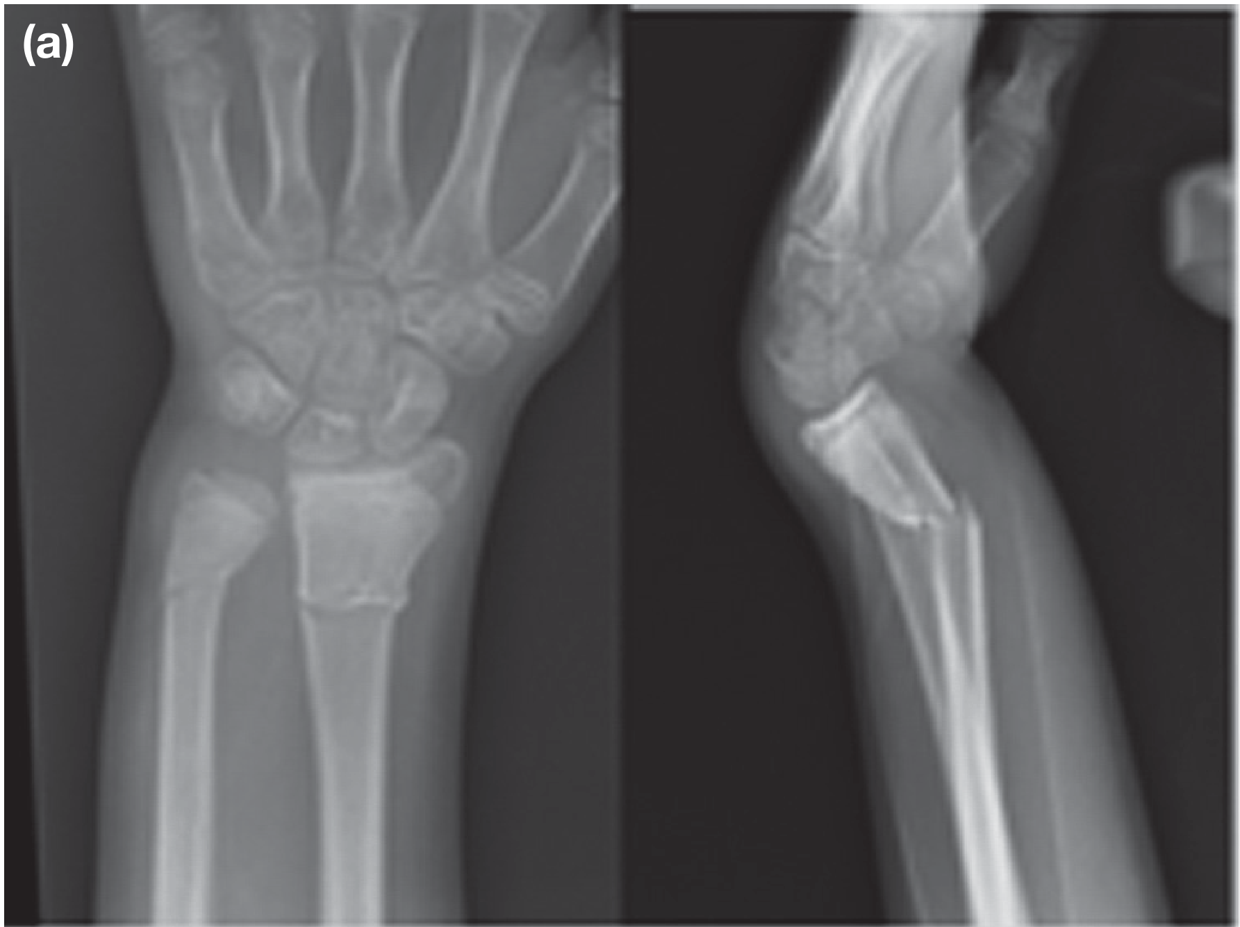
4. Supracondylar fracture of the humerus
- The most common pediatric elbow fracture; usually extension-type from a fall on an outstretched hand.
- Undisplaced fractures: collar-and-cuff or backslab, 3 weeks. Displaced fractures often need closed reduction ± percutaneous K-wire fixation if the periosteal hinge is broken.
- Feared complication: Volkmann's ischemic contracture from missed compartment syndrome of the forearm - avoid excessive elbow flexion when swelling is significant, and always document neurovascular status (radial, ulnar, median/AIN nerves; radial pulse) before and after any manipulation.
5. Nursemaid's elbow (radial head subluxation)
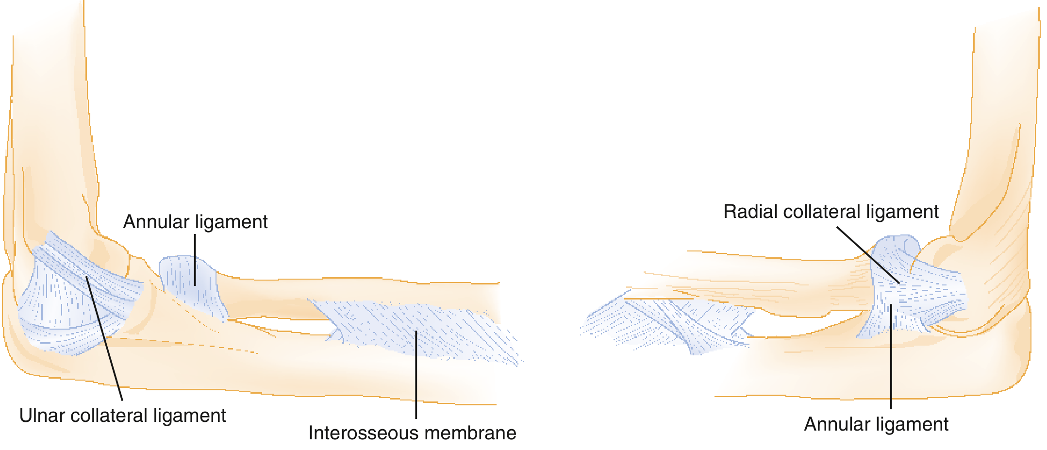
- Accounts for >20% of pediatric upper extremity injuries; peak age 1-4 years; classic mechanism is axial traction on a pronated, extended arm (e.g., a caregiver pulling the child's hand).
- Child holds the arm slightly flexed and pronated, refusing to use it, but with no swelling or deformity.
- Reduction by hyperpronation or supination-flexion; hyperpronation has a lower first-attempt failure rate per meta-analysis data. Child typically resumes normal use within 15 minutes.
6. Toddler's fracture
- Nondisplaced oblique fracture of the distal tibia from a minor fall/twist, peak age 1-4 years. History is often vague; child refuses to bear weight without obvious deformity.
- Often radiographically occult initially (thick pediatric periosteum limits displacement); if suspicion is high, immobilize and re-image in ~2 weeks for callus, or consider ultrasound.
- Managed with cast/splint/cast-boot for 3-4 weeks with weight-bearing as tolerated; outcomes are similar across immobilization methods.
7. Non-accidental trauma (NAT) - always keep on the differential
- Roughly 25% of children later diagnosed with NAT had a prior "sentinel injury" that was missed. High-risk group: children <3 years, those with disabilities, and families under socioeconomic stress.
- Skeletal survey (minimum 21 dedicated views, read by an experienced radiologist) is indicated for all suspected-abuse patients <24 months, and considered in the 24-60 month range; a single "babygram" is inadequate. A repeat survey after ≥14 days frequently reveals additional healing fractures missed on the initial study.
- Neuroimaging (CT/MRI) for suspected abusive head trauma, retinal exam + AST/ALT screening, abdominal CT if indicated, and skeletal surveys for siblings/contacts under 24 months are part of a standardized occult-injury workup (Rosen's Emergency Medicine, Ch. 172).
- Characteristic fracture patterns raising concern: classic metaphyseal lesions (corner/bucket-handle fractures), posterior rib fractures, fractures in a non-ambulatory infant, multiple fractures of different ages, and spiral/transverse femur fractures without a clear consistent mechanism.
8. Compartment syndrome
- Most often follows crush injury, long-bone fracture, or reperfusion of an ischemic limb; progressive edema raises tissue pressure above capillary pressure, halting perfusion.
- Frequent serial neurovascular exams are mandatory after any high-risk fracture (supracondylar humerus, tibial shaft) or vascular repair - pain out of proportion, pain with passive stretch, and paresthesias precede pulselessness. Do not wait for all "5 P's" to intervene; fasciotomy is time-critical.
9. Osteomyelitis (frequently co-exists with septic arthritis in children)
- Empiric coverage targets S. aureus (oxacillin/nafcillin or vancomycin if MRSA suspected); standard course is 4-6 weeks IV transitioning to oral. A recent systematic review/meta-analysis (PMID 40001163, 2025) quantifies how often concomitant osteomyelitis occurs in pediatric hip septic arthritis - relevant when deciding on imaging (MRI) and duration of antibiotics for hip infections.
Bottom line for the ED: use age to frame the differential, apply Kocher criteria (updated by recent evidence) plus synovial fluid analysis to rule out septic arthritis before dismissing a limp as benign, always check neurovascular status before/after any reduction, and keep NAT on the differential in any child under 3 with a fracture inconsistent with the stated mechanism.
If you want, I can go deeper into any one of these (e.g., detailed Kocher/CRP diagnostic algorithm, DDH screening, or a management flowchart for the limping child) - happy to expand.
This is a shared conversation. Sign in to Orris to start your own chat.