
beefy red tongue, high grade fever, sore throat, skin rash differentials
Kawasaki disease strawberry tongue rash diagnosis
strawberry tongue high fever sore throat rash differential diagnosis
strawberry tongue scarlet fever
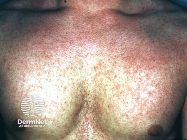
Clinical photography of the chest shows a diffuse erythematous maculopapular eruption with a rough, sandpaper-like texture covering the anterior trunk. The rash appears blanching with uniform distribution, without discrete vesicles, and involves the chest and upper abdomen. This exanthem is classically seen in scarlet fever, a streptococcal pharyngitis–associated illness, and is characterized by a diffuse erythematous rash, circumoral pallor, and sometimes strawberry tongue; desquamation can occur after several days. The image supports dermatologic assessment of an acute febrile illness with a prominent cutaneous eruption and provides a visual reference for exanthematous patterns. Differential considerations include measles, rubella, drug eruption, erythema marginatum, and, less often, Kawasaki disease, each with distinct clinical cues. Clinical correlation with fever, sore throat, tender or enlarged lymph nodes, and tongue findings is essential for accurate diagnosis. This photograph is valuable for educational purposes in dermatology and infectious disease, aiding recognition of exanthematous patterns, triage decisions, and patient counseling. Ideal applications include exam preparation, visual differential diagnostic training, and algorithm development for fever with rash workflows. It reinforces the role of rapid streptococcal testing and timely antibiotic therapy when scarlet fever is suspected. Cross-disciplinary collaboration and follow-up testing optimize patient outcomes and reduce transmission risk. Clinical documentation should include lesion mapping and progression notes.
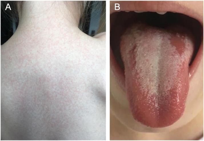
A composite of two clinical photographs (A and B) demonstrating classic physical manifestations of scarlet fever (Scarlatina) in a pediatric patient. Image A displays a characteristic scarlatiniform rash on the posterior trunk and neck. The rash is composed of diffuse, fine, erythematous maculopapular lesions that are closely spaced, giving the skin a 'sandpaper' texture. Image B shows an intraoral view of the tongue, illustrating 'white strawberry tongue.' The dorsal surface is covered by a thick, yellowish-white coating through which enlarged, hyperemic fungiform papillae project as prominent red dots. The lateral borders and the tip of the tongue appear intensely erythematous. These findings are hallmarks of a Group A Streptococcus infection. This educational material is suitable for illustrating systemic pediatric infections and dermatological-oral correlations in infectious diseases.
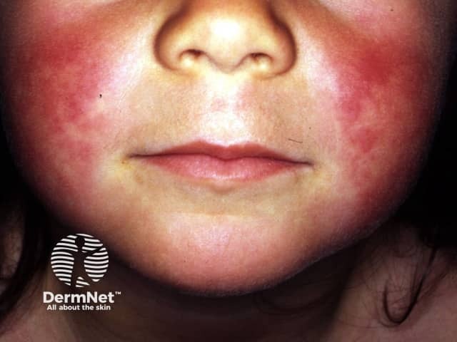
Imaging modality: Digital clinical photography of a pediatric patient's face in a frontal/anterior view. Primary subject: facial dermal eruption consistent with scarlet fever, characterized by symmetric erythematous, sandpaper-like papules on the malar cheeks with accentuation of the perioral region and circumoral pallor. The texture appears rough to the touch, with diffuse superficial vascular erythema and fine desquamation in the acute phase. The nasolabial folds are relatively spared or less reddened, a classic feature in forehead and cheek involvement. The perioral pallor contrasts with surrounding erythema, creating a striking border around the mouth. The image documents cutaneous manifestations of a systemic streptococcal infection, emphasizing the dermatologic hallmark of scarlet fever rather than a primary dermatitis. From a clinical imaging standpoint, this photograph provides documentation of rash distribution, morphology, and texture, enabling visual differentiation from other childhood exanthems such as viral illnesses or drug eruptions. Potential clinical use: bedside diagnosis, medical education, differential diagnosis training, telemedicine triage, and research into rash evolution during antibiotic therapy. Notably, accompanying signs such as fever, pharyngitis, and strawberry tongue would strengthen clinical correlation. The image underscores the value of color-rich dermatologic photography in pediatric infectious disease assessment and patient counseling.
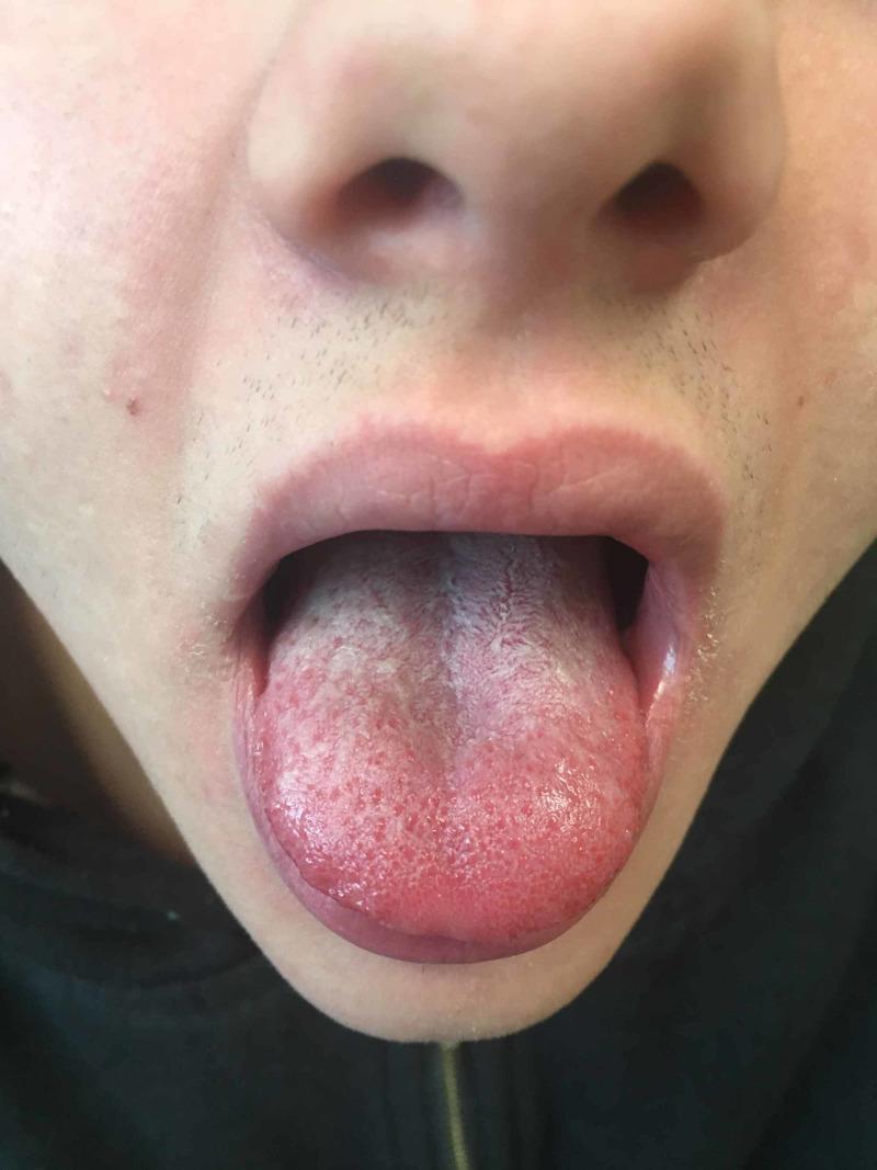
This clinical photograph shows the orofacial region of a patient, demonstrating classic intraoral and perioral signs often associated with scarlet fever or similar streptococcal infections. The primary focus is a 'strawberry tongue' appearance, characterized by an erythematous and inflamed tongue surface with prominent, enlarged fungiform papillae visible as raised red bumps. A white, pseudomembranous exudate (white coating) is present on the dorsal surface, particularly concentrated toward the posterior. Additionally, the image depicts circumoral pallor, where the skin immediately surrounding the mouth, including the upper cutaneous lip, appears noticeably paler in contrast to the surrounding malar erythema. The vermilion border of the lips appears mildly erythematous. This visual material is highly relevant for pediatric medicine and infectious disease specialties, illustrating key diagnostic physical findings used in the clinical identification of toxin-mediated exanthems.
Differential Diagnosis: Beefy/Strawberry Red Tongue + High-Grade Fever + Sore Throat + Rash
1. Scarlet Fever (Streptococcus pyogenes / Group A Strep)
- Fever, exudative pharyngitis/tonsillitis, sandpaper-textured erythematous rash starting on trunk/axillae/groin, circumoral pallor, Pastia's lines (petechial streaks in skin folds).
- Tongue: initially white-coated with red hyperemic papillae poking through ("white strawberry tongue"), which by day 4-5 becomes a uniform "red/beefy strawberry tongue" as the coating sloughs.
- Diagnosis is largely clinical; confirm with rapid strep antigen/throat culture. Treatment: penicillin/amoxicillin.
- Source: Tintinalli's Emergency Medicine, p. 2036; Sleisenger and Fordtran's GI/Liver Disease, p. 977-979; CDC clinical guidance.
2. Kawasaki Disease (Mucocutaneous Lymph Node Syndrome)
- Fever ≥5 days plus ≥4 of: bilateral non-exudative conjunctival injection, oral changes (red/cracked lips, "strawberry" tongue - classically the "red strawberry tongue" without the white coating), polymorphous truncal rash, cervical lymphadenopathy (>1.5 cm), and peripheral extremity changes (edema/erythema of palms/soles, later periungual desquamation).
- No response to antibiotics; risk of coronary artery aneurysms (~25% untreated) - needs IVIG + high-dose aspirin within 10 days.
- Distinguishing point from scarlet fever: sore throat is less prominent/exudative, conjunctivitis and extremity changes are prominent, and tongue tends to be uniformly beefy-red rather than white-then-red.
- Source: ROSEN's Emergency Medicine, p. 1956-1979; Goldman-Cecil Medicine.
3. Toxic Shock Syndrome (Staphylococcal or Streptococcal, TSST-1/superantigen-mediated)
- High fever, diffuse macular/scarlatiniform rash, mucosal hyperemia (including strawberry tongue), but additionally hypotension/shock and multiorgan dysfunction (renal, hepatic, coagulopathy), often with a source (tampon use, wound infection, post-surgical). Desquamation of palms/soles occurs 1-2 weeks later.
- This is the "can't miss" diagnosis because of rapid progression to shock.
- Source: K.J. Lee's Essential Otolaryngology; Robbins & Cotran Pathologic Basis of Disease (superantigen pathogenesis).
4. Measles (Rubeola)
- Fever, cough/coryza/conjunctivitis (3 Cs), Koplik spots, cephalocaudal morbilliform rash. Strawberry tongue is not classic here but can be confused with; the 3 Cs and Koplik spots help distinguish.
5. Multisystem Inflammatory Syndrome in Children (MIS-C)
- Kawasaki-like presentation (fever, rash, strawberry tongue, conjunctivitis) occurring 2-6 weeks after SARS-CoV-2 infection, often with more prominent GI symptoms and cardiac dysfunction/shock than classic Kawasaki disease.
6. Drug Reaction / Erythema Multiforme spectrum (SJS/TEN, DRESS)
- Fever, mucositis (including tongue/lip erosions), rash, often with a preceding new medication; distinguished by mucosal blistering/sloughing and skin pain rather than sandpaper texture.
7. Epstein-Barr Virus (Infectious Mononucleosis) / other viral pharyngitis
- Can rarely present with strawberry tongue and rash (especially if given amoxicillin, causing an ampicillin rash), alongside exudative pharyngitis, lymphadenopathy, and splenomegaly. Reported as an atypical but real cause of strawberry tongue even without prominent fever.
Key distinguishing features at a glance
| Feature | Scarlet Fever | Kawasaki Disease | TSS | MIS-C |
|---|---|---|---|---|
| Typical age | 5-15 yrs | <5 yrs | Any age | Child, post-COVID |
| Pharyngitis | Prominent, exudative | Mild | Variable | Variable |
| Rash texture | Sandpaper | Polymorphous, variable | Diffuse macular | Polymorphous |
| Tongue | White then red strawberry | Red "strawberry" early | Hyperemic/strawberry | Strawberry |
| Hypotension/shock | Rare | Rare (unless shock syndrome) | Defining feature | Common |
| Response to antibiotics | Yes (it's bacterial) | No | Partial (source control) | No (immunomodulation) |
Workup to narrow the differential
- Rapid strep antigen/throat culture (scarlet fever)
- CBC, CRP/ESR, LFTs, blood cultures, echocardiogram if Kawasaki suspected (coronary artery involvement)
- Blood pressure and perfusion status to rule out shock (TSS)
- SARS-CoV-2 antibody/PCR history if MIS-C is being considered
- Assess age (Kawasaki/MIS-C skew pediatric under 5; scarlet fever school-age children; TSS any age with a source)
Role of Levofloxacin
levofloxacin uses mechanism of action clinical role
levofloxacin respiratory tract infection efficacy
Role of Levofloxacin
Mechanism of Action
- Source: Katzung's Basic and Clinical Pharmacology; Lippincott Illustrated Reviews Pharmacology.
Spectrum of Activity
- Gram-positive: Excellent activity against Streptococcus pneumoniae (including penicillin-resistant strains), making it a key "respiratory fluoroquinolone" alongside moxifloxacin and gemifloxacin.
- Gram-negative: Good coverage of Enterobacteriaceae, Pseudomonas aeruginosa (less potent than ciprofloxacin here), Haemophilus influenzae.
- Atypicals/intracellular organisms: Accumulates in macrophages and PMNs, giving activity against Chlamydia trachomatis/pneumoniae, Mycoplasma pneumoniae, Legionella, and Listeria.
- Source: Katzung's Pharmacology, p. 1781-1790.
Clinical Indications (where it plays a defined role)
- Community-acquired pneumonia (CAP) - first-line "respiratory fluoroquinolone" option, especially for inpatients or those with comorbidities, often as monotherapy alternative to beta-lactam + macrolide combination.
- Source: Symptom to Diagnosis: An Evidence-Based Guide, p. 6425-6429; Goldman-Cecil Medicine.
- Legionella pneumonia - preferred/alternative agent alongside azithromycin and doxycycline.
- Source: Sherris & Ryan's Medical Microbiology, p. 5516-5520.
- Urinary tract infections including pyelonephritis, and chronic bacterial prostatitis - good tissue penetration into kidney and prostate.
- Skin and soft tissue infections.
- H. pylori eradication - used in levofloxacin-based triple/quadruple salvage regimens (with PPI + amoxicillin ± bismuth) after clarithromycin-based therapy failure.
- Source: Washington Manual of Medical Therapeutics, p. 3650-3674; Harrison's Principles of Internal Medicine, p. 2350-2365.
- Nosocomial pneumonia and anthrax post-exposure prophylaxis/treatment.
- Ophthalmic formulation for bacterial conjunctivitis.
Pharmacokinetics
Limitations / Why It Is Not First-Line Everywhere
- Not the drug of choice for group A streptococcal pharyngitis/scarlet fever - penicillin/amoxicillin remains standard there; fluoroquinolones are reserved for penicillin-allergic patients or specific atypical/resistant organism coverage.
- Carries boxed warnings: tendinitis/tendon rupture, peripheral neuropathy, CNS effects (seizures, confusion), and QT prolongation - use is restricted in children (except specific scenarios like cystic fibrosis exacerbations) and caution needed in the elderly and those on QT-prolonging drugs.
- Photosensitivity and risk of C. difficile colitis with broad-spectrum use.
- Source: Lippincott Illustrated Reviews Pharmacology, p. 5041-5047.