
Hematopoiesis
Reading File
Now let me search the medical library for hematopoiesis content in parallel:
Finding Sources
Finding Sources
Excellent sources found. Let me read from the best ones in parallel - Robbins (pathology), Medical Physiology, Histology, and Goodman & Gilman:
Reading File
Reading File
Reading File
Reading File
Now let me get the Robbins continuation and the Histology content, plus fetch the diagram image:
Reading File
Reading File
Loading Image
Finding Sources
Now let me get content from Harrison's on HSC and also the Histology section on erythropoiesis and granulopoiesis to complete the picture:
Reading File
Finding Sources
Finding Sources
Let me now read the erythropoiesis and granulopoiesis sections from Histology and Biochemistry:
Reading File
Reading File
I now have comprehensive content from multiple authoritative textbooks. Let me also fetch the erythropoiesis diagram:
Loading Image
I now have all the content needed for a comprehensive, textbook-based answer on hematopoiesis. Let me compile it:
Hematopoiesis
Hematopoiesis is the continuous process by which all formed elements of blood - red cells, granulocytes, monocytes, platelets, and lymphocytes - are produced from a common ancestral population of hematopoietic stem cells (HSCs). The finite life span of most mature blood cells requires their constant replacement throughout life.
1. Sites of Hematopoiesis: Ontogeny
| Developmental Period | Site | Cell Types |
|---|---|---|
| Week 3 (embryo) | Yolk sac | Embryonic red cells, tissue macrophage precursors |
| Weeks 6-7 | Aorta-gonad-mesonephros (AGM) region | Definitive HSCs first arise |
| Month 3-7 (fetal) | Liver (chief site), placenta | All blood cell lineages |
| Month 4 onward | Bone marrow | Progressive shift; dominant by birth |
| After puberty | Axial skeleton only (skull, vertebrae, pelvis, proximal long bones) | ~50% of marrow remains hematopoietically active in adults |
Blood cell progenitors first appear during the third week of embryonic development in the yolk sac... By birth, marrow throughout the skeleton is hematopoietically active, and hepatic hematopoiesis dwindles to a trickle.
- Robbins & Cotran Pathologic Basis of Disease, p. 548
Under pathologic stress (e.g., severe hemolytic anemias, myelofibrosis), the liver and spleen can resume blood cell production - this is called extramedullary hematopoiesis.
2. The Hematopoietic Stem Cell (HSC)
HSCs are the pluripotent cells that sit at the apex of the entire system. The normal adult marrow contains an estimated 50,000-200,000 HSCs. They are identified by the surface marker profile: cKIT+ / Sca-1+ / LIN- (negative for lineage-specific markers).
They have two defining properties:
- Pluripotency - a single HSC can generate all mature blood cell lineages
- Self-renewal - at least one daughter cell regenerates the stem cell pool after each division, preventing depletion
Self-renewing divisions occur within a specialized bone marrow niche where stromal cells and secreted factors (including KIT ligand, FLT3 ligand, CXCL12) nurture and protect HSCs. Under stress (e.g., severe anemia, G-CSF administration), HSCs are mobilized into peripheral blood - this is exploited therapeutically in HSC transplantation, where donors are treated with G-CSF to harvest HSCs from peripheral blood.
- Robbins & Cotran Pathologic Basis of Disease, p. 549
3. The Differentiation Hierarchy
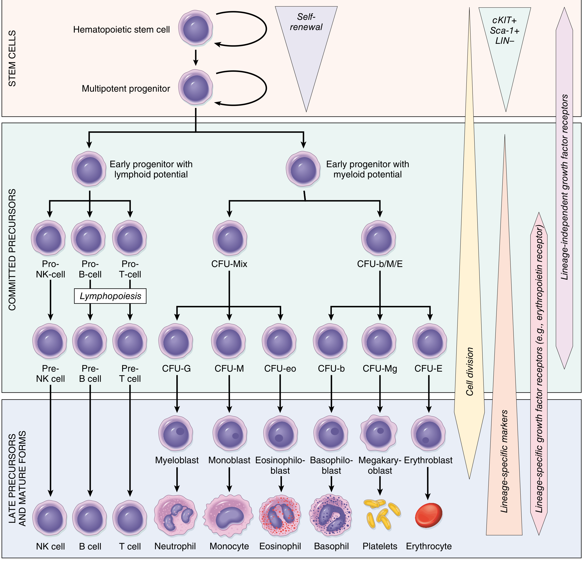
Fig. 13.1 - Differentiation of blood cells. From Robbins & Cotran Pathologic Basis of Disease.
The hierarchy proceeds as follows:
HSC (pluripotent, self-renewing)
  └─► Multipotent Progenitor (MPP) - more proliferative, less self-renewal
        ├─► Common Lymphoid Progenitor (CLP) → lymphopoiesis
        │      ├─► Pro-T → Pre-T → (thymus) → T lymphocyte
        │      ├─► Pro-B → Pre-B → B lymphocyte
        │      └─► Pro-NK → NK cell
        └─► Common Myeloid Progenitor (CMP) → myelopoiesis
               ├─► MEP (megakaryocyte-erythroid progenitor)
               │      ├─► CFU-E → Erythroblast → Reticulocyte → Erythrocyte
               │      └─► MKP → Megakaryoblast → Megakaryocyte → Platelets
               └─► GMP (granulocyte-monocyte progenitor)
                      ├─► CFU-G → Myeloblast → Neutrophil
                      ├─► CFU-M → Monoblast → Monocyte → Macrophage
                      ├─► CFU-eo → Eosinophiloblast → Eosinophil
                      └─► BaP → Basophiloblast → Basophil
- Goodman & Gilman's Pharmacological Basis of Therapeutics, p. [block 13]
4. Erythropoiesis
Erythropoiesis is driven by tissue hypoxia. When oxygen delivery falls, the kidney (peritubular fibroblasts) releases erythropoietin (EPO), which stimulates erythroid progenitor proliferation and survival.
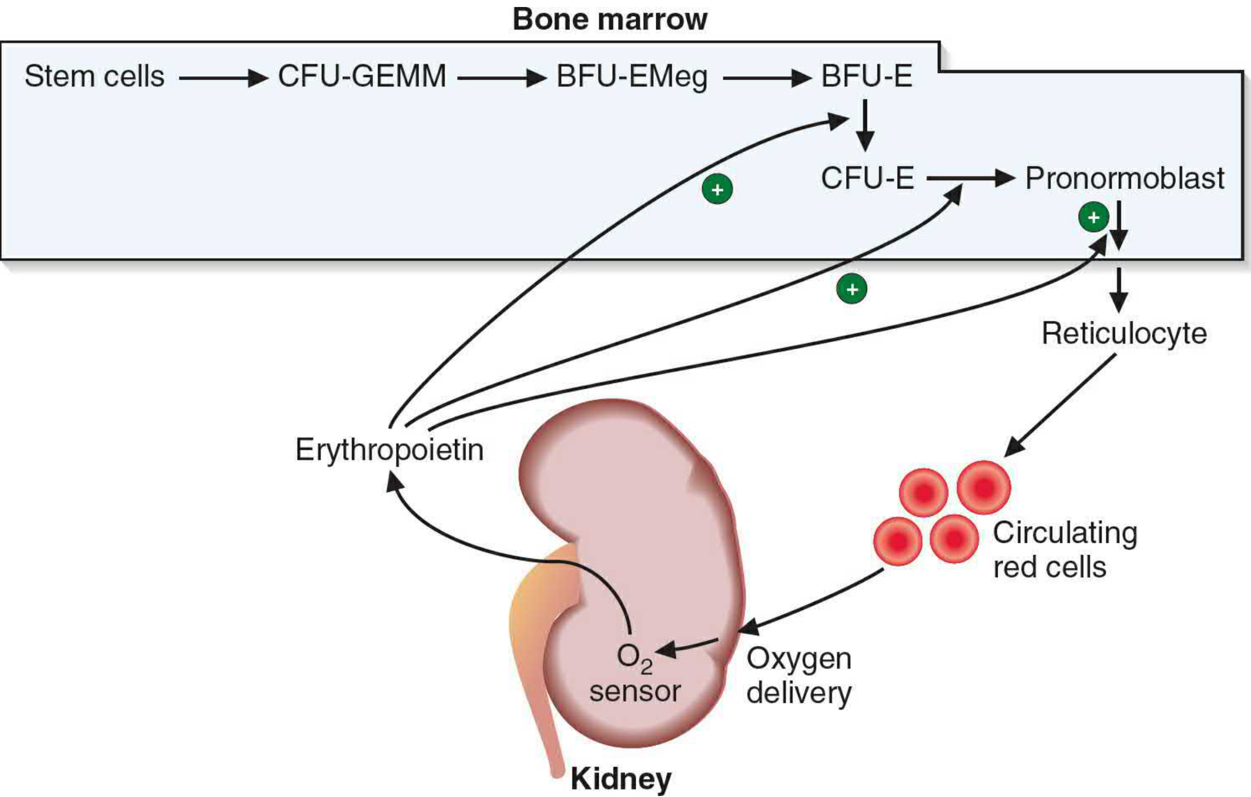
Erythropoietin stimulation of erythrocyte maturation - Basic Medical Biochemistry (6e)
Sequential stages:
| Stage | Key Features |
|---|---|
| HSC | Pluripotent origin |
| CFU-GEMM | Mixed myeloid colony-forming unit |
| BFU-E (Burst Forming Unit-Erythroid) | EPO-responsive; large burst colonies |
| CFU-E | Highly EPO-sensitive; smaller colonies |
| Pronormoblast (Proerythroblast) | First morphologically recognizable erythroid cell; large nucleus |
| Basophilic normoblast | Basophilic cytoplasm; Hb synthesis begins |
| Polychromatophilic normoblast | Mixed RNA + Hb staining |
| Orthochromatic normoblast | Condensed pyknotic nucleus |
| Reticulocyte | Nucleus extruded; retains ribosomes + mRNA; still synthesizes Hb; 1-2 days in circulation; matures in spleen |
| Mature erythrocyte | Anucleate biconcave disc; 120-day lifespan |
Each normoblast undergoes 4 mitotic divisions before nuclear extrusion. Erythrocyte production can increase more than 20-fold in response to severe anemia or hypoxemia.
- Basic Medical Biochemistry - A Clinical Approach, 6e, p. 1536
5. Granulopoiesis
Granulocytes arise from the GMP under the influence of GM-CSF, G-CSF, IL-3, and M-CSF.
Neutrophil maturation stages (6 morphologically identifiable stages):
| Stage | Key Features |
|---|---|
| Myeloblast | Earliest recognizable precursor; 14-20 µm; large euchromatic nucleus with 3-5 nucleoli; agranular basophilic cytoplasm |
| Promyelocyte | Only stage that produces azurophilic (primary) granules (myeloperoxidase, elastase, defensins) |
| Myelocyte | Specific (secondary) granules first appear; the last stage capable of mitosis |
| Metamyelocyte | Kidney-shaped nucleus; no further division; specific granules outnumber azurophilic |
| Band cell | Horseshoe-shaped nucleus; immature neutrophil seen in "left shift" |
| Mature neutrophil | Multilobed (2-5 lobes) nucleus; enters circulation |
Eosinophils and basophils follow a parallel maturation pathway but cannot be distinguished from neutrophil precursors until the myelocyte stage, when specific (secondary) granules appear.
- Eosinophils: require IL-5 (in addition to GM-CSF, IL-3)
- Basophils: arise when IL-5 is absent
- Histology: A Text and Atlas (Histology with Correlated Cell and Molecular Biology), p. 748-749
6. Thrombopoiesis (Megakaryopoiesis)
Platelets are produced from megakaryocytes in the bone marrow:
- MKP → Megakaryoblast → Megakaryocyte (endomitosis - repeated DNA replication without cell division creates polyploid cells up to 64N)
- Megakaryocyte cytoplasm fragments to release 2,000-3,000 platelets per cell
- Regulated primarily by thrombopoietin (TPO), produced by liver, kidney, and skeletal muscle
- Platelet production can increase 10-20-fold in response to thrombocytopenia
7. Lymphopoiesis
The Common Lymphoid Progenitor (CLP) differentiates under transcription factor control:
| Lineage | Key Transcription Factor | Maturation Site |
|---|---|---|
| T lymphocytes | GATA-3 | Bone marrow (pre-T) → thymus (education) → circulation |
| B lymphocytes | Pax5 (activates B-cell-specific genes) | Bone marrow, gut-associated lymphoid tissue, spleen |
| NK cells | IL-2 and IL-15 signaling | Bone marrow (primary); lymph nodes (secondary) |
Lymphocytes constitute up to 30% of all nucleated cells in the bone marrow.
- Histology: A Text and Atlas, p. 757
8. Regulation: Growth Factors and Cytokines
The marrow response is finely tuned by hematopoietic growth factors (colony-stimulating factors) acting on committed progenitors:
| Cytokine | Source | Primary Target |
|---|---|---|
| Erythropoietin (EPO) | Kidney, liver | CMP, MEP, CFU-E - erythroid lineage |
| Thrombopoietin (TPO) | Liver, kidney, skeletal muscle, bone marrow | MKP, megakaryocytes |
| GM-CSF | T cells, endothelial cells, fibroblasts | CMP, ErP, GMP, EoP, BaP, MKP - broad myeloid effects |
| G-CSF | Endothelial cells, monocytes | GMP → neutrophil lineage; also mobilizes HSCs |
| M-CSF | Monocytes, macrophages, endothelial cells | GMP, MoP → monocyte/macrophage lineage |
| IL-3 | T lymphocytes | Most progenitor cells (early multipotent effect) |
| IL-5 | CD4+ T cells, mast cells | EoP → eosinophil differentiation |
| KIT ligand / FLT3 ligand | Stromal cells | Very early committed progenitors |
- Histology: A Text and Atlas, Table 10.4, p. 754-755
Transcription factors are equally important - they are activated by these signaling molecules and control lineage-specific gene expression (e.g., PU.1 for myeloid fate, Ikaros family for lymphoid commitment, GATA-1 for erythroid/megakaryocyte lineage).
9. Requirements for Hematopoiesis
Adequate hematopoiesis requires:
- Minerals: iron, cobalt, copper
- Vitamins: folic acid, vitamin B12, pyridoxine (B6), ascorbic acid, riboflavin (B2)
Deficiencies produce characteristic anemias - iron deficiency causes microcytic/hypochromic anemia; B12/folate deficiency causes megaloblastic anemia due to impaired DNA synthesis.
- Goodman & Gilman's Pharmacological Basis of Therapeutics, p. [block 13]
10. Adult Reference Ranges
| Cell Type | Normal Range |
|---|---|
| White cells | 4.8-10.8 × 10³/µL |
| Neutrophils | 1.4-6.5 × 10³/µL |
| Lymphocytes | 1.2-3.4 × 10³/µL |
| Monocytes | 0.1-0.6 × 10³/µL |
| Eosinophils | 0-0.5 × 10³/µL |
| Basophils | 0-0.1 × 10³/µL |
| Red blood cells (male) | 4.5-5.9 × 10⁶/µL |
| Hemoglobin (male) | 13.5-17.5 g/dL |
| Platelets | 150-400 × 10³/µL |
- Robbins & Cotran Pathologic Basis of Disease, Table 13.1
11. Clinical Relevance
- Aplastic anemia: failure of HSC pool - pancytopenia; treated with HSC transplantation
- Leukemias: clonal neoplasms of hematopoietic cells that disrupt normal marrow function
- Clonal hematopoiesis of indeterminate potential (CHIP): age-related somatic mutations in HSCs; associated with cardiovascular risk
- Extramedullary hematopoiesis: return to fetal sites (liver, spleen) under chronic stress
- Left shift: appearance of band cells/metamyelocytes in peripheral blood - indicates increased granulocyte demand (sepsis, inflammation)
- G-CSF therapy: used clinically to mobilize HSCs for transplant harvest, and to shorten neutropenia after chemotherapy
Sources:
- Robbins & Cotran Pathologic Basis of Disease (10e), pp. 548-549 - Normal Hematopoiesis
- Goodman & Gilman's Pharmacological Basis of Therapeutics (14e) - Hematopoiesis chapter
- Histology: A Text and Atlas with Correlated Cell and Molecular Biology (8e), pp. 748-757 - Hemopoiesis
- Basic Medical Biochemistry: A Clinical Approach (6e), p. 1536 - Erythropoiesis
This is a shared conversation. Sign in to Orris to start your own chat.