
Explain axial skeletal system with diagram
Reading File
Now let me search the medical library for axial skeleton content and images in parallel.
Finding Sources
Finding Sources
Good - found key sources. Let me read the Gray's Anatomy for Students section on the skeletal system and also search for images.
Reading File
Finding Sources
I found an axial skeleton diagram. Let me fetch it and also read more detailed content about skull, vertebral column, ribs, and sternum.
Loading Image
Finding Sources
Finding Sources
That image is just the legend. Let me look for more detailed anatomy images and content in the imaging anatomy textbook.
Reading File
Finding Sources
Good content. Let me now read the Developing Human embryology section for more details, and also look for any full axial skeleton diagram images in the textbooks.
Reading File
Finding Sources
Let me fetch the skull diagram from Gray's Anatomy and also look for a full axial skeleton diagram image.
Reading File
Searching the Web
axial skeleton diagram labeled bones skull vertebral column ribs sternum anatomy
I found great images. Let me fetch the key axial skeleton diagrams and the skull diagram in parallel.
Loading Image
Loading Image
Loading Image
Excellent! I now have all the key diagrams and content needed. Let me also fetch the vertebral column image for completeness.
Loading Image
I have all the essential content and diagrams. Now let me compose the full explanation.
The Axial Skeletal System
The axial skeleton forms the central axis of the human body. It consists of 80 bones (out of the 206 total in an adult), while the remaining 126 form the appendicular skeleton (limbs and girdles).
Overview Diagram
The diagram below shows the axial skeleton (beige/tan) vs. the appendicular skeleton (green/teal):
Axial skeleton (beige) includes: skull, vertebral column, sternum, and ribs. Appendicular skeleton (green) includes all limb and girdle bones.
Components of the Axial Skeleton
The axial skeleton has five major divisions:
| Division | Bones | Count |
|---|---|---|
| Skull (cranial + facial bones) | Frontal, parietal, temporal, occipital, sphenoid, ethmoid + 14 facial bones | 22 |
| Auditory ossicles | Malleus, incus, stapes (x2 ears) | 6 |
| Hyoid bone | Single U-shaped bone in neck | 1 |
| Vertebral column | Cervical (7), thoracic (12), lumbar (5), sacrum (5 fused), coccyx (4 fused) | 26 |
| Thoracic cage | 12 pairs of ribs + sternum | 25 |
| Total | 80 |
1. The Skull (22 bones)
The skull has 22 bones, excluding the auditory ossicles. It is subdivided into the cranium and the mandible.
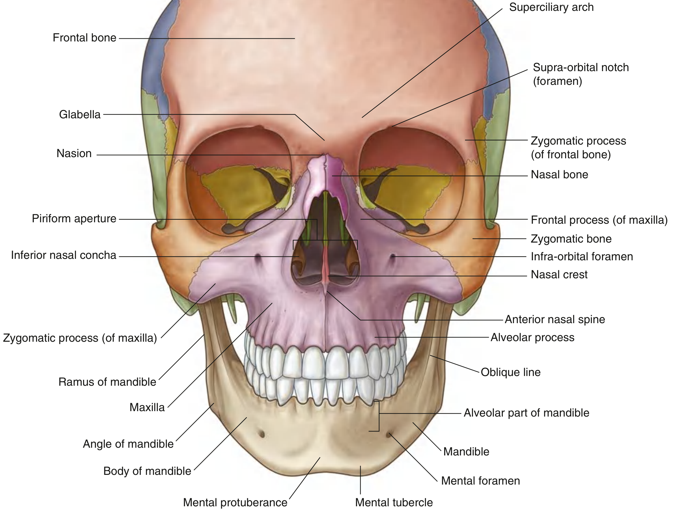
Anterior view of the skull showing frontal bone, zygomatic bones, nasal bones, maxilla, and mandible - Gray's Anatomy for Students
Cranial bones (8)
These form the calvaria (dome) and base of the cranial cavity, protecting the brain:
- Frontal bone (1) - forehead and roof of orbits
- Parietal bones (2) - sides and roof of cranium
- Temporal bones (2) - sides of skull, house the middle and inner ear
- Occipital bone (1) - base and back of skull, contains foramen magnum
- Sphenoid bone (1) - central base of skull, bat-shaped
- Ethmoid bone (1) - forms roof of nasal cavity and part of orbits
Facial bones (14)
Include the nasal bones, maxillae, zygomatic bones, palatine bones, lacrimal bones, inferior nasal conchae, vomer, and mandible. The mandible is the only movable bone of the skull.
All skull bones except the mandible are joined by sutures (immobile fibrous joints). - Gray's Anatomy for Students
2. Auditory Ossicles (6 bones)
Three tiny bones in each middle ear:
- Malleus (hammer)
- Incus (anvil)
- Stapes (stirrup) - the smallest bone in the body
They transmit sound vibrations from the tympanic membrane to the inner ear.
3. Hyoid Bone (1)
A U-shaped bone in the anterior neck, just above the larynx. It does not articulate with any other bone - it is suspended by muscles and ligaments. It supports the tongue and serves as an attachment point for muscles of swallowing and speech.
4. Vertebral Column (26 bones)
The vertebral column (spine) consists of 26 bones arranged in 5 regions:
| Region | Vertebrae | Count | Key Features |
|---|---|---|---|
| Cervical | C1-C7 | 7 | C1 = atlas (supports skull), C2 = axis (pivot for rotation) |
| Thoracic | T1-T12 | 12 | Articulate with ribs |
| Lumbar | L1-L5 | 5 | Largest, weight-bearing |
| Sacrum | S1-S5 fused | 1 | Wedge-shaped, forms posterior pelvis |
| Coccyx | 3-4 fused | 1 | Remnant tailbone |
Between each pair of adjacent vertebrae (except C1-C2) sits an intervertebral disc - a fibrocartilaginous cushion with a gelatinous nucleus pulposus at its center (derived from the notochord embryologically).
The vertebral column has four normal curves: cervical (lordosis), thoracic (kyphosis), lumbar (lordosis), and sacral (kyphosis).
5. Thoracic Cage (25 bones)
The thoracic cage (rib cage) protects the heart, lungs, and great vessels.
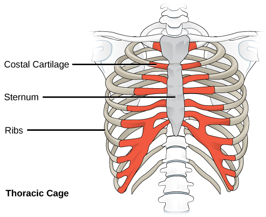
Sternum
A flat bone in the midline anterior chest, composed of three parts:
- Manubrium (superior) - articulates with clavicles and 1st rib
- Body - articulates with ribs 2-7
- Xiphoid process (inferior) - cartilaginous in youth, ossifies with age
Ribs (12 pairs = 24 bones)
- True ribs (1-7): Attach directly to the sternum via costal cartilage
- False ribs (8-10): Costal cartilage joins the cartilage of rib 7 (not directly to sternum)
- Floating ribs (11-12): No anterior attachment at all
Functions of the Axial Skeleton
- Support - forms the central structural axis of the body
- Protection - skull protects the brain; vertebral column shields the spinal cord; thoracic cage protects heart, lungs, and major vessels
- Movement - serves as attachment points for muscles of the head, neck, and trunk
- Hematopoiesis - red bone marrow in vertebrae, ribs, and sternum is a major site of blood cell production in adults
- Mineral reservoir - stores calcium and phosphorus, released when serum levels fall
Summary
The axial skeleton = skull + hyoid + auditory ossicles + vertebral column + thoracic cage (ribs + sternum). It is the central scaffolding that supports the body's vertical axis, protects the most vital neural and visceral structures, and anchors the muscles that move the head and trunk.
Sources: Gray's Anatomy for Students; Imaging Anatomy - Bones, Joints, Vessels and Nerves; The Developing Human: Clinically Oriented Embryology; Lumen Learning - Human Axial Skeleton; NCI SEER Training - Axial Skeleton
This is a shared conversation. Sign in to Orris to start your own chat.