I now have comprehensive information from multiple authoritative textbooks. Here is the complete, detailed answer:
Indications for Invasive and Non-Invasive Ventilation with Interpretation
1. Understanding the Two Approaches
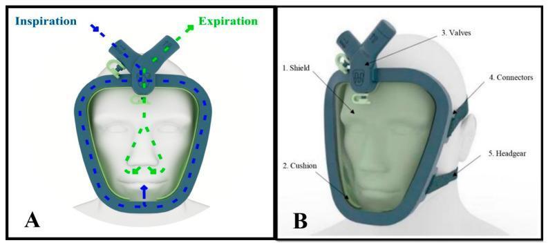
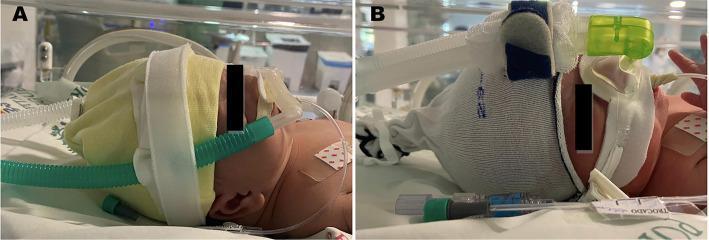
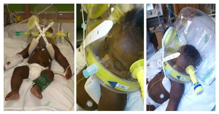
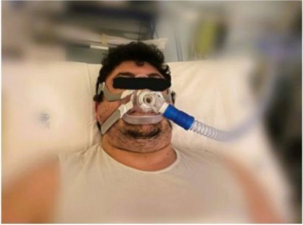
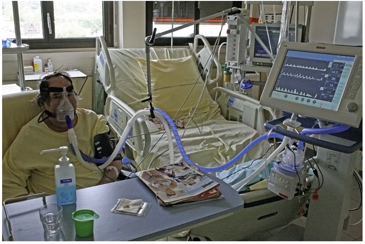
Non-Invasive Ventilation (NIV) delivers positive-pressure ventilation through an external interface (oronasal mask, nasal mask, or helmet) without an endotracheal tube. The two main modes are:
- CPAP (Continuous Positive Airway Pressure) - single fixed pressure; primarily addresses oxygenation
- BiPAP/BPAP (Bi-level Positive Airway Pressure) - separate inspiratory (IPAP) and expiratory (EPAP) pressures; addresses both oxygenation and ventilation
Invasive Mechanical Ventilation (IMV) requires endotracheal intubation or tracheostomy and is the definitive form of ventilatory support.
2. Objectives of Mechanical Ventilation (Both Invasive and Non-Invasive)
From [Fishman's Pulmonary Diseases and Disorders](Fishman's, Table 147-1):
| Category | Goal |
|---|
| Gas exchange | Reverse hypoxemia; relieve acute respiratory acidosis |
| Respiratory distress | Decrease oxygen cost of breathing; reverse respiratory muscle fatigue |
| Pressure-volume | Prevent/reverse atelectasis; improve lung compliance |
| Lung protection | Prevent further lung injury; permit lung and airway healing |
The decision to intubate is based on clinical judgment, physical signs, and physiologic data -- no single parameter is definitively diagnostic for the threshold to intubate. The most common trigger is increased work of breathing.
Physical signs assessed to judge work of breathing:
- Phasic sternomastoid contraction (gently palpate with index finger)
- Tracheal tug (finger on cricoid cartilage)
- Suprasternal fossa and intercostal recession
3. Non-Invasive Ventilation (NIV) - Indications
3A. Strong Indications (Evidence-Based, High Recommendation)
| Condition | Key Mechanism | Notes |
|---|
| COPD Exacerbation | High iPEEP, increased respiratory work, CO2 retention | Most robust evidence; first-line |
| Acute Cardiogenic Pulmonary Edema (ACPE) | Reduced compliance, increased afterload from negative intrathoracic pressure swings | CPAP alone effective; BiPAP reduces work of breathing faster |
| Facilitation of weaning/extubation in COPD | Early extubation to NIV reduces VAP and ICU days | Standard of care in COPD |
| Post-operative hypoxemia (major abdominal or thoracic surgery) | Atelectasis, splinting | Reduces re-intubation |
| Obesity-Hypoventilation Syndrome (OHS) | Chronic hypercapnia + hypoxemia | Reduces hypercapnia effectively |
| Do-Not-Intubate (DNI) patients | Ceiling-of-care ventilation | Reduces dyspnea, may prolong life |
3B. Intermediate Indications (Reasonable Evidence)
- Acute asthma exacerbation
- Hypoxemic respiratory failure (non-immunocompromised)
- Community-acquired pneumonia in COPD patients
- Neuromuscular disease with acute decompensation
- Preventive use during procedures (upper endoscopy, intubation preoxygenation)
3C. Weak/Uncertain Indications
- Mild ARDS (P/F ratio 200-300)
- Acute respiratory failure in immunocompromised patients (controversial)
- Community-acquired pneumonia (non-COPD)
- Trauma with chest wall injury
- Extubation failure (non-COPD patients)
- Post-operative esophageal/lung surgery (low pressures only)
3D. Conditions Where NIV is NOT Recommended / Contraindicated
| Condition | Reason |
|---|
| Severe ARDS (P/F <200) | High rate of NIV failure; delays definitive intubation |
| ARDS with multiple organ failure | NIV insufficient |
| End-stage pulmonary fibrosis | No benefit; may delay palliative care |
| Non-drained pneumothorax | Risk of worsening |
| Respiratory arrest | Cannot maintain airway |
| Hemodynamic instability | May require immediate intubation |
| Loss of consciousness | Aspiration risk; cannot protect airway |
| Abdominal distension / vomiting | High aspiration risk |
| Non-cooperative patient | Mask compliance impossible |
| Recent facial/gastroesophageal surgery | Mask contraindicated |
| Craniofacial trauma / burns | Interface cannot be fitted |
(Sources: Murray & Nadel Table 136.1; Rosen's Emergency Medicine Table 60.3; GOLD 2021 criteria)
4. ABG Criteria for NIV - Interpretation Thresholds
For COPD exacerbation (GOLD criteria), NIV is indicated when all three of the following are present:
| Parameter | NIV Threshold |
|---|
| pH | ≤ 7.35 |
| PaCO2 | ≥ 45 mmHg (6 kPa) |
| Respiratory rate | > 25 breaths/min |
| Dyspnea | Moderate-severe, with accessory muscle use |
Persistent hypoxemia despite supplemental O2 (SpO2 persistently below target range of 88-92% in COPD) is also an indication.
(Source: [Washington Manual Table 9-8](Washington Manual of Medical Therapeutics, p.322); Rosen's Table 60.3)
5. Invasive Mechanical Ventilation (IMV) - Indications
5A. Absolute / Immediate Indications
| Indication | Interpretation |
|---|
| Respiratory or cardiac arrest | No ventilatory effort; immediate intubation |
| NIV failure | Failure to improve on NIV within 1-2 hours |
| Unable to tolerate NIV | Patient agitation, mask intolerance |
| Persistent diminished consciousness | Cannot protect airway; aspiration risk |
| Massive aspiration | Airway protection required |
| Persistent inability to clear secretions | Suction requires direct airway access |
5B. Physiologic / ABG-Based Indications (COPD context)
For patients with COPD failing NIV (GOLD criteria):
| Parameter | Invasive Ventilation Threshold |
|---|
| pH | < 7.25 |
| PaCO2 | > 60 mmHg (8 kPa) |
| PaO2 | < 40 mmHg (5.3 kPa) |
| Respiratory rate | > 35 breaths/min |
(Source: [Washington Manual Table 9-9](Washington Manual of Medical Therapeutics, p.322))
5C. Additional Clinical Indications for IMV
- Hemodynamic instability not responding to fluids and vasopressors
- Life-threatening hypoxemia not correctable by NIV or high-flow O2
- Severe ARDS (P/F < 200, especially < 150)
- Status asthmaticus not responding to bronchodilators
- Coexisting conditions requiring intubation: cardiovascular collapse, metabolic crisis, septic shock, large pleural effusion, pneumothorax, pulmonary embolism with cardiovascular compromise
(Sources: Tintinalli's Table 70-5; Rosen's Table 60.3; Fishman's p.2605)
6. Quick Comparison Summary
| Feature | NIV (BiPAP/CPAP) | Invasive MV |
|---|
| Airway | External interface (mask) | Endotracheal tube / tracheostomy |
| Sedation | Usually not required | Usually required |
| VAP risk | Low | High |
| Secretion clearance | Limited | Direct suction possible |
| Monitoring intensity | Lower | Higher |
| Best for | Cooperative patient, reversible cause, intact airway | Arrest, NIV failure, airway compromise |
| Key advantage | Avoids intubation complications | Full airway control |
| Key limitation | Aspiration risk if vomiting; face seal needed | Nosocomial infection, prolonged weaning |
7. NIV Predictors of Success vs. Failure
Favorable predictors (NIV likely to succeed):
- Conscious and cooperative patient
- Hypercapnic failure (rather than pure hypoxemic failure)
- Hemodynamically stable
- No other organ failures
- Improvement in gas exchange, respiratory rate, and heart rate within first 1-2 hours of NIV
Red flags - consider early intubation if:
- No improvement in pH, RR, or PaCO2 within 1-2 hours of NIV
- Worsening consciousness
- Hemodynamic deterioration
- Increasing secretions
8. Interpretation Framework: Which Ventilation to Choose?
Respiratory Failure
        |
   Assess: Is the patient able to protect airway? Cooperative?
        |
   NO (arrest, coma, uncontrollable secretions, hemodynamic collapse)
        --> INVASIVE MV immediately
        |
   YES (conscious, cooperative, hemodynamically stable)
        |
   Classify failure:
        |
   TYPE I (Hypoxemic) PaO2<60 on O2:
        - Mild-moderate: try NIV (CPAP for pulmonary edema; BiPAP for ARDS/pneumonia)
        - Severe ARDS (P/F<150-200): consider early intubation
        |
   TYPE II (Hypercapnic) PaCO2>45 + pH<7.35:
        - COPD exacerbation, OHS, NMD: NIV is FIRST LINE
        - pH<7.25 or failing NIV: Invasive MV
        |
   MIXED (Type I + II):
        - NIV first if stable and cooperative
        - Reassess at 1-2 hours
Key References:
- [Fishman's Pulmonary Diseases & Disorders](Fishman's Pulmonary Diseases and Disorders, Chapter 147 & 148) - objectives and indications for MV
- [Murray & Nadel's Respiratory Medicine](Murray & Nadel's Textbook of Respiratory Medicine, Table 136.1) - NIV recommendations by disease and clinical status
- [Rosen's Emergency Medicine](Rosen's Emergency Medicine, Table 60.3) - side-by-side NIV vs. IMV criteria
- [Washington Manual of Medical Therapeutics](Washington Manual, Tables 9-8 & 9-9) - specific GOLD-based ABG thresholds
- [Tintinalli's Emergency Medicine](Tintinalli's Emergency Medicine, Table 70-5) - indications for intubation