
Cardiomyopathy
Reading File
Cardiomyopathy is a broad topic. Let me search the medical library systematically.
Searching sources...
Finding Sources
Searching PubMed
"cardiomyopathy"[MeSH Terms] AND ("classification" OR "management")
Good sources found. Let me pull from the most authoritative textbooks - Goldman-Cecil Medicine, Robbins Pathology, and Tintinalli's Emergency Medicine simultaneously.
Reading File
Reading File
Reading File
Good foundations. Now let me get detailed content on each major type - DCM, HCM, and Restrictive - from the best sources.
Finding Sources
Finding Sources
Finding Sources
Excellent. Now let me read the detailed content from Robbins Basic Pathology (the best source for pathology), Braunwald's Heart Disease (cardiology gold standard), and Robbins & Kumar Pathologic Basis for each type simultaneously.
Reading File
Reading File
Reading File
Excellent - I have very rich content from Robbins. Let me now get the HCM and Restrictive detailed sections from Robbins Basic Pathology, plus ARVC content:
Reading File
Finding Sources
I now have comprehensive content. Let me fetch the key histological images to include in the answer:
Loading Image
Loading Image
Reading File
Now let me get the HCM image and the Robbins Pathologic Basis of Disease detailed HCM section:
Reading File
Excellent - I now have comprehensive information on all major cardiomyopathies. Let me fetch the HCM and ARVC images from this section:
Loading Image
I now have all the content I need for a comprehensive response. Let me compose the full answer.
Cardiomyopathy
Cardiomyopathies are intrinsic diseases of cardiac muscle - distinct from myocardial dysfunction caused by coronary artery disease, hypertension, valvular disease, or congenital defects. They are classified into three major functional patterns based on pathophysiology.
Classification Overview
| Type | LV Ejection Fraction | Dysfunction | Prevalence |
|---|---|---|---|
| Dilated (DCM) | <40% | Systolic (contractile) | ~90% of cardiomyopathies |
| Hypertrophic (HCM) | 50-80% | Diastolic (relaxation) | ~1 in 500 |
| Restrictive | Near-normal | Diastolic (compliance) | Least common |
- Robbins, Cotran & Kumar Pathologic Basis of Disease, Table 12.5
1. Dilated Cardiomyopathy (DCM)
Definition & Pathophysiology
DCM is characterized by progressive cardiac dilation and contractile (systolic) dysfunction, usually with concomitant hypertrophy. All four chambers dilate, but LV failure dominates. The heart appears flabby and hypocontracting.
Causes / Pathogenesis
Several pathways converge on the same end-stage phenotype:
- Genetic (20-50% of cases): Over 50 genes implicated, predominantly autosomal dominant inheritance. Loss-of-function mutations in cytoskeletal, sarcolemmal, or nuclear envelope proteins. The most common are titin (TTN) truncation mutations, accounting for 10-20% of all DCM. Other genes: β-myosin heavy chain, cardiac troponin T, desmin, lamin A/C, dystrophin (X-linked; associated with Duchenne/Becker muscular dystrophy).
- Viral myocarditis: Coxsackievirus B, adenovirus, parvovirus B19, HHV-6. Sequential biopsies have documented progression from myocarditis to DCM.
- Alcohol/toxins: Ethanol and its metabolite acetaldehyde are directly cardiotoxic. Chronic alcohol use may also cause thiamine deficiency (beriberi heart disease). Chemotherapy agents (notably doxorubicin/anthracyclines) are important toxic causes.
- Peripartum cardiomyopathy: Occurs in the last trimester or up to 5 months postpartum; likely multifactorial (genetic susceptibility, volume overload, nutritional factors).
- Other: Hemochromatosis, sarcoidosis, chronic anemia, cobalt toxicity, idiopathic.
Morphology
- All four chambers dilated; heart is heavy (up to 900 g)
- Mural thrombi common, especially at the LV apex (risk of systemic embolism)
- Histology: myocyte hypertrophy, nuclear enlargement, interstitial fibrosis - all nonspecific
Gross and histological appearance of DCM:
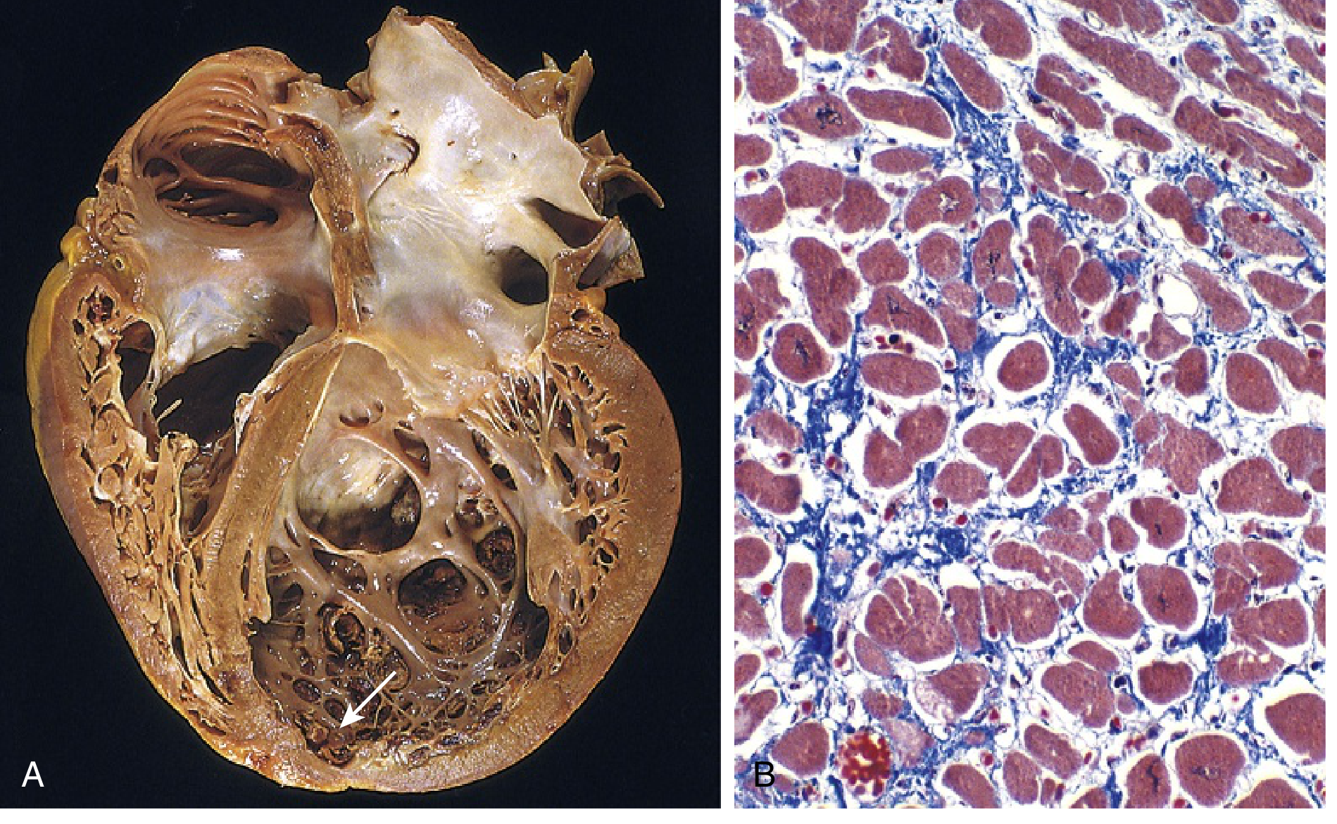
(A) Four-chamber dilation; mural thrombus at LV apex (arrow). (B) Myocyte hypertrophy and interstitial fibrosis (collagen = blue, Masson trichrome) - Robbins Basic Pathology, Fig. 9.25
Clinical Features
- Symptoms of progressive heart failure: dyspnea, fatigue, peripheral edema
- Dilated ventricle promotes dispersion of ventricular depolarization/repolarization - substrate for ventricular tachyarrhythmias and sudden cardiac death
- Mural thrombi - embolic stroke risk
- NYHA class II-III patients: higher risk of sudden cardiac death than pump failure; class IV: more likely to die of pump failure
2. Hypertrophic Cardiomyopathy (HCM)
Definition & Pathophysiology
HCM is characterized by massive myocardial hypertrophy without ventricular dilation, defective diastolic filling, and - in approximately one-third of cases - ventricular outflow obstruction (obstructive HCM). Systolic function is preserved (EF 50-80%), but diastolic dysfunction dominates. Prevalence is approximately 1 in 500 - making it the most common inherited cardiac disorder.
Pathogenesis
- Almost all cases are autosomal dominant, gain-of-function mutations in sarcomeric proteins
- More than 400 mutations in at least 9 genes identified
- Most commonly affected genes: β-myosin heavy chain (MYH7), myosin-binding protein C (MYBPC3), and cardiac troponin T (TNNT2) - together accounting for 70-80% of cases
- These are gain-of-function mutations causing myofilament hypercontractility, increased energy consumption, and net negative energy balance
- Note: some of the same genes (e.g., β-myosin) carry loss-of-function mutations in DCM
Morphology
- Massive myocardial hypertrophy; heart weight often 600-1000 g
- 90% of cases: Disproportionate thickening of the ventricular septum relative to the LV free wall - "asymmetric septal hypertrophy"
- Remaining 10%: concentric hypertrophy
- The thickened septum can bulge into the LV outflow tract
- Histology (pathognomonic): myocyte disarray - haphazardly oriented, interlocking hypertrophic myocytes, with interstitial and replacement fibrosis
Outflow Obstruction (Obstructive HCM)
- The hypertrophied septum narrows the LV outflow tract
- Dynamic obstruction created by systolic anterior motion (SAM) of the anterior mitral leaflet
- SAM causes both outflow obstruction AND mitral regurgitation
- Obstruction is dynamic: worsens with decreased preload (dehydration, Valsalva, standing), decreased afterload, or increased contractility (exercise, inotropes)
Clinical Features
- Dyspnea, angina, syncope (classic triad)
- Harsh systolic ejection murmur along the left sternal border (increases with Valsalva, decreases with squatting)
- Most common cardiovascular cause of sudden cardiac death in young athletes (accounts for ~1/3 of such events)
- Ventricular tachyarrhythmias arise from the substrate of myocyte disarray and fibrosis
- Atrial fibrillation is common; preferred rate/rhythm control: disopyramide + beta-blocker, or verapamil/diltiazem; amiodarone if needed
ICD Indications in HCM
ICD is recommended for patients with:
- Prior cardiac arrest or VF
- First-degree relative with sudden cardiac death
- Unexplained syncope
- LV wall thickness ≥ 30 mm
- Abnormal BP response to exercise with other risk factors
- Hemodynamically significant or non-sustained VT
- High-risk children with unexplained syncope, massive LVH, or family history of SCD
- Tintinalli's Emergency Medicine, p. 95; Braunwald's Heart Disease
3. Restrictive Cardiomyopathy
Definition & Pathophysiology
Restrictive cardiomyopathy is characterized by a decrease in ventricular compliance, resulting in impaired ventricular filling during diastole. The wall is stiff - the ventricle cannot relax and fill normally. Systolic function is typically preserved; EF may be near-normal. Atria dilate due to high filling pressures.
Morphology
- Ventricles approximately normal in size; cavities not dilated; myocardium is firm
- Both atria markedly dilated (due to high filling pressures)
- Microscopy: variable interstitial fibrosis; etiology-specific features on biopsy
Major Causes
1. Cardiac Amyloidosis
- Extracellular deposition of proteins with β-pleated sheet conformation
- Can occur in systemic amyloidosis (e.g., AL amyloid from multiple myeloma/plasma cell dyscrasias) or isolated cardiac form (ATTR - transthyretin amyloidosis)
- ATTR: deposition of normal or mutant transthyretin in older adults
- A specific TTR mutation (Val122Ile) is found in ~4% of African Americans, increasing cardiac amyloidosis risk >4-fold
- AL amyloid light chains are also directly cardiotoxic beyond just mechanical deposition
2. Endomyocardial Fibrosis
- Most common worldwide form of restrictive cardiomyopathy
- Children and young adults in Africa and other tropical regions
- Diffuse fibrosis of ventricular endocardium and subendocardium, involving tricuspid and mitral valves
- Associated with helminthic infections and nutritional deficiencies
3. Loeffler Endomyocarditis
- No geographic predilection
- Peripheral hypereosinophilia + eosinophilic tissue infiltrates
- Eosinophil major basic protein causes endocardial/myocardial necrosis, then scarring, mural thrombus formation, and thrombus organization
- Endocardial fibrosis with large mural thrombi
Other causes: Radiation fibrosis, sarcoidosis, mucopolysaccharide and sphingolipid storage diseases (Fabry, Gaucher, Hurler)
4. Arrhythmogenic Right Ventricular Cardiomyopathy (ARVC)
ARVC is an autosomal dominant disorder with variable penetrance, classically manifesting with:
- Right-sided heart failure
- Ventricular arrhythmias of RV origin (VT with left bundle branch block morphology)
- Syncope and sudden cardiac death (especially in young athletes)
Pathogenesis
Most causal mutations involve genes encoding desmosomal junctional proteins at the intercalated disk - e.g., plakoglobin, plakophilin-2, desmoplakin, desmoglein - or proteins interacting with the desmosome (desmin). Disrupted cell-cell junctions lead to myocyte death and replacement by fat and fibrosis.
- Naxos syndrome: ARVC + palmoplantar keratoderma (skin hyperkeratosis) - associated with plakoglobin mutations
Morphology
- Right ventricular wall severely attenuated by myocyte loss
- Near-transmural replacement of RV free wall by fat and fibrosis
- LV involvement possible but less prominent
- Mononuclear inflammation may surround degenerating myocytes
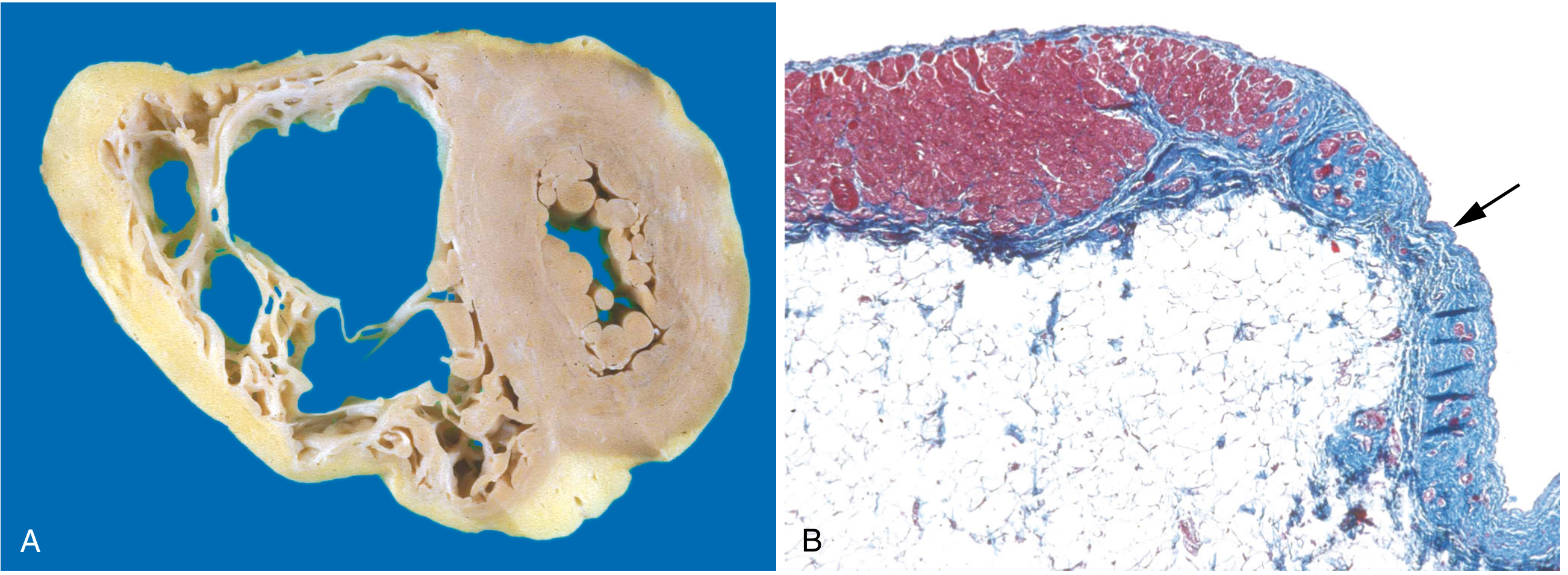
(A) Marked RV dilation; near-transmural RV free wall replacement by fat and fibrosis. (B) Masson trichrome: myocardium (red) replaced by fibrosis (blue, arrow) and fat. - Robbins, Cotran & Kumar Pathologic Basis of Disease, Fig. 12.31
ECG Features
ECG typically shows: epsilon wave (small positive deflection after QRS in right precordial leads), T-wave inversions in V1-V3, right bundle branch block morphology VT.
Summary Comparison Table
| Feature | DCM | HCM | Restrictive | ARVC |
|---|---|---|---|---|
| Ventricular size | Dilated | Normal/small cavity | Normal/small cavity | RV dilated |
| Wall thickness | Thin | Thick | Normal | RV thinned |
| EF | Reduced (<40%) | Normal-high (50-80%) | Normal | Variable |
| Dysfunction | Systolic | Diastolic | Diastolic | Arrhythmia/RHF |
| Key genetics | Titin (TTN), dystrophin, lamin A/C | β-myosin, MYBPC3, TnT | TTR mutations (amyloid) | Desmosomal proteins |
| Mutation type | Loss-of-function | Gain-of-function | - | Desmosome disruption |
| SCD risk | Moderate | High (young athletes) | Lower | High (young athletes) |
| Key finding | Mural thrombi | Myocyte disarray, SAM | Bilateral atrial dilation | Epsilon wave on ECG |
Key Points for Exams
- DCM: titin (TTN) is the most common single gene mutated; loss-of-function sarcomere mutations
- HCM: gain-of-function sarcomere mutations; asymmetric septal hypertrophy in 90%; myocyte disarray is pathognomonic; #1 cause of SCD in young athletes
- HCM murmur: increases with Valsalva (decreased preload), decreases with squatting (increased preload)
- Restrictive: both atria dilated; biventricular filling pressures elevated; amyloid is the key cause in the Western world
- ARVC: desmosomal protein mutations; fatty replacement of RV; epsilon wave; VT with LBBB morphology; Naxos syndrome (+ palmoplantar keratoderma)
- Peripartum cardiomyopathy: a form of DCM; up to 5 months postpartum; generally reversible in ~50% of cases
Sources: Robbins & Kumar Basic Pathology, 10e; Robbins, Cotran & Kumar Pathologic Basis of Disease, 10e; Goldman-Cecil Medicine; Tintinalli's Emergency Medicine, 9e; Braunwald's Heart Disease, 12e; Washington Manual of Medical Therapeutics
Cardiomyopathy
Reading File
Searching sources...
Finding Sources
Searching PubMed
"cardiomyopathy"[MeSH Terms]
Now pulling the core content from the best sources in parallel:
Reading File
Reading File
Finding Sources
Reading File
Reading File
Reading File
I have comprehensive content from the textbooks. Now let me fetch the key pathology images to include:
Loading Image
Loading Image
Loading Image
All images and content loaded. Here is the full, comprehensive response:
Cardiomyopathy
Cardiomyopathies are intrinsic diseases of cardiac muscle - distinct from myocardial dysfunction caused secondarily by coronary artery disease, hypertension, valvular disease, or congenital defects. There are three major pathophysiologic categories.
Classification at a Glance
| Type | Prevalence | LV EF | Core Dysfunction | Key Genetics |
|---|---|---|---|---|
| Dilated (DCM) | ~90% | <40% | Systolic (contractile) | Loss-of-function sarcomere/cytoskeletal mutations |
| Hypertrophic (HCM) | 1 in 500 | 50-80% | Diastolic (relaxation) | Gain-of-function sarcomere mutations |
| Restrictive | Least common | Near-normal | Diastolic (compliance) | Amyloid (TTR), infiltrative diseases |
| ARVC | Rare | Variable (RV failure) | Arrhythmia + RHF | Desmosomal protein mutations |
- Robbins, Cotran & Kumar Pathologic Basis of Disease
1. Dilated Cardiomyopathy (DCM)
Definition
DCM is characterized by progressive cardiac dilation and contractile (systolic) dysfunction, usually with concomitant hypertrophy. All four chambers dilate; the heart appears flabby and hypocontracting. It accounts for roughly 90% of all cardiomyopathies.
Pathogenesis
Genetic causes (20-50% of cases)
- Autosomal dominant inheritance is predominant, with variable penetrance
- Over 50 genes implicated; all involve loss-of-function mutations in cytoskeletal, sarcolemmal, or nuclear envelope proteins
- Titin (TTN) truncation mutations are the single most common cause, accounting for 10-20% of all DCM
- Other key genes: β-myosin heavy chain, cardiac troponin T, desmin, lamin A/C
- Dystrophin mutations (X-linked DCM): couples cytoskeleton to ECM; also mutated in Duchenne and Becker muscular dystrophies. X-linked DCM typically presents after puberty
- Mitochondrial gene deletions (oxidative phosphorylation defects) affect the pediatric population
Viral myocarditis
- Coxsackievirus B, adenovirus, parvovirus B19, HHV-6
- Sequential endomyocardial biopsies have documented progression from myocarditis to DCM
- Many "idiopathic" DCM cases are attributed to prior unrecognized viral infections
Alcohol and toxins
- Ethanol and especially acetaldehyde have direct toxic effects on the myocardium
- Chronic alcohol use causes thiamine deficiency - beriberi heart disease (a form of DCM)
- No morphologic features distinguish alcoholic DCM from other DCM causes
- Chemotherapy agents (doxorubicin/anthracyclines) are important toxic causes
- Cobalt (heavy metal) can cause DCM (historic contamination of beer production)
Peripartum cardiomyopathy
- Occurs late in pregnancy or up to 5 months postpartum
- Mechanism is multifactorial: genetic susceptibility, volume overload, hypertension, nutritional deficiency
- Recovers spontaneously in approximately 50% of cases
Other causes: hemochromatosis, sarcoidosis, chronic anemia, idiopathic
Morphology
- All four chambers dilated; heart weight markedly increased (up to 900 g)
- Mural thrombi common, especially at the LV apex - risk of systemic thromboembolism
- Histology: myocyte hypertrophy, nuclear enlargement, interstitial fibrosis - nonspecific findings
Fig. 9.25 - Dilated cardiomyopathy. (A) Four-chamber dilation with mural thrombus at LV apex (arrow). (B) Myocyte hypertrophy and interstitial fibrosis; collagen = blue on Masson trichrome. - Robbins Basic Pathology
Clinical Features
- Symptoms of progressive heart failure: dyspnea, fatigue, peripheral edema
- Dilated ventricle creates dispersion of depolarization/repolarization - substrate for ventricular tachyarrhythmias and sudden cardiac death
- Mural thrombi - risk of embolic stroke
- NYHA class II-III: higher SCD risk than pump-failure death; class IV: more likely to die of pump failure
2. Hypertrophic Cardiomyopathy (HCM)
Definition
HCM is characterized by massive myocardial hypertrophy without ventricular dilation, defective diastolic filling, and - in approximately one-third of cases - ventricular outflow obstruction. The heart is thick-walled, heavy, and hypercontracting, in striking contrast to DCM. Prevalence is 1 in 500, making it the most common inherited cardiac disorder. It is the leading cause of unexplained LV hypertrophy.
Pathogenesis
- Almost all cases are autosomal dominant with variable penetrance
- Over 400 mutations in 9 different genes; all are gain-of-function mutations in sarcomeric proteins, causing hypercontractility, increased energy consumption, and net negative energy balance
- This insight has led to novel therapeutics targeting myosin inhibition (mavacamten)
- Most commonly mutated genes (accounting for 70-80% of cases):
- β-myosin heavy chain (MYH7)
- Myosin-binding protein C (MYBPC3)
- Cardiac troponin T (TNNT2)
- Key contrast: the same β-myosin gene carries loss-of-function mutations in DCM but gain-of-function mutations in HCM
Morphology
- Massive myocardial hypertrophy; heart weight 600-1000 g
- 90% of cases: asymmetric septal hypertrophy - disproportionate thickening of the ventricular septum vs. LV free wall
- Remaining 10%: concentric hypertrophy
- Histology (pathognomonic): myocyte disarray - haphazardly oriented, bizarrely shaped, interlocking hypertrophic myocytes with interstitial fibrosis
Outflow Obstruction (Obstructive HCM)
- Hypertrophied septum narrows the LV outflow tract
- Dynamic obstruction by systolic anterior motion (SAM) of the anterior mitral leaflet
- SAM produces both outflow obstruction AND mitral regurgitation
- Obstruction worsens with: decreased preload (dehydration, Valsalva, standing), decreased afterload, or increased contractility (exercise, inotropes)
- Obstruction improves with: increased preload (squatting, leg raise), increased afterload
Clinical Features
- Classic triad: dyspnea, angina, syncope
- Harsh systolic ejection murmur along the left sternal border
- Increases with Valsalva (decreases preload)
- Decreases with squatting (increases preload)
- #1 cause of sudden cardiac death in young athletes (accounts for ~1/3 of such events)
- VT substrate: myocyte disarray + fibrosis create re-entry circuits
- Atrial fibrillation is common
ICD Indications in HCM
- Prior cardiac arrest or VF
- First-degree relative with sudden cardiac death
- Unexplained syncope
- LV wall thickness ≥ 30 mm
- Non-sustained or hemodynamically significant VT
- Abnormal blood pressure response to exercise (with other risk factors)
- High-risk children with massive LVH, unexplained syncope, or family history of SCD
- Tintinalli's Emergency Medicine
3. Restrictive Cardiomyopathy
Definition
Restrictive cardiomyopathy is defined by a decrease in ventricular compliance - the wall is stiff and cannot relax or fill normally during diastole. Systolic function is typically preserved (EF near-normal). Both atria dilate markedly due to elevated filling pressures transmitted backward.
Morphology
- Ventricles approximately normal in size; cavities not dilated; myocardium is firm
- Both atria markedly dilated - a hallmark finding
- Microscopy shows variable interstitial fibrosis; endomyocardial biopsy often identifies the specific cause
Major Causes
Cardiac Amyloidosis
- Extracellular deposition of proteins forming insoluble β-pleated sheets
- AL amyloid: immunoglobulin light chains from multiple myeloma/plasma cell dyscrasias; light chains are also directly cardiotoxic beyond mere deposition
- ATTR amyloid (transthyretin): deposition of normal (wild-type) or mutant transthyretin in older adults - increasingly recognized and treatable (tafamidis)
- ~4% of African Americans carry a TTR Val122Ile mutation, increasing cardiac amyloidosis risk >4-fold
Endomyocardial Fibrosis
- Most common restrictive cardiomyopathy worldwide
- Children and young adults in Africa and other tropical regions
- Diffuse fibrosis of ventricular endocardium and subendocardium, often involving tricuspid and mitral valves
- Associated with helminthic infections and nutritional deficiencies
Loeffler Endomyocarditis
- No geographic predilection
- Peripheral hypereosinophilia + eosinophilic tissue infiltrates
- Eosinophil major basic protein causes endocardial/myocardial necrosis → scarring → mural thrombus formation → thrombus organization → fibrosis
- Large mural thrombi are characteristic
Other causes: radiation fibrosis, sarcoidosis, hemochromatosis, storage diseases (mucopolysaccharidoses, Fabry disease, Gaucher disease)
4. Arrhythmogenic Right Ventricular Cardiomyopathy (ARVC)
ARVC is an autosomal dominant disorder with variable penetrance, classically presenting with:
- Right-sided heart failure
- Ventricular arrhythmias of RV origin - VT with left bundle branch block morphology
- Syncope and sudden cardiac death, especially in young athletes
Pathogenesis
- Mutations in genes encoding desmosomal junctional proteins at the intercalated disk: plakoglobin, plakophilin-2, desmoplakin, desmoglein-2, or proteins interacting with the desmosome (desmin)
- Disrupted cell-cell adhesion leads to myocyte death and replacement by fat and fibrosis
Naxos syndrome - ARVC + palmoplantar keratoderma (hyperkeratosis of palms and soles) - caused by plakoglobin mutations
Morphology
- RV wall severely attenuated by myocyte loss
- Near-transmural replacement of RV free wall by fat and fibrosis (fatty infiltration is the hallmark)
- LV can also be involved but less severely
- Mononuclear inflammation may surround degenerating myocytes (but ARVC is not a primarily inflammatory disorder)
Fig. 12.31 - Arrhythmogenic cardiomyopathy. (A) Marked RV dilation; near-transmural fatty/fibrotic replacement of RV free wall. (B) RV free wall: myocardium (red) replaced by fibrosis (blue, arrow) and fat - Masson trichrome. - Robbins, Cotran & Kumar Pathologic Basis of Disease
ECG Features
- Epsilon wave - small positive deflection after the QRS in right precordial leads (V1-V3)
- T-wave inversions in V1-V3
- VT with left bundle branch block morphology (arising from RV)
5. Myocarditis (Related Entity)
Myocarditis is not strictly a cardiomyopathy, but it is clinically important because it can evolve into DCM.
- Most common cause (USA): Viral - coxsackieviruses A and B, enteroviruses; increasingly parvovirus B19 and HHV-6
- Less common: CMV, HIV, influenza
- Clinical spectrum: asymptomatic → acute heart failure → chronic DCM
- Giant cell myocarditis (autoimmune, rapidly fatal without immunosuppression) must be distinguished from lymphocytic viral myocarditis
Comparison Summary
| Feature | DCM | HCM | Restrictive | ARVC |
|---|---|---|---|---|
| Chamber size | All dilated | Small LV cavity | Normal | RV dilated |
| Wall thickness | Thin | Thick | Normal/firm | RV thinned |
| EF | Reduced (<40%) | Normal-high | Near-normal | Variable |
| Dysfunction | Systolic | Diastolic | Diastolic | Arrhythmia/RHF |
| Key mutation type | Loss-of-function | Gain-of-function | TTR (amyloid) | Desmosomal |
| Key genes | TTN, dystrophin, lamin A/C | MYH7, MYBPC3, TNNT2 | TTR, light chain | Plakoglobin, PKP2 |
| Pathognomonic histology | Nonspecific fibrosis | Myocyte disarray | Amyloid/fibrosis | Fatty/fibrotic RV |
| SCD risk | Moderate | High (young athletes) | Lower | High (young athletes) |
| Mural thrombi | Common (LV apex) | Less common | Common (Loeffler) | Less common |
High-Yield Exam Points
- DCM = loss-of-function mutations; titin (TTN) most common single gene; all-chamber dilation; flabby heart
- HCM = gain-of-function mutations; asymmetric septal hypertrophy (90%); myocyte disarray (pathognomonic); #1 SCD cause in young athletes; SAM causes dynamic obstruction
- HCM murmur increases with Valsalva/standing (reduced preload); decreases with squatting/passive leg raise (increased preload)
- Restrictive = bilateral atrial dilation; amyloid (#1 in West); endomyocardial fibrosis (#1 worldwide); Congo red + apple-green birefringence for amyloid
- ARVC = desmosomal mutations; fatty RV replacement; epsilon wave; VT with LBBB morphology; Naxos syndrome (+ palmoplantar keratoderma)
- Peripartum cardiomyopathy = form of DCM; last trimester to 5 months post-delivery; ~50% recover
- Same gene, opposite mutation effect: β-myosin loss-of-function → DCM; β-myosin gain-of-function → HCM
Sources: Robbins Basic Pathology, 10e; Robbins, Cotran & Kumar Pathologic Basis of Disease, 10e; Goldman-Cecil Medicine; Tintinalli's Emergency Medicine, 9e; Braunwald's Heart Disease, 12e
This is a shared conversation. Sign in to Orris to start your own chat.