
Submandibular salivary gland Larynx Sub occipital region Para nasal sinuses
What would you like to know about these four topics (Submandibular gland, Larynx, Suboccipital region, Paranasal sinuses)?
submandibular gland anatomy relations mylohyoid
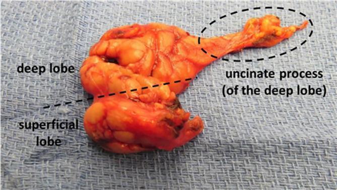
Clinical photograph of a gross submandibular gland resection specimen, demonstrating its complex lobular anatomy. The specimen is displayed on a blue surgical drape and is annotated to show the division between the larger superficial lobe and the smaller deep lobe, separated by a dashed line representing the anatomical plane typically defined by the mylohyoid muscle. Extending anteriorly from the deep lobe is the uncinate process, highlighted within a dotted oval. This process appears as an elongated, thinner extension of parenchymal tissue that normally wraps around the posterior border of the mylohyoid muscle to enter the sublingual space. The glandular tissue exhibits a characteristic yellowish-orange, lobulated appearance. This image serves as an educational reference for head and neck surgery, specifically illustrating the 'uncinate process' which can be mistaken for sublingual gland tissue or involved in pathologies like ranulas. The specimen represents an extended resection that includes both the deep lobe and Wharton's duct.
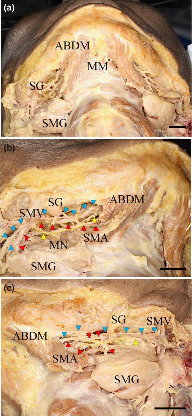
This composite of three clinical photographs (a-c) displays the gross anatomical dissection of the human submental region and mandibular floor. Figure (a) provides a wide superior view showing the mylohyoid muscle (MM), the anterior belly of the digastric muscle (ABDM), the submandibular gland (SMG), and the submandibular lymphatic ganglion (SG). Figures (b) and (c) offer higher magnification views of the right and left sides, respectively, detailing the neurovascular bundle situated between the MM and SMG. The submental nerve (MN), highlighted with yellow arrowheads and dotted lines, is seen coursing alongside the submental artery (SMA, red arrowheads) and the submental vein (SMV, blue arrowheads). These microscopic views emphasize the spatial relationship of the MN as it provides motor innervation to the mylohyoid muscle. The images illustrate the clinical anatomy relevant to submandibular surgery and local anesthesia, specifically identifying fine nerve branches that pierce the mylohyoid muscle to potentially communicate with the lingual nerve.
larynx anatomy cartilages supraglottis glottis subglottis
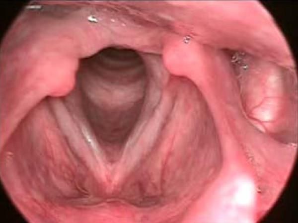
This diagnostic endoscopic photograph displays the human larynx from a superior view, highlighting the anatomy of the glottis and vocal folds. The primary pathology is a sulcus vocalis, visible as a distinct, linear groove or indentation running longitudinally along the mucosal cover of the right vocal fold (anatomical left of the image), parallel to its free edge. This groove disrupts the normally smooth surface of the vocal fold mucosa. In contrast, the contralateral vocal fold appears more uniform. The surrounding structures include the arytenoid cartilages positioned posteriorly, appearing as rounded pinkish masses, and the epiglottis superiorly. The space between the vocal folds reveals the entrance to the subglottis and trachea. The clinical significance of this finding relates to dysphonia, as the sulcus increases mucosal stiffness and prevents complete glottic closure during phonation. This image serves as an educational reference for Otolaryngology (ENT) residents and medical students studying laryngeal disorders and voice pathology.
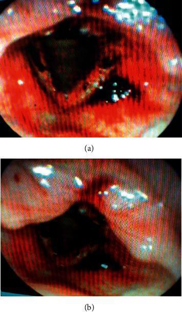
This diagnostic endoscopic image, labeled Figure 2(a) and 2(b), displays the laryngeal anatomy under nasopharyngolaryngoscopy. The visual shows significant pathology of the supraglottis, false vocal folds, and interarytenoid region. There is generalized erythema and intense inflammation across the mucosal surfaces. Key clinical findings include multiple scattered petechiae (small, red hemorrhagic spots) on the supraglottic surface and false vocal folds. Darker, focal areas of scabbing or crusting are prominent, particularly within the interarytenoid region and central glottic area. The glottis appears inflamed with associated mucosal edema. These findings are characteristic of laryngeal mucosal injury or hemorrhage. The images provide a clinical example of upper airway irritation or systemic bleeding manifestations within the larynx, relevant for Otolaryngology (ENT) training and diagnostic classification of vocal fold lesions and laryngeal trauma.
suboccipital triangle muscles vertebral artery
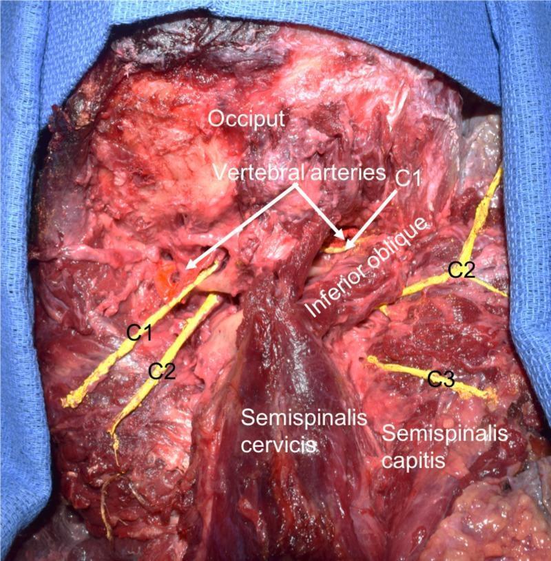
This clinical photograph displays a posterior anatomical dissection of the suboccipital region in a human cadaver, highlighting key neurovascular and muscular structures. Labels identify the occiput, vertebral arteries, inferior oblique muscle, semispinalis cervicis, and semispinalis capitis (reflected laterally). Crucially, the image demonstrates a comparative view of the suboccipital nerves (dorsal rami of C1). On the specimen's left side, a significantly enlarged and elongated dorsal ramus of C1 is visible, coursing medially to the vertebral artery as it crosses the posterior arch of the atlas. In contrast, the right-sided C1 nerve exhibits typical morphology. The dorsal rami of the C2 and C3 spinal nerves are also identified bilaterally, positioned inferior to the suboccipital triangle. The dissection illustrates the spatial relationship between the vertebral artery and the suboccipital muscles, specifically showing the artery's proximity to the inferior oblique muscle. This specimen serves as an educational example of anatomical variation in cervical spinal nerve morphology and suboccipital neuroanatomy.
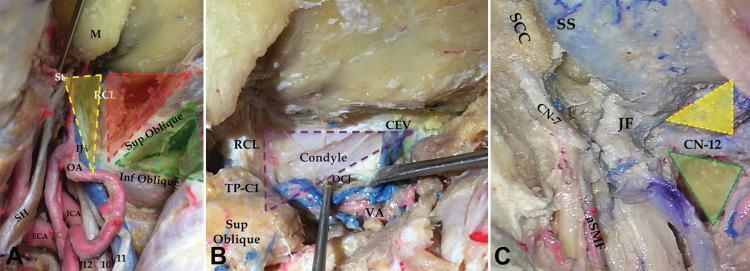
This composite of three anatomical photographs (A-C) illustrates deep neurosurgical triangles of the suboccipital and condylar regions, essential for approaching the jugular foramen (JF). Panel A shows the suboccipital triangle (green), condylar triangle (red), and jugular triangle (yellow), bounded by the superior oblique, inferior oblique, and rectus capitis lateralis (RCL) muscles. Notable vascular structures include the external and internal carotid arteries (ECA, ICA). Panel B highlights the deep condylar and trans-condylar triangles (purple), bounded by the condylar emissary vein (CEV), RCL, and the transverse process of the atlas (TP-C1). The occipital condyle and atlanto-occipital joint (OCJ) are visible after mobilizing the vertebral artery (VA). Panel C demonstrates the supra-hypoglossal (yellow) and infra-hypoglossal (green) triangles after condyle drilling. These triangles serve as surgical corridors to the infrajugular area and OCJ. Key neural landmarks include the facial nerve (CN-7) and hypoglossal nerve (CN-12) near the jugular foramen and the artery of the stylomastoid foramen (aSMF).
paranasal sinuses anatomy diagram frontal maxillary ethmoid sphenoid
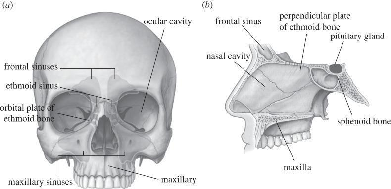
This medical anatomical diagram displays the human skull in two views, focusing on the paranasal sinuses and internal cranial structures. View (a) is an anterior (frontal) perspective of the skull with semi-transparent overlays illustrating the paranasal sinuses. Labeled structures include the frontal sinuses located superior to the orbits, the ethmoid sinus situated medially between the eyes, the orbital plate of the ethmoid bone forming the medial orbital wall, the ocular cavity (orbit), and the large maxillary sinuses within the maxilla. View (b) provides a mid-sagittal section of the facial and mid-cranial region. It highlights the spatial relationship between the nasal cavity and surrounding structures, including the frontal sinus, the perpendicular plate of the ethmoid bone (part of the nasal septum), and the maxilla. Posteriorly, it shows the sphenoid bone containing the sella turcica, where the pituitary gland is seated. This illustration is designed for medical education regarding craniofacial anatomy, specifically for procedures such as Functional Endoscopic Sinus Surgery (FESS) and maxillofacial osteotomies.
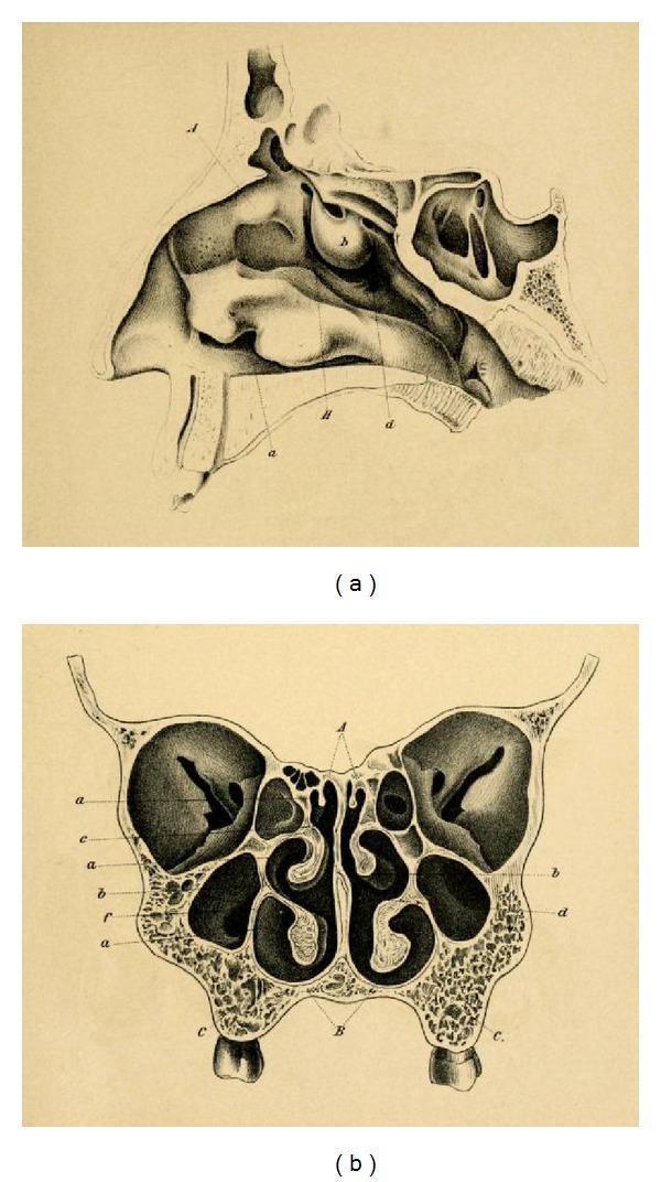
This historical anatomical diagram by Emil Zuckerkandl (1882) illustrates the paranasal sinuses and nasal anatomy in two planes. Image (a) is a sagittal section of the nasal cavity, detailing the complex arrangement of the superior, middle, and inferior turbinates (conchae) along the lateral wall. It visualizes the frontal sinus superiorly, the sphenoid sinus posteriorly, and the relationship between the nasal cavity and the hard palate. Image (b) presents a frontal (coronal) section, showcasing the bilateral symmetry of the mid-facial anatomy. Key features include the central nasal septum, the convoluted turbinates projecting into the nasal passages, and the expansive maxillary sinuses situated laterally within the maxilla bones. Small ethmoid air cells are visible medially to the orbits and superior to the maxillary sinuses. This anatomical illustration serves as a foundational reference for otorhinolaryngology and neurosurgical approaches, particularly regarding the development of transsphenoidal surgery for pituitary lesions.
1. Submandibular Salivary Gland
Overview
- Size: approximately that of a walnut
- Type: mixed gland, but predominantly serous
- Structure: a larger superficial part and a smaller deep part, continuous around the posterior border of the mylohyoid muscle
- Enclosed within a well-defined capsule derived from the deep cervical fascia (splits to enclose it)
Location
- The superficial part lies in the digastric triangle (submandibular triangle)
- Extends medial to the body of the mandible above; below, it overlaps the intermediate tendon of the digastric muscle and the insertion of the stylohyoid muscle
Relations
| Surface | Relation |
|---|---|
| Inferior surface | Skin, platysma, deep fascia; crossed by the facial vein and cervical branch of facial nerve |
| Lateral surface | Submandibular fossa on medial surface of mandible; medial pterygoid muscle attachment |
| Medial (deep) surface - anterior | Mylohyoid muscle |
| Medial surface - intermediate | Hyoglossus muscle (separated by styloglossus, lingual nerve, submandibular ganglion, hypoglossal nerve, deep lingual vein) |
| Medial surface - posterior | Styloglossus muscle |
| Deep part | Lies between mylohyoid (inferolateral) and hyoglossus/styloglossus (medial); extends forward to posterior end of sublingual gland |
Wharton's Duct (Submandibular Duct)
- Length: approximately 5 cm
- Emerges from the medial surface of the superficial part, behind the posterior border of mylohyoid
- Passes through the deep part of the gland
- Runs between the sublingual gland and genioglossus muscle
- Opens on the summit of the sublingual papilla at the side of the frenulum of the tongue
- On the hyoglossus muscle, it is crossed laterally by the lingual nerve (the lingual nerve winds under the duct)
Blood Supply
- Facial artery: enters deep to the posterior belly of digastric and stylohyoid muscles, passes through or superficial to the gland, then grooves the mandible at its lower border. Glandular branches must be ligated when removing the gland.
- Facial vein: lies superficial to the gland
Nerve Supply
- Secretomotor (parasympathetic):
- Preganglionic fibres travel via the facial nerve → chorda tympani → joins lingual nerve → submandibular ganglion (a small fusiform body on the upper part of hyoglossus, suspended from the lingual nerve)
- Postganglionic fibres from the ganglion are distributed to the submandibular and sublingual glands
- Sympathetic: vasomotor fibres via the plexus on the facial artery
- Marginal mandibular branch of facial nerve: lies in the superficial fascia, traverses over the facial vessels (important in surgery - Martin's technique uses ligation and flipping of these vessels superiorly to protect this nerve during neck dissection)
Calculus (Salivary Stone / Sialolithiasis)
- More common in the submandibular gland than in the parotid (secretion is more viscous/alkaline; duct is longer, tortuous, and ascends against gravity)
- Composition: calcium and magnesium phosphates (similar to dental tartar)
- May form within the gland or its duct
- Pathognomonic feature: swelling of the gland during meals (salivary colic), often preceded by a colicky pain
- Diagnosis: lemon juice test (triggers swelling and reveals absence of salivation from affected duct); bimanual palpation of duct; plain X-ray (80-90% radiopaque); ultrasound
Tumours
- Tumours are uncommon compared to parotid tumours; enlargement is more often due to calculus
- Most common tumour: pleomorphic adenoma (mixed tumour) - slow growing, moderately sized, hard but not stony hard
- Carcinoma: extremely rare
- Mikulicz's disease: symmetrical enlargement of all salivary + lacrimal glands (lymphocytic replacement); dry mouth (xerostomia)
Clinical Note
- Swelling in the submandibular region is more often due to lymph node enlargement than salivary gland tumour
- The hypoglossal nerve must be identified and preserved during gland excision
2. Larynx
Overview
- Location: anterior to the hypopharynx; extends from the tip of the epiglottis (level of lower border of C3) to the lower border of the cricoid cartilage (level of C6)
- Functions: airway protection, phonation, respiration
Cartilaginous Skeleton
Hyaline Cartilages (begin to ossify ~20 years of age - radiopaque)
- Thyroid cartilage: largest; two laminae fused anteriorly forming the laryngeal prominence; vocal cords attach at the "figure-of-eight" on lateral X-ray
- Cricoid cartilage: only complete ring in the airway; narrowest point of the paediatric airway
- Arytenoid cartilages (most of each): paired; sit on the superior surface of the cricoid posteriorly; vocal processes for vocal cord attachment
Elastic Cartilages (do NOT ossify - radiolucent)
- Epiglottis: leaf-shaped; attached to posterior thyroid at its inferior end
- Corniculate cartilages (of Santorini): tips of arytenoids
- Cuneiform cartilages (of Wrisberg): in aryepiglottic folds
- Vocal process and apex of arytenoids
Anatomical Subdivisions
| Region | Boundaries | Structures |
|---|---|---|
| Supraglottis | From tip of epiglottis to lateral wall of ventricle | Suprahyoid + infrahyoid epiglottis, aryepiglottic folds, arytenoids, false vocal cords (ventricular folds), ventricle (of Morgagni) |
| Glottis | True vocal cords + anterior and posterior commissures; lower boundary = 5 mm below free margin of cords (or 1 cm below apex of ventricle) | True vocal cords; anterior commissure (approaches within 1 cm of skin) |
| Subglottis | From lower boundary of glottis to inferior margin of cricoid | Air column; begins transition to trachea |
Key Spaces
- Preepiglottic space: fat-filled space anterior to epiglottis; easily identified on CT/MRI
- Paraglottic (paralaryngeal) space: fat-filled; surrounds the ventricle; important for assessing deep invasion by tumour
- Reinke's space: superficial lamina propria of vocal cord - site of Reinke's oedema (polypoidal degeneration)
Muscles of the Larynx
- Only abductor of vocal cords: Posterior cricoarytenoid (PCA) - opens the airway
- Adductors: Lateral cricoarytenoid, interarytenoid (transverse + oblique)
- Tensors: Cricothyroid (lengthens/tenses cord - extrinsic; supplied by external branch of superior laryngeal nerve)
- Relaxer: Thyroarytenoid (vocalis)
Nerve Supply
- Recurrent laryngeal nerve (RLN) (branch of vagus): supplies all intrinsic muscles EXCEPT cricothyroid; also sensory below vocal cords
- Right RLN: loops around right subclavian artery
- Left RLN: loops around aortic arch (longer course; more vulnerable)
- Superior laryngeal nerve (SLN) (branch of vagus):
- Internal branch: sensory above vocal cords and to epiglottis
- External branch: motor to cricothyroid muscle
Blood Supply
- Superior laryngeal artery (from superior thyroid artery)
- Inferior laryngeal artery (from inferior thyroid artery)
Lymphatic Drainage
- Supraglottic: rich bilateral lymphatics → upper deep cervical nodes (Level II, III)
- Glottic: sparse lymphatics (true cords have almost none - explains low nodal spread in early glottic cancer)
- Subglottic: drains to Level IV and Level VI (pretracheal/paratracheal)
Laryngocele
- Internal type: air sac stays within paralaryngeal fat (supraglottic region)
- External (mixed) type: pierces the thyrohyoid membrane → neck mass
- Cause: increased intraglottic pressure (horn players, glass blowers) or obstruction of laryngeal ventricle
Important Clinical Points
- Epiglottitis (supraglottitis): thickens epiglottis and aryepiglottic folds; "thumbprint sign" on lateral X-ray; life-threatening airway compromise
- Laryngeal fracture on CT: linear lucency in ossified cartilage; best seen on bone windows
- Imaging: CECT and MRI are relatively insensitive to superficial mucosal lesions; fat planes in preepiglottic and paraglottic spaces are key landmarks
3. Suboccipital Region
Overview
- A deep region at the base of the occipital bone in the upper cervical area
- Contains the suboccipital muscles (4 pairs) that move the head at the atlantooccipital and atlantoaxial joints
- All innervated by the suboccipital nerve = posterior ramus of C1 (purely motor)
The Four Suboccipital Muscles
| Muscle | Origin | Insertion | Function |
|---|---|---|---|
| Rectus capitis posterior major | Spinous process of axis (C2) | Lateral part of occipital bone (below inferior nuchal line) | Extension of head; rotation of face to same side |
| Rectus capitis posterior minor | Posterior tubercle of atlas (C1) | Medial part of occipital bone (below inferior nuchal line) | Extension of head |
| Obliquus capitis superior | Transverse process of atlas (C1) | Occipital bone (between superior and inferior nuchal lines) | Extension of head; bends head to same side |
| Obliquus capitis inferior | Spinous process of axis (C2) | Transverse process of atlas (C1) | Rotation of face to same side (largest of the four) |
Suboccipital Triangle
| Border | Muscle |
|---|---|
| Medial | Rectus capitis posterior major |
| Lateral (superior) | Obliquus capitis superior |
| Inferior | Obliquus capitis inferior |
- Posterior ramus of C1 (suboccipital nerve) - emerges above the posterior arch of the atlas between the vertebral artery and the arch
- Vertebral artery - runs through its groove on the superior surface of the posterior arch of the atlas (third part of vertebral artery, V3 segment)
- Suboccipital venous plexus
- Greater occipital nerve (C2) - winds posteriorly as it passes the lower margin of the obliquus capitis inferior → sensory to posterior scalp
- Third occipital nerve (C3) - also passes at this level
Vascular Supply
- Branches of the vertebral artery and occipital artery
- The deep cervical artery (branch of costocervical trunk) runs between semispinalis capitis and cervicis muscles nearby
Overlying Muscles (superficial to suboccipital region, removed during dissection)
- Trapezius
- Sternocleidomastoid
- Splenius capitis
- Semispinalis capitis
Key Landmark
- The spinous process of the axis (C2) is the most important landmark in the deep nuchal region (the largest palpable spinous process in this area)
Clinical Significance
- Suboccipital nerve block / greater occipital nerve block: used in management of cervicogenic headache and occipital neuralgia
- Vertebral artery injury: the V3 segment in the suboccipital triangle is vulnerable during posterior cervical surgery and atlantoaxial surgery
- Suboccipital approach: a neurosurgical/otological approach used to access the posterior cranial fossa and cerebellopontine angle (e.g., for acoustic neuroma)
4. Paranasal Sinuses
Overview
Individual Sinuses
Maxillary Sinus (Antrum of Highmore)
- Largest paranasal sinus
- Located in the body of the maxilla
- Drainage: into the middle meatus via the maxillary ostium → infundibulum (worst drainage - ostium is on the superomedial wall, not the floor)
- Relations: floor = alveolar process (roots of upper molar teeth may project into it); roof = orbital floor; medial wall = lateral nasal wall; posterior wall = infratemporal fossa
- Nerve supply: superior alveolar nerves (branches of maxillary V2)
- First sinus to develop; present at birth as small pouches; adult size by puberty
Frontal Sinus
- Located in the frontal bone, above the orbits
- Drainage: into the middle meatus via the frontonasal duct / nasofrontal recess
- Absent at birth; begins to develop at age 2; reaches adult size by late teens
- Often asymmetric; may be absent unilaterally in 5% of people
- Anterior wall is the thicker; posterior wall is thin (separates from anterior cranial fossa)
Ethmoid Sinuses (Labyrinth)
- Multiple small air cells within the ethmoid bone
- Divided into: anterior ethmoid cells (drain into middle meatus) and posterior ethmoid cells (drain into superior meatus)
- Medial wall: lateral wall of nasal cavity; lateral wall: lamina papyracea (paper-thin - orbital wall; easily breached in trauma or disease)
- Key structure: ethmoid bulla (largest anterior ethmoid cell; landmark for middle meatus)
- Important: most commonly involved in sinusitis, and most commonly implicated in orbital complications of sinusitis
Sphenoid Sinus
- Located in the body of the sphenoid bone
- Drainage: into the sphenoethmoidal recess (above superior turbinate)
- Important relations:
- Superior: pituitary gland in sella turcica, optic chiasm
- Lateral: cavernous sinus (internal carotid artery, oculomotor, trochlear, ophthalmic, abducens nerves)
- Posterior: pons/brainstem
- Inferior floor: nasopharynx
- Surgical importance: transsphenoidal approach to pituitary tumours
The Ostiomeatal Complex (OMC)
- The functional unit of sinus drainage in the middle meatus
- Components: maxillary sinus ostium, infundibulum, uncinate process, ethmoid bulla, hiatus semilunaris, middle turbinate
- Uncinate process forms the medial wall of the infundibulum; ethmoid bulla forms the lateral wall
- Haller cells (infraorbital ethmoid cells): anatomic variant that can narrow the infundibulum → predisposes to sinusitis
- Obstruction of the OMC → impaired mucociliary clearance → sinusitis
Drainage Summary
| Sinus | Drains Into |
|---|---|
| Maxillary | Middle meatus (via hiatus semilunaris) |
| Frontal | Middle meatus (via frontonasal duct) |
| Anterior ethmoid | Middle meatus |
| Posterior ethmoid | Superior meatus |
| Sphenoid | Sphenoethmoidal recess |
Imaging
- CT (NCCT): investigation of choice for sinuses; essential for pre-operative planning before FESS (Functional Endoscopic Sinus Surgery)
- Coronal plane: best for OMC anatomy and preoperative planning
- CT distinguishes: aerated sinus (normal), mucosal thickening (chronic sinusitis), air-fluid level (acute sinusitis), complete opacification (mucocele, tumour)
- MRI: superior for distinguishing tumour from obstructed secretions; sinusonasal tumours show low-to-intermediate signal on T2WI; obstructed inspissated mucus shows high T1 and variable T2
Pathological Conditions
- Sinusitis: Acute (air-fluid level), Chronic (mucosal thickening + bony sclerosis), Invasive (mucormycosis - destroys bone rapidly; in immunocompromised)
- Mucocele: complete obstruction → slow expansion; remodels bone (not destroys it); most common in frontal sinus
- Nasal polyps: eosinophilic; "cascading" enhancement pattern on CECT; arise from ethmoid mucosa
- Malignancy: bone destruction on CT; low-to-intermediate T2 signal on MRI; adenoid cystic carcinoma and squamous cell carcinoma are most common
Quick Reference Summary
| Topic | Key Numbers/Facts |
|---|---|
| Submandibular gland | Wharton's duct = 5 cm; opens at sublingual papilla; lingual nerve crosses over duct |
| Larynx | C3 (epiglottis tip) to C6 (cricoid lower border); only abductor = PCA; only intrinsic muscle with SLN = cricothyroid |
| Suboccipital triangle | Medial = rectus capitis major; lateral = obliquus superior; inferior = obliquus inferior; contents = C1 ramus, vertebral artery, veins |
| Paranasal sinuses | Posterior ethmoid + sphenoid DO NOT drain into middle meatus; OMC = key functional unit; lamina papyracea = orbital wall of ethmoid |
Inner cavity of larynx
inner cavity larynx vestibule ventricle rima glottidis vocal folds diagram
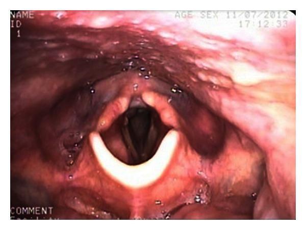
This clinical photograph displays an endoscopic view of the human larynx, specifically a fiberoptic nasoendoscopy. The image highlights the glottis and surrounding supraglottic structures. Centrally, the true vocal folds (vocal cords) appear as bright, pearly-white, crescent-shaped bands. The edges of the folds are smooth and symmetrical, framing the dark, patent rima glottidis (airway opening). Above the vocal folds, the vestibular folds (false vocal cords) and the laryngeal vestibule are visible, exhibiting a healthy, reddish-pink mucosal lining with normal vascularity. The epiglottis is partially visible at the top of the frame. Small droplets of clear mucus are scattered across the mucosal surface, which is a normal finding. This image serves as an educational example of normal post-traumatic recovery, demonstrating preserved laryngeal anatomy and unobstructed airway patency following a penetrating neck injury.
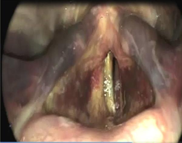
This clinical photograph displays an endoscopic view (videolaryngoscopy) of the larynx, highlighting significant laryngeal trauma. The image demonstrates a widespread laryngeal hematoma, characterized by prominent purplish-red and ecchymotic discoloration across the laryngeal mucosa. Marked edema and swelling are visible throughout the supraglottic and glottic regions, leading to structural distortion and partial obscuration of the true vocal folds. The rima glottidis is significantly narrowed and compromised by the surrounding inflammatory changes and blood accumulation. There is also evidence of superficial fibrin or discharge on the vocal fold surfaces. This visual serves as a critical example of postoperative or post-intubation laryngeal complications, illustrating the physical manifestations of airway trauma that may lead to vocal symptoms like hoarseness or respiratory distress in an otolaryngology context.
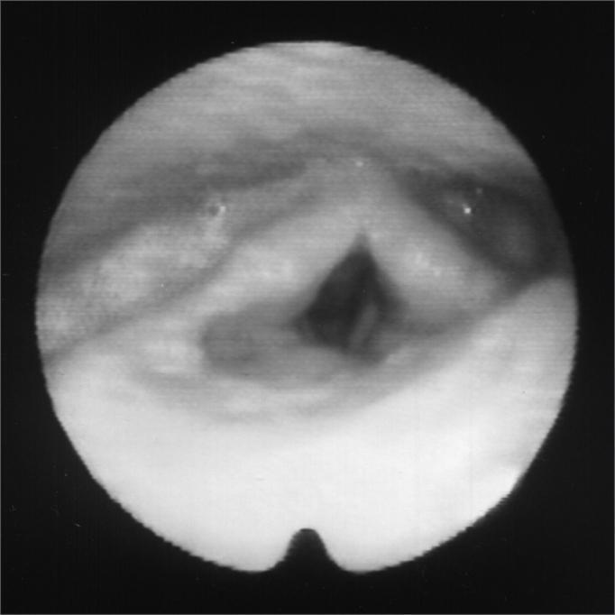
This clinical diagnostic image shows an endoscopic view of the larynx, specifically a fiberoptic laryngoscopy capturing the glottis at rest following botulinum toxin (Botox) injection. Centrally, the true vocal folds are visible, appearing relatively relaxed and positioned in a neutral or slightly adducted state. The glottic opening (rima glottidis) is seen as a dark, roughly diamond-shaped space between the vocal folds. Posteriorly, the arytenoid eminences and the interarytenoid area are discernible as soft tissue contours. The image demonstrates the pharmacological effect of Botox on the laryngeal musculature, likely the thyroarytenoid or lateral cricoarytenoid muscles, used to treat conditions such as adductor spasmodic dysphonia or paradoxical vocal fold motion. The mucosa appears healthy without evidence of gross lesions, masses, or acute inflammatory exudate. This visual serves as an educational reference for the post-procedural appearance of the laryngeal framework during medical management of laryngeal dystonia.
laryngeal saccule ventricle of Morgagni conus elasticus quadrangular membrane cross section
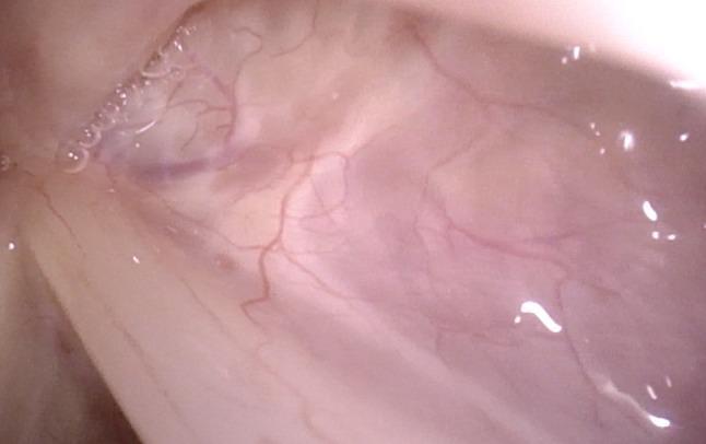
This clinical photograph displays a high-magnification endoscopic view of a normal human larynx, specifically targeting the anterior region of the right sinus Morgagni (laryngeal ventricle). The image, captured via a specialized rotation maneuver during flexible laryngoscopy, shows the healthy mucosal lining of the ventricle. The tissue is characterized by a healthy pinkish hue with a smooth, moist texture. Fine, branching subepithelial blood vessels are clearly visible, illustrating the normal vascular pattern of the laryngeal mucosa. Small fluid droplets and light reflections on the surface indicate the physiological presence of mucus. This perspective is educationally significant for demonstrating the ability to visualize deep laryngeal recesses, such as the ventricle and the inferior aspect of the vocal folds, which are often obscured during standard distal laryngoscopy.
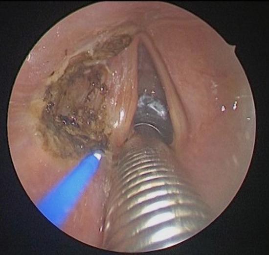
This endoscopic clinical photograph captures an intraoperative view of the larynx during a posterior ventriculo-cordectomy. The image demonstrates a transverse incision of the left ventricular fold (false vocal cord), which has been performed using a contact diode laser. The surgical site on the left lateral aspect shows significant tissue alteration, characterized by a dark, irregular, and charred surface consistent with laser thermal coagulation and vaporization. This procedure aims to remove a sail-shaped portion of the fold to reveal the superior aspect of the true vocal fold and open Morgagni's ventricle. Centrally positioned in the foreground is a metallic, corrugated endotracheal tube, likely a laser-protected tube required for airway management under general anesthesia. To the left, a laser fiber tip is visible, emitting a characteristic blue light, indicating active thermal application. The surrounding laryngeal mucosa appears hyperemic, a typical response to surgical trauma. This visual is used to teach endoscopic laryngeal surgery techniques and the management of narrowed airways via laser resection.
Inner Cavity of the Larynx (Laryngeal Cavity)
Definition
The Laryngeal Inlet (Aditus Laryngis)
| Boundary | Structure |
|---|---|
| Anterior | Free upper margin of the epiglottis |
| Posterior | Interarytenoid notch (mucosa between the two arytenoids) |
| Lateral (each side) | Aryepiglottic fold (runs from lateral margin of epiglottis to apex/corniculate cartilage of arytenoid) |
- Cuneiform cartilage (of Wrisberg): anterosuperiorly - produces the cuneiform tubercle
- Corniculate cartilage (of Santorini): posteriorly at the tip of arytenoid - produces the corniculate tubercle
Three Compartments of the Laryngeal Cavity
I. Laryngeal Vestibule (Upper Part)
- Extends from the laryngeal inlet down to the vestibular (false) vocal folds
- Anterior wall: laryngeal surface of the epiglottis (4-5 cm long; connected to base of tongue by glossoepiglottic folds and to hyoid by hyoepiglottic ligament)
- Posterior wall: flat, at the level of the interarytenoid area
- Lateral walls: medial surfaces of the aryepiglottic folds
II. Intermediate (Middle) Laryngeal Cavity
- The smallest compartment
- Extends from the vestibular (false) folds above to the true vocal folds below
- On each side, the wall bulges out laterally to form the laryngeal ventricle (sinus of Morgagni)
Laryngeal Ventricle (Sinus / Ventricle of Morgagni)
- A deep horizontal mucosal recess between the false cord above and the true vocal cord below
- Saccule (appendix of the ventricle): a conical, blind-ending pouch that ascends anteriorly from the ventricle into the paralaryngeal fat
- Contains numerous minor seromucous salivary glands (lubricates and moistens the vocal cords)
- A distended saccule = laryngocele (internal type stays within paralaryngeal fat; external/mixed type pierces the thyrohyoid membrane)
III. Infraglottic (Subglottic) Cavity (Lower Part)
- Extends from the undersurface of the true vocal folds to the inferior margin of the cricoid cartilage
- Gradually widens craniocaudally as the cricoid ring expands
- Its wall is formed almost entirely by the conus elasticus (cricovocal membrane)
- Continues inferiorly as the trachea
The Two Pairs of Folds (Valves)
Vestibular Folds (False Vocal Cords)
- Lie above the true vocal cords
- Contains the vestibular ligament = free inferior margin of the quadrangular membrane (the upper elastic membrane of the larynx)
- Also contains numerous seromucous glands
- Do not vibrate during normal phonation
- Space between the two vestibular folds = rima vestibuli (wider than the rima glottidis below)
- Function: accessory sphincter; close tightly during swallowing, coughing, and Valsalva manoeuvre
True Vocal Folds (Vocal Cords)
- Lie below the vestibular folds
- Pearly white appearance (avascular lamina propria)
- Layers (medial to lateral):
- Stratified squamous epithelium (only part of laryngeal cavity NOT lined by respiratory epithelium)
- Lamina propria (3 layers - the "body-cover" model):
- Superficial layer (Reinke's space): very loose, rich in hyaluronic acid - allows mucosal wave to vibrate freely; site of Reinke's oedema (polypoid degeneration from smoking)
- Middle layer: elastic fibres (denser)
- Deep layer: collagen fibres (densest)
- Vocal ligament = fused middle + deep layers of lamina propria
- Vocalis muscle (medial part of thyroarytenoid muscle)
Attachments of True Vocal Cords
- Anteriorly: anterior commissure - attached to inner thyroid cartilage at the anterior commissure tendon (Broyles' ligament); comes within ~1 cm of the skin here
- Posteriorly: vocal process of the arytenoid cartilage
Parts of Each Vocal Cord
| Part | Extent | Importance |
|---|---|---|
| Membranous (anterior) part | Anterior commissure → vocal process of arytenoid (approx. 3/5 of length) | Vibrates to produce voice |
| Cartilaginous (posterior) part | Vocal process + body of arytenoid (approx. 2/5) | Moved by intrinsic muscles; involved in abduction/adduction |
Rima Glottidis
| State | Shape |
|---|---|
| Quiet respiration | Triangular (wider anteriorly, narrower posteriorly) |
| Deep inspiration / abduction | Rhomboid (teardrop/diamond) |
| Phonation | Slit-like |
| Swallowing/Valsalva/cough | Completely closed |
- Intermembranous part (glottic chink): between the true vocal cords anteriorly
- Intercartilaginous part (respiratory chink): between the arytenoids posteriorly (triangular when breathing)
The rima glottidis is the narrowest point of the adult larynx (in the child, the subglottis at the cricoid is narrowest - relevant to croup and subglottic stenosis)
Membranes and Ligaments of the Laryngeal Cavity
| Membrane | Location | Forms |
|---|---|---|
| Quadrangular membrane | Upper part - between epiglottis and arytenoid | Free lower border = vestibular ligament (in false cord); upper border = aryepiglottic fold |
| Conus elasticus (cricovocal membrane) | Lower part - from cricoid to vocal cord | Free upper border = vocal ligament (in true cord) |
Spaces of Clinical Importance
| Space | Boundaries | Clinical Significance |
|---|---|---|
| Preepiglottic space | Anterior: thyroid cartilage + thyrohyoid membrane; Posterior: epiglottis; Superior: vallecula | Fat-filled; invaded early in supraglottic cancers; not a barrier to tumour spread |
| Paraglottic space | Thyroid lamina laterally, conus elasticus medially/inferiorly, quadrangular membrane medially/superiorly | Fat-filled; pathway for transglottic cancer spread; contains the laryngeal ventricle |
Mucosa of the Laryngeal Cavity
| Area | Epithelium |
|---|---|
| Vestibule, ventricular folds, subglottis | Ciliated pseudostratified columnar (respiratory) epithelium |
| True vocal cords (free edges) | Stratified squamous epithelium (non-keratinising) - due to mechanical stress |
| Laryngeal vestibule and vestibular folds | Contains numerous mixed seromucous glands |
Summary Diagram (Text)
LARYNGEAL INLET (epiglottis + aryepiglottic folds + interarytenoid notch)
        |
   VESTIBULE (I)
        |
   VESTIBULAR FOLDS (false cords) ←── Rima vestibuli (wider)
        |
   VENTRICLE of MORGAGNI (saccule extends up from it)
        |
   TRUE VOCAL FOLDS ←── Rima glottidis (narrowest in adult)
        |
   INFRAGLOTTIC / SUBGLOTTIC CAVITY (III)
        |
   (continuous with TRACHEA at lower cricoid border)
As a mind map
A detailed medical mind map titled "Inner Cavity of the Larynx" at the center in a bold circle. The map radiates outward with the following main branches and sub-branches, using different colors for each main branch: BRANCH 1 (Red): "Laryngeal Inlet (Aditus)" with sub-branches: Epiglottis (anterior), Aryepiglottic Folds (lateral), Interarytenoid Notch (posterior), Corniculate Tubercle, Cuneiform Tubercle, Piriform Recess (lateral to inlet) BRANCH 2 (Orange): "Vestibule (Upper)" with sub-branches: Inlet → Vestibular Folds, Anterior wall = Epiglottis, Posterior wall = Interarytenoid area, Lined by respiratory epithelium, Contains seromucous glands BRANCH 3 (Green): "Intermediate Cavity (Middle)" with sub-branches: Smallest compartment, Between false & true vocal folds, Ventricle of Morgagni → Saccule (blind pouch anteriorly) → Minor salivary glands → Laryngocele if distended, Rima Vestibuli (between false cords - wider) BRANCH 4 (Blue): "Infraglottic/Subglottic Cavity (Lower)" with sub-branches: Vocal folds → Lower cricoid border, Wall = Conus Elasticus, Widens craniocaudally, Continuous with trachea, Narrowest in children (cricoid level) BRANCH 5 (Purple): "Vestibular Folds (False Cords)" with sub-branches: Contain vestibular ligament, Lower free edge of quadrangular membrane, Contain seromucous glands, NOT for phonation, Close in swallow/cough/Valsalva BRANCH 6 (Teal): "True Vocal Folds" with sub-branches: Pearly white (avascular), Layers → Squamous epithelium → Reinke's space (superficial LP) → Vocal ligament (middle+deep LP) → Vocalis muscle, Membranous part (ant 3/5) = phonation, Cartilaginous part (post 2/5) = movement, Rima Glottidis → triangular (breathing) / slit (phonation) / closed (swallow) BRANCH 7 (Brown): "Key Spaces" with sub-branches: Preepiglottic space (fat, invaded in supraglottic Ca), Paraglottic space (transglottic spread), Reinke's space (oedema site) BRANCH 8 (Pink): "Mucosa" with sub-branches: Respiratory epithelium (most of cavity), Stratified squamous (true cord free edge only), Glands in vestibule and false cords Clean white background, professional medical illustration style, all text clearly legible, branches connected with smooth curved lines, color-coded nodes.
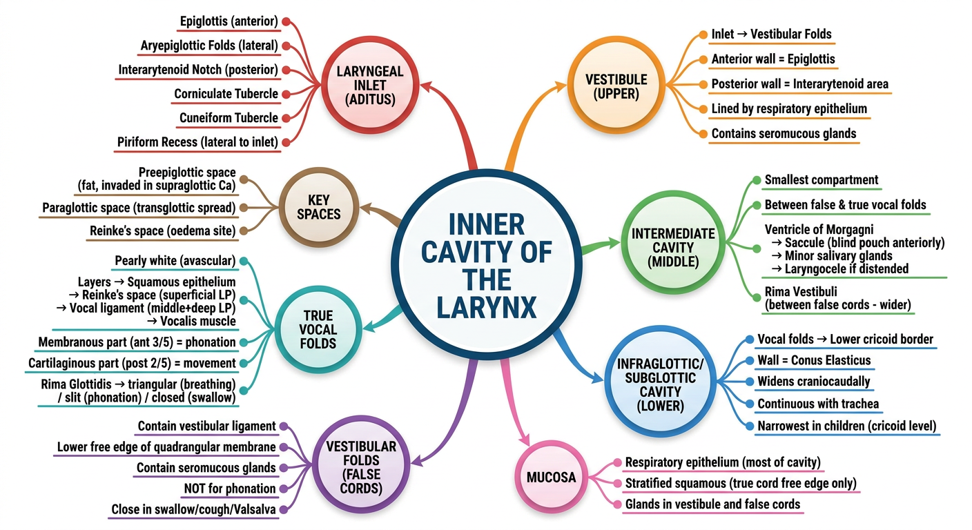
| Branch | Color | Content |
|---|---|---|
| Laryngeal Inlet | Red | Epiglottis, aryepiglottic folds, interarytenoid notch, tubercles, piriform recess |
| Vestibule | Orange | Boundaries, epithelium, glands |
| Intermediate Cavity | Green | Ventricle of Morgagni, saccule, laryngocele, rima vestibuli |
| Infraglottic Cavity | Blue | Conus elasticus, tracheal continuity, narrowest in children |
| False Cords | Purple | Vestibular ligament, quadrangular membrane, no phonation |
| True Vocal Cords | Teal | Layers (Reinke's space, vocal ligament, vocalis), rima glottidis shapes |
| Key Spaces | Brown | Preepiglottic, paraglottic, Reinke's space |
| Mucosa | Pink | Respiratory vs squamous epithelium distribution |