
What is the treatment modality for hyperpigmented scars on extremeties
hyperpigmented scars treatment
hyperpigmented scar treatment laser skin
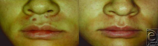
This comparison clinical photograph demonstrates a side-by-side progression of a post-burn hypertrophic scar on the philtrum and upper lip before and after Nd:YAG laser treatment. In the 'before' image (left), multiple raised, erythematous (red) nodules and plaque-like lesions are visible between the nasal base and the vermilion border. The scar tissue exhibits uneven texture, significant elevation, and distinct hyperpigmented and erythematous coloration compared to the surrounding skin. In the 'after' image (right), following five laser treatments, there is a marked clinical improvement. The previously elevated lesions show significant flattening (reduction in hypertrophy), and the associated erythema has notably diminished, leading to a more uniform skin tone. The overall texture of the perioral area appears smoother, with the scar tissue becoming less distinct and blending more naturally with the adjacent healthy tissue. This clinical case illustrates the efficacy of laser therapy in reducing the vascularity and volume of hypertrophic scarring in high-visibility facial regions.
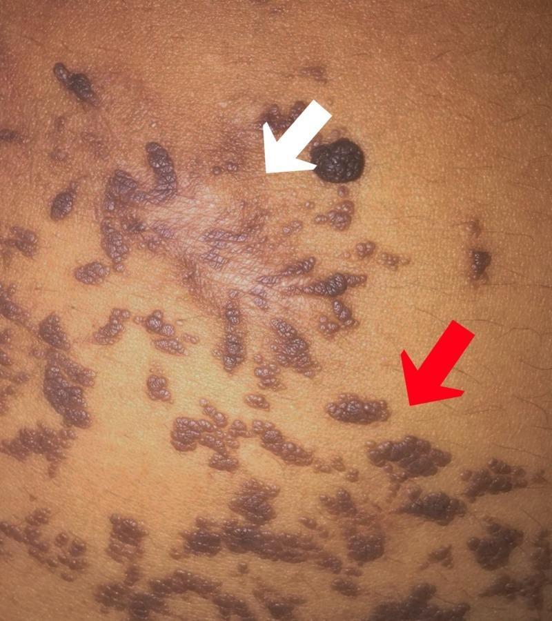
Clinical photograph of the abdominal skin in a patient with an epidermal verrucous nevus, illustrating a side-by-side comparison of untreated lesions and post-treatment sequelae. The untreated lesions (marked by a red arrow) consist of multiple, grouped, hyperpigmented dark-brown to black plaques with a distinct verrucous or 'wart-like' texture. They are irregularly shaped and distributed across the skin surface. The area marked by a white arrow demonstrates the results of previous electrofulguration and CO2 laser treatment, characterized by a flattened, hypopigmented to slightly erythematous scar with an altered skin texture and fine wrinkling. Within the treated field, a single raised, dark verrucous nodule remains. This image is used in dermatology to demonstrate the typical morphology of verrucous nevi and the potentially unaesthetic scarring associated with invasive therapeutic modalities like laser or electrofulguration on extensive lesions.
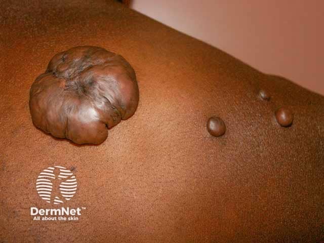
This dermatologic image presents a clinical macro photograph of a keloid scar on the upper back. Imaging modality: Clinical photography, high‑resolution color capture, macroscopic view, posterior dorsal aspect of the thoracic region. Anatomical localization: integumentary system, skin on the upper back (posterior trunk; scapular/toracic area) showing a markedly raised fibroproliferative lesion extending beyond the original wound margin. Visual features: large, rounded, lobulated nodule with a broad base, dense fibrous tissue, smooth to slightly shiny surface, hyperpigmented brown tone, and adjacent smaller nodules along the scar band. The lesion demonstrates classic keloid morphology with prominent induration and subtle overlying epidermal hyperpigmentation; minimal surrounding erythema; no obvious ulceration. Pathological implications (inferred): excessive type III collagen deposition and disorganized dermal matrix from fibroblast proliferation, resulting in an exuberant scar extending into surrounding dermis. Diagnostic significance: characteristic of keloid disease; helps differentiate from hypertrophic scar, scar tissue from minor trauma; influences treatment planning (steroid injections, silicone, radiotherapy, laser, surgical considerations). Clinical use cases: dermatology consults, scar management planning, educational material on abnormal scar formation, research datasets for scar biology, machine learning image annotation. Keywords: keloid, scar, dermis, fibroproliferation, upper back, thoracic skin, cosmetic concern, recurrence risk, treatment planning.
Treatment Modalities for Hyperpigmented Scars on Extremities
Step 1: Identify the Type of Hyperpigmentation
- Wood's light examination is the first step - epidermal melanin enhances under Wood's light; dermal melanin (gray-blue hue) does not. This guides treatment selection.
- Epidermal PIH responds better to topical agents; dermal melanosis is more refractory and may need laser.
A. Topical Therapies (First-Line)
| Agent | Mechanism | Notes |
|---|---|---|
| Hydroquinone (2-4%) | Inhibits tyrosinase, blocks melanin synthesis | First-line for epidermal PIH; enhances with Wood's light |
| Tretinoin (topical retinoid) | Accelerates epidermal turnover, disperses melanosomes | Enhances effect of hydroquinone; use with sun protection |
| Triple combination cream | Hydroquinone + tretinoin + mild steroid (e.g., fluocinolone) | Synergistic depigmentation |
| Azelaic acid | Selective cytotoxicity to hyperactive melanocytes | Useful in darker skin types |
| Kojic acid, Niacinamide, Vitamin C | Various anti-melanogenic mechanisms | Adjuncts, used in combination |
B. Chemical Peels
- Used with caution in darker skin types (Fitzpatrick IV-VI) as they can paradoxically worsen pigmentation (PIH from the peel itself).
- Superficial peels: glycolic acid, salicylic acid - suitable for extremity scars with epidermal pigmentation.
- Must be combined with pre-treatment hydroquinone for 4-6 weeks in pigmented skin.
C. Laser Therapies (For Refractory Cases)
1. Q-Switched Lasers (First Choice for Refractory Pigmentation)
- Q-switched Nd:YAG (1064 nm) - preferred for darker skin types (IV-VI); targets dermal and epidermal melanin.
- Q-switched Ruby (694 nm) and Alexandrite (755 nm) - effective but higher PIH risk in darker skin.
- Mechanism: Selective photothermolysis targeting melanosomes.
2. Non-Ablative Fractional Lasers
- 1550 nm erbium-doped fractionated laser (e.g., Fraxel) - treats both pigmentation and textural irregularity.
- Safer in darker skin phototypes compared to ablative options.
- Lower densities preferred to minimize PIH risk.
3. Ablative Fractional CO2 Laser
- More powerful remodeling; reserved for hypertrophic/contracted scars.
- Can also address dyspigmentation but higher PIH risk on extremities and in darker phototypes.
- Typically 3+ sessions at 4-6 week intervals.
4. Pulsed Dye Laser (PDL, 585/595 nm)
- Targets erythematous component; also useful when scar has both erythema and hyperpigmentation.
Key principle from Dermatology 5e (Elsevier): "Scar hyperpigmentation is most commonly due to deposition of melanin... Hyperpigmentation refractory to traditional topical therapy may be treated with Q-switched lasers (694, 755, and 1064 nm) or non-ablative fractional resurfacing. Because darker skin phototypes are more susceptible to PIH with high-density non-ablative fractional resurfacing, lower densities are preferable." (Dermatology 2-Volume Set 5e, p. 2861)
D. Fat Grafting
E. Combination / Multimodal Approaches
F. Special Considerations for Extremities
- Extremities (especially lower legs) heal more slowly and have poorer vascularity - higher risk of hypertrophic scarring and prolonged PIH.
- Pre-treatment sun avoidance and sunscreen for several weeks prior to any laser/peel is mandatory.
- Compression garments are a useful adjunct for hypertrophic/keloid scars with pigmentation on limbs.
- Silicone gel sheets reduce hypertrophic scar formation and associated pigmentation.
- In darker skin types (common in South Asian, African populations), start conservatively: topical therapy first, then Q-switched Nd:YAG if needed.
Treatment Algorithm Summary
Hyperpigmented Scar on Extremity
        |
        ├─ Epidermal pigmentation (Wood's light enhances)
        │        └─ Topical: Hydroquinone ± Tretinoin ± Sunscreen
        │                 If refractory → Superficial chemical peel or Q-switched laser
        │
        └─ Dermal/mixed pigmentation (gray-blue hue, does not enhance)
                 └─ Q-switched Nd:YAG (1064 nm) [especially darker skin]
                          + Non-ablative fractional laser (1550 nm)
                          ± Topical agents (adjunct)
                          ± Fat grafting (for extensive burn scars)
- Dermatology 2-Volume Set 5e, "Treatment of Scars," p. 2861
- Andrews' Diseases of the Skin, "Postinflammatory Hyperpigmentation," p. 992
- Fat grafting meta-analysis: Alessandri Bonetti et al., 2024 (PMID 38286897)
- Scar-modulating combination review: Bernabe et al., 2024 (PMID 37979279)
Can you suggest me few creams containing triple combined therapy Creams commonly available as brand names but fir extremeties
triple combination cream hydroquinone tretinoin fluocinolone brand names available India hyperpigmentation
triple combination cream brands India hydroquinone tretinoin fluocinolone alternatives Melamet Cosmelite Pigmanorm extremities body use
Triple Combination Creams for Hyperpigmented Scars
- Hydroquinone - inhibits tyrosinase, reduces melanin synthesis
- Tretinoin - accelerates epidermal turnover, enhances hydroquinone penetration, protects HQ from oxidation
- Fluocinolone acetonide (mild Class V steroid) - reduces inflammation caused by the other two agents, also has mild depigmenting effect
Brand Names Available
| Brand Name | Composition | Manufacturer | Region |
|---|---|---|---|
| Tri-Luma | HQ 4% + Tretinoin 0.05% + Fluocinolone 0.01% | Galderma | India, USA, Global (gold standard, FDA-approved) |
| Triderma | HQ 4% + Tretinoin 0.05% + Fluocinolone 0.01% | Generic | USA |
| Melamet | HQ 2% + Tretinoin 0.025% + Mometasone 0.1% | Menarini India | India (commonly available, uses mometasone instead of fluocinolone) |
| Cosmelite | HQ 2% + Tretinoin 0.025% + Fluocinolone 0.01% | Elder Pharma | India |
| Pigmanorm | HQ 5% + Tretinoin 0.1% + Hydrocortisone 1% | Almirall | Europe/Middle East |
| Melanocyl | HQ + Tretinoin + Betamethasone | Various Indian pharma | India |
| Lustra-AF | HQ 4% + Retinol + Glycolic acid | Taro Pharma | USA (no steroid, different variant) |
Note: Melamet is very widely used in India and is more accessible than Tri-Luma. It substitutes mometasone (a medium-potency steroid) for fluocinolone.
Important Cautions for Use on Extremities Specifically
-
Steroid absorption risk - Extremity skin (especially flexures like the antecubital fossa, popliteal fossa) has moderate absorption. Avoid occlusion. Do NOT use on large body surface areas continuously.
-
Duration limit - These creams should NOT be used for more than 8-12 weeks continuously on body areas. Prolonged use on extremities risks:
- Skin atrophy
- Striae
- Telangiectasia
- Paradoxical rebound hyperpigmentation on stopping
-
Tretinoin irritation - Extremity skin, especially on the legs, can be drier and more prone to retinoid dermatitis. Start with every other night application, then build up.
-
Sun protection - Tretinoin and hydroquinone both cause photosensitivity. On arms, this is a real risk. Apply only at night; use SPF 30+ in the daytime.
-
Avoid on knees/elbows - These are naturally darker due to friction and thickened skin; triple combination can be used but results are slower. Consider combining with a gentle exfoliant (AHA/urea-based moisturizer).
Practical Tip for Extremities
- Use Melamet (lower steroid potency with mometasone) as it is gentler than the higher-HQ Tri-Luma
- Apply at night only, thin layer, no occlusion
- Combine with a sunscreen SPF 50 during the day
- Limit to 6-8 weeks, then take a break and reassess
- Consider rotating with plain hydroquinone 2% cream to avoid steroid-related side effects on longer treatment courses