
Alopecia areata clear pathophysiology with diagram and flowchart
Reading File
Now let me search the medical library and web in parallel for alopecia areata pathophysiology.
Finding Sources
Searching the Web
alopecia areata pathophysiology immune privilege collapse mechanism
Searching PubMed
"alopecia areata"[MeSH Terms] AND pathophysiology
Now let me read the main textbook sections in parallel:
Reading File
Reading File
I found the diagram image URL. Let me fetch it and also read more of the textbook content:
Loading Image
Reading File
Reading File
I now have comprehensive textbook material and the official diagram. Let me now compose the full answer with the flowchart and diagram.
Finding Sources
Reading File
I now have all the information needed for a comprehensive answer. Let me compose the full pathophysiology explanation with the textbook diagram and a detailed flowchart.
Alopecia Areata - Pathophysiology
Overview
Alopecia areata (AA) is a non-scarring autoimmune alopecia driven by collapse of hair follicle immune privilege, T-cell-mediated cytotoxic attack on anagen hair bulbs, and activation of the JAK-STAT signaling cascade. Follicular stem cells in the bulge are spared, which is why the disease is reversible.
Textbook Diagram (Fitzpatrick's Dermatology)
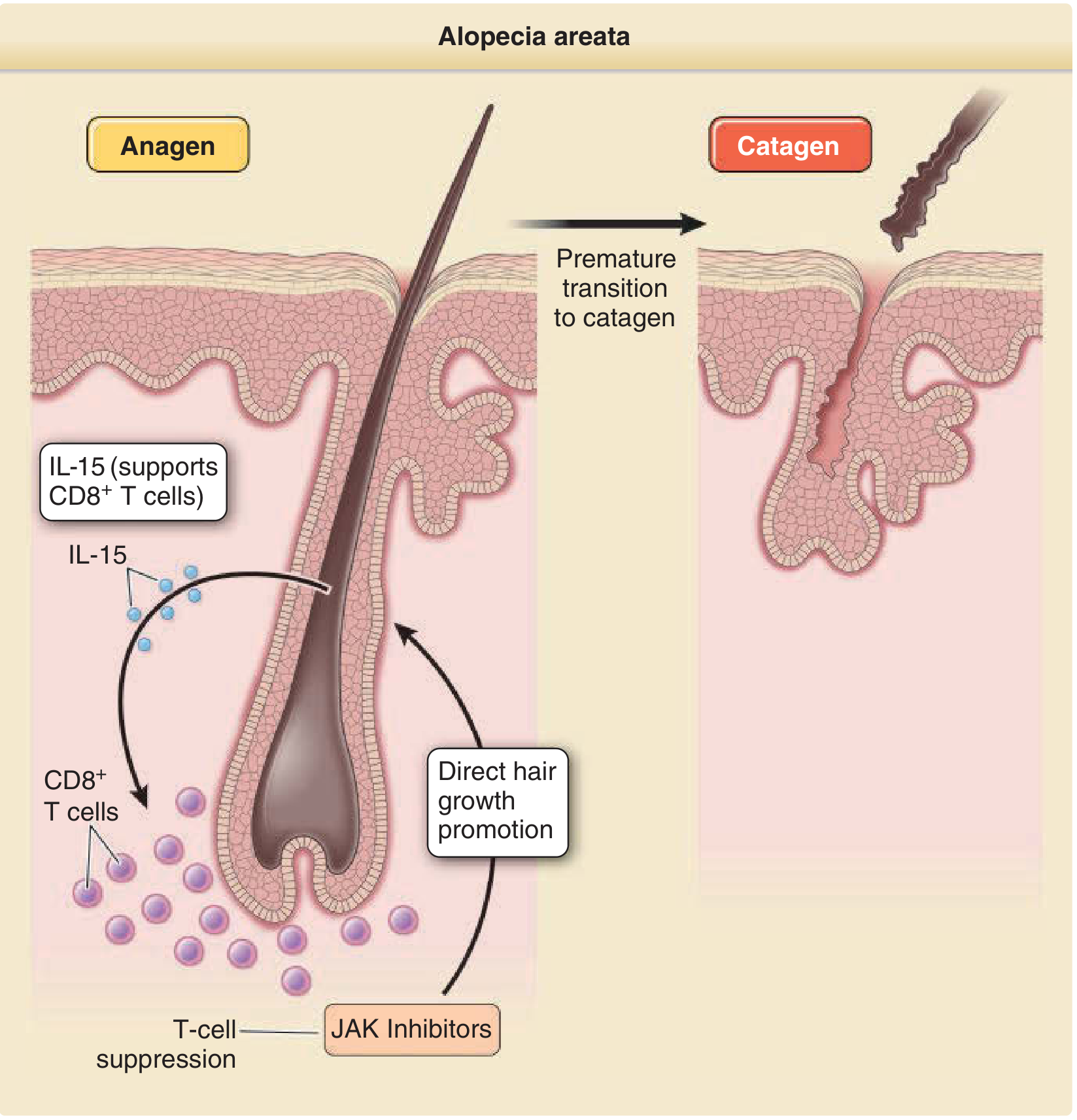
Figure: IL-15 activates CD8+ T cells that infiltrate the hair bulb during anagen, forcing premature transition to catagen and hair loss. JAK inhibitors block this process at multiple levels. (Fitzpatrick's Dermatology, Fig. 13-4)
Pathophysiology - Step by Step
1. Genetic Susceptibility
| Gene/Locus | Role |
|---|---|
| HLA-DQB1*03, HLA-DR4/DR11/DQ7 | Antigen presentation to autoreactive T cells |
| ULBP gene cluster (chr 6q25) | Encodes NKG2D receptor ligands - marks follicle for attack |
| CTLA-4, IL-2/IL-21, IL-2RA | T-cell regulation susceptibility loci |
| PTPN22 | Encodes phosphatase that suppresses T-cell proliferation (loss = overactive T cells) |
2. Normal Hair Follicle Immune Privilege (What Gets Broken)
The anagen hair bulb normally maintains local immune privilege through:
- Markedly reduced MHC Class I and Class II expression on follicular keratinocytes
- Local production of immunosuppressive cytokines and neuropeptides
- Expression of "no danger" signals (e.g., CD200)
- Perifollicular regulatory T cells (Tregs) and mast cells
- Low or absent NKG2D ligand expression
This immune privilege prevents autoreactive CD8+ cytotoxic T cells from recognizing follicular self-antigens - particularly melanogenesis-associated antigens in the hair matrix.
3. Triggers - Immune Privilege Collapse
Environmental triggers (viral infection, stress, medications, vaccines) - combined with the genetic background above - disrupt these protective mechanisms:
- MHC Class I upregulation on follicular keratinocytes
- NKG2D ligands (ULBP3/MICA) become expressed on the follicle surface
- IFN-γ release further amplifies MHC Class I expression
- Immunosuppressive milieu is lost
4. T-Cell Recruitment and Attack
Once immune privilege collapses:
- Autoreactive CD8+ NKG2D+ cytotoxic T cells recognize follicular autoantigens
- CD4+ Th1 helper T cells aggravate the response (peribulbar infiltrate)
- The resulting perifolliular lymphocytic infiltrate has the classic "swarm of bees" pattern on histology
- NKG2D receptor on CD8+ T cells binds ULBP ligands on the follicle surface - a direct cytotoxic trigger
5. Cytokine Amplification Loop (The Core Engine)
This is the self-sustaining positive feedback loop:
- Stressed/attacked follicle keratinocytes upregulate IL-15
- IL-15 activates and sustains CD8+ NKG2D+ T cells
- CD8+ T cells secrete IFN-γ and IL-2
- IFN-γ further upregulates MHC Class I and NKG2D ligands on follicles (more antigen presentation)
- IFN-γ also stimulates more IL-15 production by keratinocytes
- IL-15 and IFN-γ both signal through the JAK-STAT pathway (JAK1/JAK3 - STAT1/STAT3/STAT5)
- This loop self-perpetuates and drives ongoing follicular damage
6. Hair Follicle Damage - Anagen to Catagen Transition
- CD8+ T cell-mediated cytotoxicity + cytokine environment causes apoptosis of hair matrix cells and melanocytes
- This triggers premature anagen-to-catagen transition
- The hair shaft tapers proximally (dystrophic anagen hair) and fractures at the surface - producing the classic "exclamation point hair"
- The lower follicle involutes; the bulb involutes upward in the dermis
- Repeated cycles cause follicular miniaturization
- Follicular stem cells in the bulge region are NOT targeted - hence no scarring and potential for regrowth
7. Why White/Gray Hair is Spared
Follicular melanocytes are the primary autoantigen target. White/gray hairs lack active melanocytes - therefore the immune attack finds no target and these hairs are largely spared.
Pathophysiology Flowchart
GENETIC SUSCEPTIBILITY
(HLA-DQB1*03, ULBP genes, CTLA-4, PTPN22 mutations)
         │
         ▼
ENVIRONMENTAL TRIGGER
(Infection / Stress / Drug / Unknown)
         │
         ▼
COLLAPSE OF HAIR FOLLICLE IMMUNE PRIVILEGE
• MHC Class I ↑ on follicular keratinocytes
• NKG2D ligands (ULBP3, MICA) expressed
• CD200 ↓ / Immunosuppressive signals lost
• Treg dysfunction
         │
         ▼
FOLLICULAR AUTOANTIGEN PRESENTATION
(Melanogenesis-related peptides displayed on MHC I)
         │
         ▼
AUTOREACTIVE T-CELL RECRUITMENT
• CD8+ NKG2D+ cytotoxic T cells infiltrate peribulbar region
• CD4+ Th1 cells support & amplify (peribulbar)
• Classic "swarm of bees" lymphocytic infiltrate
         │
         ▼
CYTOKINE AMPLIFICATION LOOP ←──────────────────┐
• Keratinocytes release IL-15                           │
• IL-15 → JAK1/JAK3-STAT5 → activates CD8+ T cells  │
• CD8+ T cells secrete IFN-γ + IL-2                   │
• IFN-γ → JAK1/JAK2-STAT1 → ↑ MHC I, ↑ NKG2D Lig. │
• IFN-γ → more IL-15 by keratinocytes ─────────────────┘
         │
         ▼
HAIR MATRIX CELL & MELANOCYTE APOPTOSIS
(CD8+ cytotoxicity + cytokine-induced cell death)
         │
         ▼
PREMATURE ANAGEN → CATAGEN TRANSITION
• Dystrophic anagen hair → tapered fracture
• "Exclamation point hairs" at patch margin
• Follicle involutes; bulb rises in dermis
         │
         ▼
HAIR SHAFT FRACTURE & LOSS
(Non-scarring alopecia - bulge stem cells intact)
         │
         ▼
FOLLICULAR MINIATURIZATION (if chronic)
         │
    ┌────┴────┐
    ▼         ▼
REGROWTH   PROGRESSION
(Treg       (→ Alopecia Totalis
recovery /   → Alopecia Universalis)
treatment)


━━━━━━━━━━━━━━━━━━━━━━━━━━━━━━━━━━━━━
TREATMENT TARGET: JAK-STAT PATHWAY
━━━━━━━━━━━━━━━━━━━━━━━━━━━━━━━━━━━━━
JAK Inhibitors (baricitinib, tofacitinib, ruxolitinib)
  → Block IL-15 and IFN-γ signaling
  → Suppress CD8+ T cell activation/proliferation
  → Directly promote hair follicle growth
  → Restore immune privilege (partial)
Key Players Summary Table
| Component | Role in AA |
|---|---|
| CD8+ NKG2D+ T cells | Primary effectors - directly kill hair matrix cells |
| CD4+ Th1 cells | Amplify/sustain the immune attack (peribulbar) |
| Regulatory T cells (Tregs) | Protective - deficiency promotes AA |
| IL-15 | Activates and sustains CD8+ T cells; upregulated in follicles |
| IFN-γ | Upregulates MHC I, amplifies IL-15; Th1 signature cytokine |
| IL-2 | Supports T-cell proliferation |
| TNF-α | Early mediator of hair matrix damage |
| NKG2D receptor | On CD8+ T cells; binds ULBP/MICA on follicle - direct cytotoxic trigger |
| MHC Class I | Absent in normal follicle (immune privilege); upregulated in AA |
| JAK1/JAK2/JAK3 | Kinases mediating IL-15 and IFN-γ signaling |
| STAT1/STAT3/STAT5 | Downstream transcription factors; promote T-cell survival/proliferation |
Key Histological Features
- "Swarm of bees" peribulbar lymphocytic infiltrate (predominantly CD8+ intrafollicular, CD4+ perifollicular)
- Dystrophic anagen hairs, follicular miniaturization
- Melanin pigment in fibrous tract remnants (from destroyed melanocytes)
- Fibrous streamer (stelae) beneath miniaturized bulbs
- No scarring of the follicular unit (bulge intact)
Sources: Fitzpatrick's Dermatology (8e), p. 229-230 | Andrews' Diseases of the Skin, p. 868-869 | Dermatology 2-Volume Set (5e), Pathogenesis section
This is a shared conversation. Sign in to Orris to start your own chat.