
Hookworm -etiology, pathogenesis, lifecycle, diagnosis, treatment, important points
Reading File
Now let me search the medical library for hookworm content across multiple angles simultaneously.
Finding Sources
Finding Sources
Excellent - multiple rich sources found. Let me read the most comprehensive ones in parallel.
Reading File
Reading File
Reading File
Reading File
Excellent content obtained. Let me also read the Park's textbook entry and Red Book, and fetch the lifecycle image.
Reading File
Reading File
Loading Image
Loading Image
I now have comprehensive, multi-source data. Let me compile the complete answer.
Hookworm - Complete Overview
1. Etiology (Causative Agents)
Primary human hookworms:
| Species | Attachment Organ | Geographic Predominance | Eggs/day (female) |
|---|---|---|---|
| Necator americanus | Cutting plates | Tropics/subtropics worldwide; Americas, sub-Saharan Africa, Asia | 5,000-10,000 |
| Ancylostoma duodenale | Biting teeth (2 pairs) | Mediterranean, Middle East, North India, China, Japan | 10,000-30,000 |
| Ancylostoma ceylanicum | - | Southeast Asia (zoonotic - cats/dogs) | - |
Zoonotic species (do NOT complete life cycle in humans):
- A. caninum (dog hookworm) - causes eosinophilic enteritis (Australia) and cutaneous larva migrans
- A. braziliense (cat hookworm) - classic cause of cutaneous larva migrans
- Bunostomum phlebotomum, Uncinaria stenocephala - occasional CLM
Adults measure 7-13 mm, with a dorsally curved anterior end ("hook" shape). Adults reside in the proximal small intestine (mainly jejunum).
2. Lifecycle
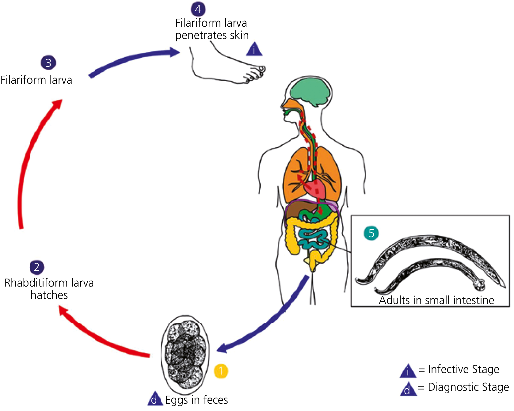
Step-by-step:
- Egg stage: Adult female worms in the small intestine produce eggs that are passed out in feces (diagnostic stage - eggs are 40x60 μm, oval, thin-shelled)
- Rhabditiform larva (L1/L2): In warm, moist soil (23-33°C), eggs hatch within 1-2 days into non-infective rhabditiform larvae, which undergo two molts over 5-10 days
- Filariform larva (L3 - infective stage): Third-stage filariform larvae develop, migrate upward on grass blades, can survive in soil for up to 1 month
- Skin penetration: Infective filariform larvae penetrate unprotected skin (usually feet/hands), triggering "ground itch"
- A. duodenale can also be transmitted orally (ingested larvae survive and develop)
- Migration: Larvae enter venous circulation → pulmonary vasculature → penetrate alveoli → ascend bronchial tree to larynx → swallowed
- Intestinal development: In the small intestine, larvae molt twice more and mature into sexually mature adults
- Prepatent period: 6-8 weeks from skin penetration to eggs in feces (can be longer with Ancylostoma spp.)
Adult worm survival: A. duodenale ~1-14 years; N. americanus ~2-5 years
3. Pathogenesis
3a. Blood Loss - The Core Mechanism
- Adult worms attach to intestinal villi, aspirate a plug of tissue, secrete anticoagulant and antiplatelet enzymes, and ingest blood
- A. duodenale extracts ~0.2 mL blood/worm/day
- N. americanus extracts ~0.03 mL blood/worm/day
- Worms move frequently, leaving bleeding points at old attachment sites
- Accumulated blood loss in heavy/prolonged infection leads to iron-deficiency anemia and hypoalbuminemia
3b. Three Phases of Disease
Phase 1 - Skin Entry ("Ground Itch"):
- Pruritic erythematous maculopapular rash at site of larval penetration
- More common with N. americanus; typically between toes or on ankles
- Resolves spontaneously within days
Phase 2 - Pulmonary Migration (Larval Phase):
- Dry cough, sore throat, wheezing, low-grade fever
- Less frequent and less severe than in ascariasis
- Transient eosinophilia
- Loeffler-like syndrome is uncommon
Phase 3 - Intestinal/Chronic Phase:
- Epigastric pain, abdominal bloating, abnormal peristalsis
- Chronic iron-deficiency anemia (hypochromic microcytic) - severity depends on worm burden, dietary iron intake, concurrent blood loss
- Hypoalbuminemia (protein-losing enteropathy) in heavy infections
- Fatigue, weakness, exertional dyspnea
- In children: growth retardation, cardiac failure (with severe anemia), kwashiorkor-like picture
- In pregnant women: increased maternal/fetal morbidity (~44 million pregnant women infected globally)
3c. Immune Response
- Elicits humoral antibody response and immediate hypersensitivity
- Peripheral eosinophilia and gut eosinophilia
- Eosinophils may play a role in worm destruction; however, evidence that immune responses significantly limit infection is lacking
4. Epidemiology
- Global burden: Over 400-700 million people infected worldwide (figures vary by source)
- Estimated 50,000-60,000 deaths/year; collectively extract over 1 million liters of blood/day from infected individuals
- Distributed between 45°N and 30°S latitude; greatest burden in sub-Saharan Africa, Asia, and rural Americas
- Transmission requires: feces-contaminated shady/moist soil + adequate rainfall + high temperature (23-33°C) + direct skin contact
- Risk factors: poverty, no footwear, use of human excrement as fertilizer, poor sanitation
- Prevalence increases through childhood, plateauing by age ~10 years; intensity peaks by age ~20 and may rise again in elderly
- Male sex more commonly infected; women and children more vulnerable to anemia/sequelae
5. Diagnosis
| Method | Details |
|---|---|
| Stool microscopy (gold standard) | Characteristic 40 x 60 μm oval, thin-shelled eggs in feces; stool concentration techniques (e.g., formalin-ether) needed for light infections |
| Fresh vs. old stool | Old (not fresh) stool may show hatched rhabditiform larvae - must be differentiated from Strongyloides stercoralis larvae |
| Species differentiation | Eggs of all three species are indistinguishable by light microscopy |
| PCR | Species-specific; high sensitivity/specificity but remains a research tool, not commercially available |
| CBC | Hypochromic microcytic anemia; eosinophilia in larval migration phase |
| Biochemistry | Hypoalbuminemia in heavy infections |
| Stool egg count | Quantitative Kato-Katz technique - estimates worm burden (correlates with intensity of disease) |
6. Treatment
| Drug | Dose | Notes |
|---|---|---|
| Albendazole | 400 mg once (single dose) OR 400 mg/day x 3 days | 3-day course preferred for N. americanus (lower single-dose efficacy) |
| Mebendazole | 500 mg single dose OR 100 mg BD x 3 days | Effective for both species |
| Pyrantel pamoate | 11 mg/kg (max 1g) x 3 days | Second-line; less effective for N. americanus |
| Iron supplementation | Ferrous sulfate | Concurrent treatment of iron-deficiency anemia |
- For cutaneous larva migrans (zoonotic hookworm): albendazole 400 mg/day x 3-5 days or ivermectin 200 μg/kg x 1-2 days
- Albendazole single dose has lower cure rates for N. americanus than 3-day regimens
- Treat symptomatic disease even when worm burden is light in vulnerable populations (pregnant women, children)
7. Important Points (High-Yield Summary)
-
"Hook" shape of worm = dorsally curved anterior end; name derives from this
-
Two cutting mechanisms: A. duodenale uses biting teeth (2 pairs), N. americanus uses cutting plates - both enable mucosal attachment and blood ingestion
-
Infective stage = filariform (L3) larva - the diagnostic stage in stool = eggs
-
A. duodenale is unique: larvae can also infect orally (swallowed, survive, develop directly)
-
Ground itch = pruritic rash at skin entry site, typically between toes, more common with N. americanus
-
A. duodenale extracts ~7x more blood per worm than N. americanus (0.2 vs 0.03 mL/day) - more anemia-inducing
-
Eggs in old/stale stool may hatch - must distinguish rhabditiform larvae from Strongyloides (S. stercoralis has a longer buccal cavity)
-
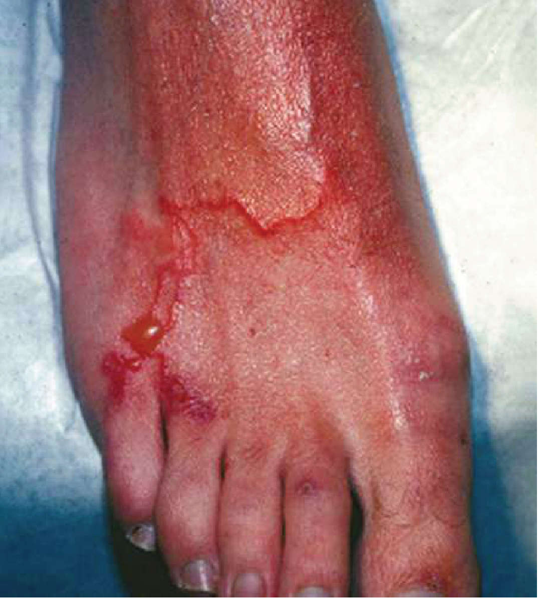
Cutaneous larva migrans = serpiginous, creeping track in skin from dog/cat hookworm larvae; a self-limited condition
-
Most infections are asymptomatic - disease develops only with heavy worm burden + prolonged infection + poor dietary iron
-
Pulmonary symptoms less severe than ascariasis - remember this distinction for exams
-
PCR for species differentiation - eggs are morphologically identical between species
-
WHO mass drug administration (MDA) targets school-age children in endemic areas using albendazole or mebendazole
-
Prevention: wearing footwear, sanitary disposal of feces, avoiding use of human feces as fertilizer
-
Hyperinfection does NOT occur with hookworm (unlike Strongyloides stercoralis) - key distinguishing feature in immunocompromised patients
-
Wakana disease (A. duodenale oral ingestion): nausea, vomiting, pharyngeal irritation, cough - follows oral ingestion of larvae
Sources: Harrison's Principles of Internal Medicine 22E (2025) - Goldman-Cecil Medicine - Sherris & Ryan's Medical Microbiology 8E - Yamada's Textbook of Gastroenterology 7E - Park's Textbook of Preventive and Social Medicine - Sleisenger & Fordtran's GI and Liver Disease
This is a shared conversation. Sign in to Orris to start your own chat.