
Critically discuss the role of Point-of-Care Ultrasound (eFAST) in the management of blunt abdominal trauma with hemodynamic instability.
eFAST ultrasound blunt abdominal trauma hemodynamic instability
FAST ultrasound trauma accuracy sensitivity
PMID: 38185679
eFAST extended FAST ultrasound trauma free fluid views
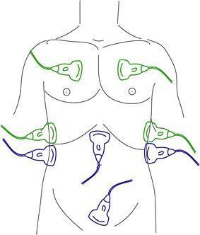
An anatomical diagram illustrating the transducer placement for the FAST (Focused Assessment with Sonography in Trauma) and eFAST (extended FAST) examinations on a human torso. The diagram uses color-coded ultrasound probes to distinguish between protocols. Blue probes indicate the standard FAST positions: the subxiphoid view (pericardial assessment), the right upper quadrant (Morison's pouch/hepatorenal space), the left upper quadrant (splenorenal space), and the suprapubic view (pelvic/rectovesical or recto-uterine pouch assessment) to detect free intraperitoneal fluid. Green probes represent the eFAST extensions: bilateral anterior thoracic views at the second to fourth intercostal spaces and mid-axillary views to assess for pneumothorax and hemothorax. This illustration is an educational tool for emergency medicine, trauma surgery, and radiology, demonstrating the systematic sonographic approach used to identify life-threatening hemorrhage or air in the thoracic and abdominal cavities during the primary survey of trauma patients.
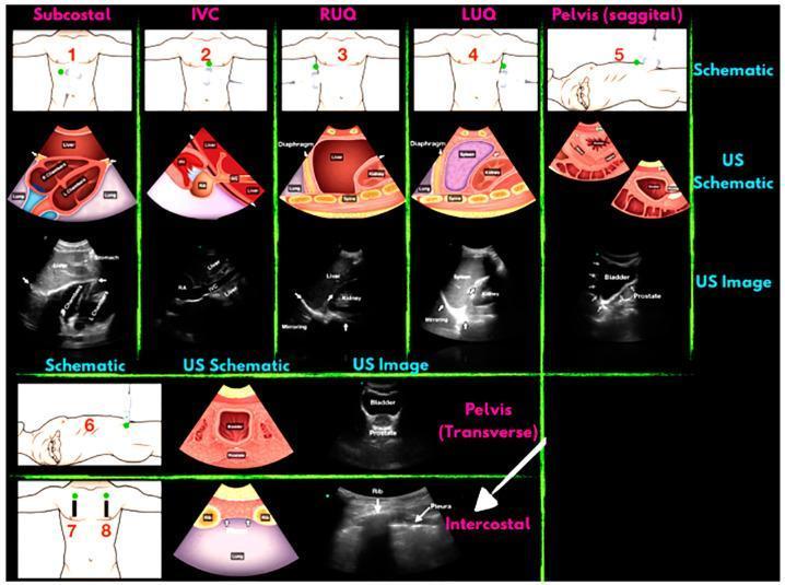
A comprehensive medical educational schematic illustrating the Focused Assessment with Sonography for Trauma (FAST) and Extended FAST (eFAST) examination protocols. The infographic is organized into a grid mapping specific anatomical sites to their corresponding ultrasound (US) views. For each location—Subcostal, IVC, Right Upper Quadrant (RUQ), Left Upper Quadrant (LUQ), Pelvis (Sagittal and Transverse), and Intercostal—the diagram provides three levels of visualization: 1) a torso schematic showing probe placement marked by green dots; 2) an anatomical US schematic cross-section detailing internal structures; and 3) a representative grayscale clinical ultrasound image. Key structures identified include the four heart chambers (Subcostal), the Inferior Vena Cava (IVC), the liver and kidney interface (RUQ), the spleen and kidney interface (LUQ), and the bladder and prostate (Pelvic views). The intercostal view focuses on identifying the pleura and rib for pneumothorax assessment. This educational resource is designed for emergency medicine training to assist in the rapid detection of free fluid (hemoperitoneum, hemopericardium) and pneumothorax in trauma patients.
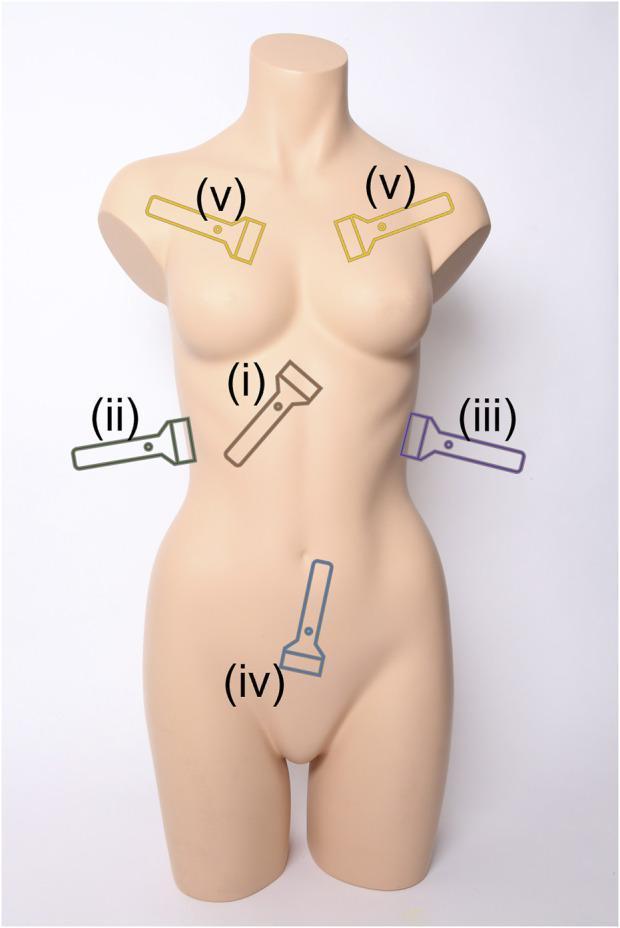
This clinical diagram illustrates the standard anatomical scan points for an extended Focused Assessment with Sonography for Trauma (eFAST) examination using a female torso phantom. The visual provides a topographic guide for probe placement during emergency triage to detect free fluid or air. Five primary views are labeled with color-coded ultrasound transducer icons: (i) Subxiphoid view (brown) positioned centrally below the sternum to evaluate for pericardial effusion; (ii) Right Upper Quadrant (RUQ) view (green) located at the mid-axillary line to assess Morrison’s pouch and the hepatorenal space; (iii) Left Upper Quadrant (LUQ) view (purple) at the posterior-axillary line to evaluate the splenorenal recess; (iv) Pelvic view (blue) positioned over the suprapubic region to assess the rectovesical or rectouterine pouch; and (v) Intercostal views (yellow) placed bilaterally at the upper anterior chest to assess for pneumothorax by identifying lung sliding. The diagram serves as an educational tool for training in rapid ultrasonographic assessment for internal hemorrhage and thoracic injury.
"point of care ultrasound" trauma hemodynamic instability management
Point-of-Care Ultrasound (eFAST) in Blunt Abdominal Trauma with Hemodynamic Instability: A Critical Discussion
1. Background and Conceptual Basis
2. The eFAST Protocol: Views and Findings
| Window | Target | Positive Finding |
|---|---|---|
| Subxiphoid/subcostal | Pericardium | Pericardial effusion (tamponade) |
| Right upper quadrant (Morrison's pouch) | Hepatorenal space | Anechoic free fluid |
| Left upper quadrant (splenorenal recess) | Splenorenal space | Anechoic free fluid |
| Suprapubic (sagittal + transverse) | Rectovesical/rectouterine pouch | Anechoic free fluid |
| Bilateral anterior thorax | Pleura/lung | Absent lung sliding, A-lines, lung point sign (pneumothorax) |
| Bilateral mid-axillary (thoracic) | Pleural space | Anechoic fluid above diaphragm (hemothorax) |
3. Diagnostic Performance: What the Evidence Actually Shows
3.1 Sensitivity and Specificity
- Abdominal trauma: sensitivity 68%, specificity 95%
- Thoracic trauma: sensitivity 96%, specificity 99%
3.2 The Hemodynamic Instability Context
-
In a hemodynamically unstable patient with a POSITIVE eFAST: the finding of free intraperitoneal fluid in the context of shock identifies the abdomen as the likely source of hemorrhage. This is an immediate indication for emergent laparotomy or damage control surgery. Physical examination alone identifies intra-abdominal hemorrhage only approximately half the time; eFAST raises this to over 90% sensitivity when the pre-test probability is high and the volume of free fluid is significant. - Fischer's Mastery of Surgery, 8e
-
In a hemodynamically unstable patient with a NEGATIVE eFAST: the sensitivity limitation becomes the clinical problem. A negative eFAST in this context should not reassure the clinician. Retroperitoneal injury, hollow viscus injury, solid organ injuries without significant free fluid (e.g., subcapsular hematoma), and small-volume hemorrhage are all frequently missed. The clinician must pursue other diagnostic or therapeutic strategies.
4. Key Clinical Role: Triage and Decision-Making
Hemodynamically UNSTABLE blunt trauma
        ↓
    eFAST
   /       \
POSITIVE   NEGATIVE
   ↓            ↓
Emergent    Seek other source
laparotomy  (pelvis, retroperitoneum,
or DCR      thorax, cardiac tamponade)
            Consider DPL if eFAST
            remains equivocal
5. eFAST vs. Competing Modalities
5.1 CT Scanning
- Requires patient transport to the scanner
- Contraindicated in hemodynamic instability (risk of cardiovascular collapse)
- Time delay is unacceptable in the actively bleeding patient
- Provides anatomical detail that does not change the immediate decision in the unstable patient
5.2 Diagnostic Peritoneal Lavage (DPL)
- When eFAST is technically inadequate (morbid obesity, subcutaneous emphysema, bowel gas interference)
- When the eFAST result is equivocal in a deteriorating patient
- In centers without eFAST capability
5.3 eFAST vs. FAST
- Tension pneumothorax and massive hemothorax are immediately life-threatening
- Both cause hemodynamic instability that mimics hemorrhagic shock
- eFAST for pneumothorax has high sensitivity/specificity (approaching 100% in experienced hands in some series)
- Chest radiograph, the traditional tool, is inferior to eFAST for detecting hemothorax - Tintinalli's Emergency Medicine
6. Limitations and Critical Appraisal
6.1 The Sensitivity Problem
- Blood volume is insufficient to collect in dependent spaces (often <200-250 mL is undetectable)
- Injuries are retroperitoneal (e.g., renal pedicle injury, duodenal, pancreatic injury) - eFAST cannot evaluate the retroperitoneum
- Solid organ injuries are contained within the capsule (subcapsular hematoma without hemoperitoneum)
- Hollow viscus injury: the hallmark is bowel wall edema and minimal free fluid, and a direct sign is difficult to detect
6.2 Operator Dependency
6.3 The Pelvic Fracture Problem
6.4 The Problem of Positive eFAST Without Hemodynamic Instability
6.5 Pediatric Considerations
6.6 Interoperator Variability and False Negatives
7. Integration into ATLS: When eFAST Drives Immediate Operative Decision
8. Emerging Developments
- Automated AI-assisted eFAST: A 2025 systematic review (PMID: 40820074, Ultrasound in Medicine and Biology) assessed AI-assisted POCUS for automated detection of abdominal hemorrhage - a promising development that may reduce operator dependency and improve sensitivity in high-volume or resource-limited settings.
- Resuscitative transesophageal echocardiography (rTEE): Increasingly used in intubated, critically injured patients where transthoracic eFAST windows are poor. rTEE provides superior cardiac views and can detect intra-cardiac thrombus, ventricular dysfunction, and pericardial pathology that standard eFAST misses.
- Prehospital POCUS: The 2024 meta-analysis (Lin et al.) confirms high prehospital specificity, supporting the use of FAST by paramedics and pre-hospital physicians to expedite decision-making and reduce time to definitive care.
9. Summary: Strengths and Weaknesses
| Dimension | Strength | Limitation |
|---|---|---|
| Speed | 2-3 minutes at bedside | - |
| Safety | No radiation, no contrast, no transport | - |
| Positive predictive value | High (95-99% specificity) | - |
| Sensitivity | Moderate overall (68-83%) | Misses retroperitoneal, hollow viscus, small bleeds |
| Hemodynamic instability | Directly drives operative decision | Operator dependent |
| Thoracic assessment | Excellent for pneumo/hemothorax | Needs eFAST extension |
| Retroperitoneum | Cannot assess | Major limitation in pelvic fractures |
| Pediatric performance | Good for thoracic injury | Poor sensitivity for intra-abdominal injury (~35%) |
| Equipment/access | Available in most trauma bays | Requires training and QA |
Conclusion
- Current Surgical Therapy, 14e - FAST sensitivity/specificity data and imaging algorithm
- Fischer's Mastery of Surgery, 8e - eFAST in primary survey of IPSO injuries
- Rosen's Emergency Medicine, 9e - FAST in pelvic fracture and hemoperitoneum
- Mulholland & Greenfield's Surgery, 7e - eFAST in solid organ injuries, sonographic signs of pneumothorax
- Tintinalli's Emergency Medicine - eFAST in chest trauma
- Lin KT et al. (2024). Prehospital ultrasound for abdominal free fluid detection: systematic review and meta-analysis. BMC Emergency Medicine. PMID: 38185679.