
Ca endometrium Everything In detail from Dutta
Reading File
Now let me search the library for Dutta's content on carcinoma endometrium.
Finding Sources
Finding Sources
The user specifically wants from "Dutta" - which refers to DC Dutta's Textbook of Gynecology. Let me search for that book specifically.
Finding Sources
Finding Sources
DC Dutta's Textbook of Gynecology is not in the library. The closest comprehensive gynecology textbook available is Berek & Novak's Gynecology, which has extensive coverage of endometrial cancer. Let me read all the relevant sections from Berek & Novak's as well as other sources to compile the most comprehensive answer possible, and supplement with Robbins pathology.
Reading File
Reading File
Reading File
Reading File
Reading File
Reading File
Reading File
Reading File
Reading File
Reading File
Reading File
Reading File
I now have extensive data. Let me also quickly fetch the histology images to include them, then compile the comprehensive answer.
Loading Image
Loading Image
Note: DC Dutta's Textbook of Gynecology is not available in this library. The following comprehensive answer is compiled from Berek & Novak's Gynecology (the most authoritative gynecology textbook available) and Robbins & Cotran Pathologic Basis of Disease, covering every topic Dutta addresses in his gynecology text on Ca endometrium.
Carcinoma of the Endometrium - Comprehensive Notes
1. Introduction & Epidemiology
Endometrial carcinoma is the most common invasive cancer of the female genital tract in the United States, accounting for 7% of all invasive cancers in females (excluding skin). It occurs more frequently than cervical cancer because of widespread prevention and early detection of cervical precancerous lesions combined with an increasing incidence of endometrial carcinoma in younger women.
Peak incidence: 55-65 years of age (postmenopausal). However, incidence in younger women is rising. Uncommon before 40 years.
2. Risk Factors
All major risk factors share a common mechanism: prolonged unopposed estrogen stimulation of the endometrium.
| Risk Factor | Mechanism/Notes |
|---|---|
| Obesity | Peripheral conversion of androgens to estrone in adipose tissue; most important risk factor |
| Nulliparity / Infertility | Chronic anovulation, excess estrogen |
| Late menopause | Prolonged estrogen exposure |
| Diabetes mellitus | Frequently coexists; associated with obesity |
| Hypertension | Associated with obesity; the classic triad |
| Unopposed exogenous estrogen | 4-8x increased risk; dose- and duration-dependent |
| Tamoxifen use | Partial estrogenic agonist on endometrium |
| Estrogen-secreting tumors | Granulosa-theca cell tumors |
| PCOS | Chronic anovulation |
| Lynch II syndrome (HNPCC) | Lifetime risk 32-60%; autosomal dominant; mutations in hMSH2, hMLH1, PMS1, PMS2, hMSH6 |
Protective factors: Combined OCP use, multiparity, smoking (reduces bioavailable estrogen).
3. Classification: Type I vs Type II
| Feature | Type I (Estrogen-dependent) | Type II (Estrogen-independent) |
|---|---|---|
| Histology | Endometrioid | Serous, clear cell |
| Grade | Low (1, 2) | High (3) |
| Background endometrium | Hyperplasia | Atrophic |
| Patient profile | Obese, perimenopausal | Older, thin |
| Molecular | PTEN, PIK3CA, KRAS, ARID1A, MSI | TP53 mutations |
| Prognosis | Favorable | Poor |
4. Pathogenesis and Molecular Biology
Endometrioid (Type I) Carcinoma - Key Mutations:
- PTEN tumor suppressor: Mutated in 30-80% of endometrioid carcinomas (most common); loss of function increases PI3K/AKT signaling
- PIK3CA: Activating mutations in ~40%; encodes catalytic subunit of PI3K
- KRAS: Activating mutations in ~25%
- ARID1A: Loss-of-function mutations in ~one-third; regulates chromatin structure; also mutated in ovarian endometrioid and clear cell carcinomas
- DNA mismatch repair (MMR) genes: Defects in ~20% of sporadic tumors (MLH1 epigenetically silenced); Lynch syndrome-associated tumors have germline MMR mutations
- POLE mutations: <10% of cases; ultramutated phenotype with remarkably high burden of somatic mutations; paradoxically have favorable prognosis
Serous (Type II) Carcinoma - Key Mutation:
- TP53: Mutated in >90% of serous endometrial carcinomas; missense mutations causing protein accumulation
Four TCGA Molecular Subtypes (clinically important):
- POLE ultramutated - excellent prognosis
- MSI hypermutated - intermediate prognosis
- Copy number low / nonspecific - intermediate prognosis
- Copy number high / TP53 mutated - worst prognosis (corresponds to serous/high-grade endometrioid)
5. Precursor Lesion: Endometrial Hyperplasia
Endometrial hyperplasia is the established precursor to endometrioid carcinoma.
WHO 2014 Classification:
- Hyperplasia without atypia (simple/complex) - low risk of progression (<5%)
- Atypical hyperplasia / Endometrial Intraepithelial Neoplasm (EIN) - up to 25-29% harbor concurrent carcinoma; high risk of progression
Histology of hyperplasia:
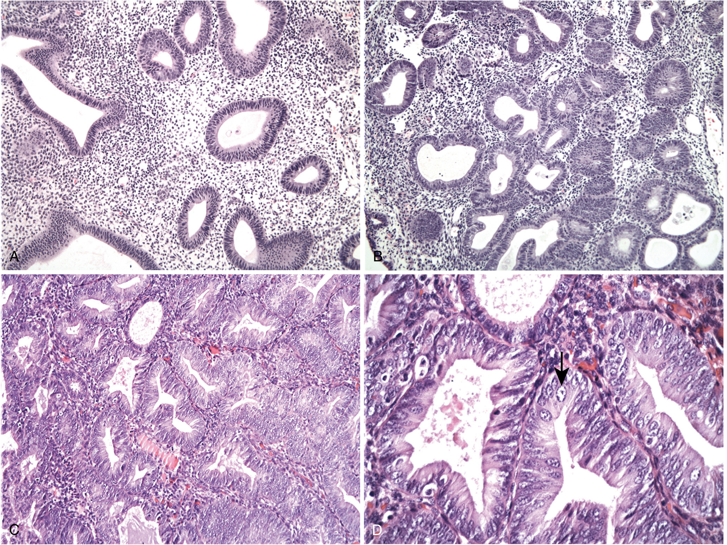 A: Hyperplasia without atypia - mild glandular crowding and cystic dilation. B: Increased glandular crowding with back-to-back glands. C: Atypical hyperplasia with nuclear atypia. D: High magnification showing rounded vesicular nuclei with prominent nucleoli.
6. Pathology
Gross Morphology
- May present as a localized polypoid mass or diffuse involvement of endometrial lining
- Typically arises in the fundus
- Spreads by myometrial invasion, then to adjacent structures, broad ligament, regional lymph nodes, and eventually distant metastases (lungs, liver, bones)
Histological Types (Classification):
| Type | Frequency | Notes |
|---|---|---|
| Endometrioid adenocarcinoma | 80-85% | Most common; variants include villoglandular, secretory, with squamous differentiation |
| Mucinous carcinoma | Rare | >50% mucinous cells; usually low-grade |
| Papillary serous carcinoma | ~10-15% | High-grade; TP53 mutated; arises in atrophic endometrium |
| Clear cell carcinoma | ~5% | High-grade; poor prognosis |
| Squamous carcinoma | Very rare | |
| Undifferentiated carcinoma | Very rare | |
| Mixed carcinoma | Present | Two or more types |
FIGO Histologic Grading (for endometrioid and mucinous carcinomas):
| Grade | Solid Growth | Significance |
|---|---|---|
| Grade 1 | ≤5% solid | Well-differentiated |
| Grade 2 | 6-50% solid | Moderately differentiated |
| Grade 3 | >50% solid | Poorly differentiated |
Note: Significant nuclear atypia upgrades the tumor by one grade. Serous and clear cell carcinomas are always considered high-grade - grading unnecessary.
Diagnostic criteria for invasion (differentiating carcinoma from atypical hyperplasia):
- Desmoplastic stroma
- Back-to-back glands without intervening stroma
- Extensive papillary pattern
- Squamous epithelial differentiation
- Area of involvement ≥ half of a low-power field (>4.2 mm)
Endometrioid Adenocarcinoma - Histology
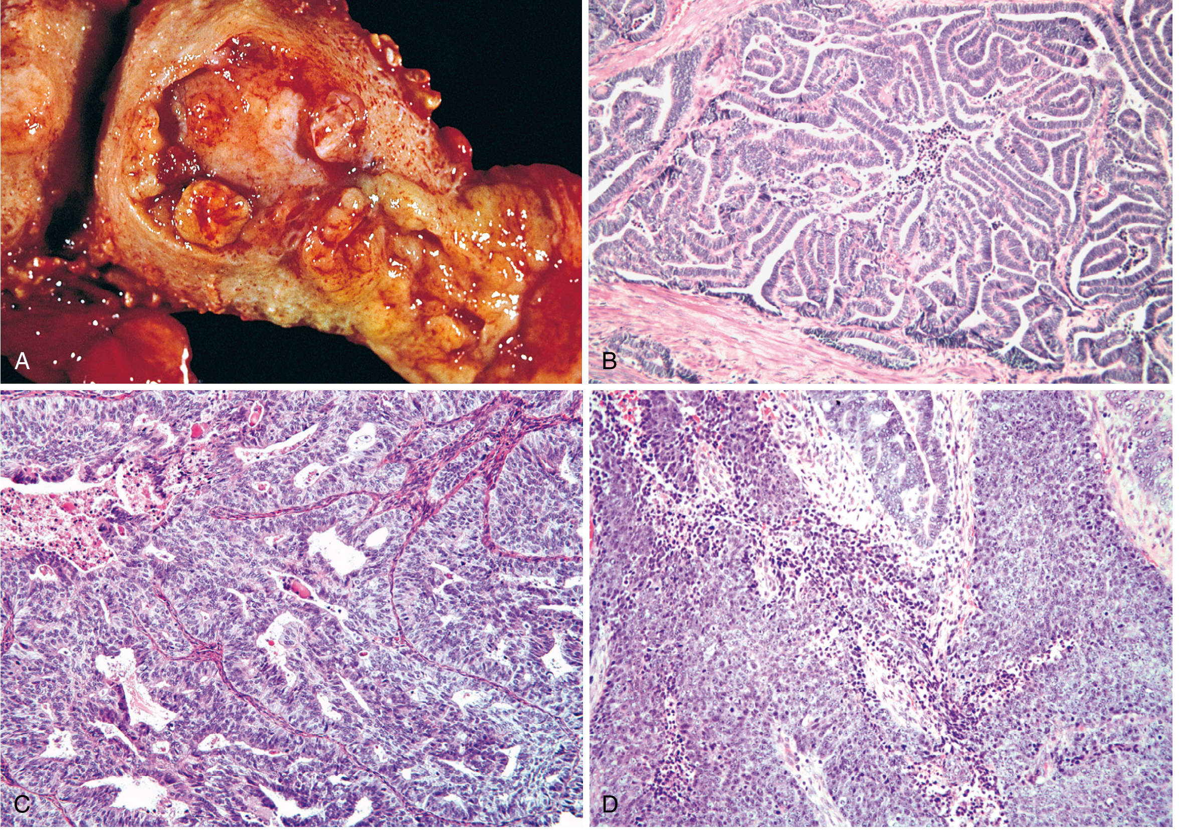
- Up to 20% of endometrioid carcinomas contain foci of squamous differentiation
- When squamous elements look benign = "adenoacanthoma" (old term); when malignant = "adenosquamous carcinoma" (old term)
- Graded by glandular component only regardless of squamous areas
Serous Endometrial Carcinoma
- Occurs ~10 years later than endometrioid carcinoma
- Arises in atrophic endometrium
- Precursor: Endometrial Intraepithelial Carcinoma (EIC) - serous cells confined to epithelium
- Papillary growth with marked cytologic atypia: high N:C ratio, atypical mitoses, hyperchromasia, prominent nucleoli
- Despite superficial endometrial involvement, may have extensive peritoneal disease (spreads via tubal regurgitation)
- 5-year survival: only 18-27%; even when confined to uterus, recurrence rate 80%
- More common in women of African descent (2-fold higher mortality)
Clear Cell Carcinoma
- High-grade tumor; arises in postmenopausal women
- Back-to-back glands lined by polygonal to columnar cells with clear cytoplasm (glycogen-rich), distinct cell membranes, prominent nucleoli
- Poor prognosis
7. Clinical Features
Symptoms
- Postmenopausal uterine bleeding - cardinal symptom; present in majority
- Important: <10% of patients with postmenopausal bleeding have endometrial cancer
- Most common cause of postmenopausal bleeding is endometrial atrophy (60-80%)
- Perimenopausal irregular bleeding - menometrorrhagia, oligomenorrhea, or cycles extending past usual age of menopause
- Watery or blood-stained vaginal discharge (earlier)
- Pyometra in elderly
- Pain is a late feature (pelvic/lower abdominal)
Causes of Postmenopausal Bleeding:
| Cause | Percentage |
|---|---|
| Endometrial atrophy | 60-80% |
| Estrogen replacement therapy | 15-25% |
| Endometrial polyps | 2-12% |
| Endometrial hyperplasia | 5-10% |
| Endometrial cancer | ~10% |
Signs
- Obesity (common constitutional finding)
- Usually normal pelvic examination in early disease
- Uterus may be normal in size or slightly enlarged
- Bimanual examination: assess uterine size/mobility, adnexal masses, parametrial induration, cul-de-sac nodularity
- Check: inguinal/peripheral lymph nodes, hepatomegaly, ascites, omental masses (in advanced disease)
- Vagina and cervix: carefully inspect for metastatic deposits
8. Diagnosis
Step 1: Endometrial Biopsy (First-line)
- Office endometrial aspiration biopsy is the accepted first step
- Adequate tissue obtained in most patients; sensitivity ~90%
- If biopsy insufficient or negative but suspicion remains high → proceed to D&C
Transvaginal Ultrasound (TVS)
- Endometrial thickness >4-5 mm in postmenopausal women warrants biopsy
- Useful for assessing myometrial invasion preoperatively
Fractional Curettage (D&C)
- Separate curettings from endocervix and endometrium
- Endocervical involvement indicates stage II disease
- Still considered gold standard if office biopsy inconclusive
Hysteroscopy + Directed Biopsy
- Better visualization; useful for polyps and focal lesions
- Concern about possible dissemination (not fully proven)
Other Investigations (Pretreatment Evaluation)
- Chest X-ray: Exclude pulmonary metastasis; assess cardiorespiratory status
- ECG, CBC, blood group and screen
- Serum CA-125: May be elevated in advanced disease; useful for monitoring
- CT abdomen and pelvis: Consider for type II tumors; assess for extrauterine disease
- MRI: Best modality for assessing myometrial invasion and cervical involvement preoperatively
- Cystoscopy, colonoscopy, IVP: only if symptoms dictate
- Pap smear: Inadequate for screening; only positive in ~50% of cases
9. Staging
FIGO Staging (Surgical Staging - Current):
| Stage | Description |
|---|---|
| Stage I | Confined to corpus uteri |
| IA | No or <50% myometrial invasion |
| IB | ≥50% myometrial invasion |
| Stage II | Cervical stromal invasion (not just glandular) |
| Stage III | Local/regional spread outside uterus |
| IIIA | Uterine serosa and/or adnexa |
| IIIB | Vaginal and/or parametrial involvement |
| IIIC1 | Pelvic lymph node metastasis |
| IIIC2 | Para-aortic lymph node metastasis |
| Stage IV | Extends beyond true pelvis or mucosal involvement of bladder/bowel |
| IVA | Bladder/bowel mucosal involvement |
| IVB | Distant metastasis (including inguinal LN) |
Note: Staging is surgical - not clinical. Positive peritoneal cytology alone no longer changes the stage (as per 2009 FIGO revision), though it remains prognostically important.
5-Year Survival by Stage:
- Stage I (grade 1/2): ~90%
- Stage I (grade 3): ~75%
- Stage II/III: ~50%
- Stage IV: <20%
10. Prognostic Variables
The most important adverse prognostic factors:
| Factor | Detail |
|---|---|
| Age | Older age = worse prognosis; younger patients have more favorable tumors |
| Histologic type | Serous/clear cell much worse than endometrioid |
| Histologic grade | Grade 3 significantly worse; most important for endometrioid |
| Myometrial invasion | Deep invasion (≥50%) → increased lymphatic spread and recurrence |
| Lymphovascular space invasion (LVSI) | Independent predictor of lymph node metastasis and recurrence |
| Cervical/isthmus extension | Stage II; worse prognosis |
| Lymph node metastasis | Pelvic > para-aortic |
| Tumor size | >2 cm: 15% LN mets; entire cavity: 35% LN mets; 2 cm: 4% |
| Hormone receptor status | ER+/PR+ = better prognosis; PR levels stronger predictor than ER |
| Peritoneal cytology | Positive = worse outcome |
| DNA ploidy | Aneuploid = worse; ~2/3 are diploid |
| Adnexal involvement | Stage IIIA |
| Molecular markers | POLE mutated = best; TP53 mutated = worst |
Lymph Node Metastasis Risk by Myometrial Invasion:
- No invasion: 1% pelvic LN mets
- Inner 1/3: 5%
- Middle 1/3: 6%
- Outer 1/3: 25%
11. Patterns of Spread
- Direct extension: Downward to cervix, outward to myometrium, then uterine serosa and adnexa
- Lymphatic spread: To pelvic (obturator, external iliac, internal iliac) and para-aortic nodes
- Hematogenous: Lungs (most common), liver, bones, brain
- Transperitoneal: Especially serous carcinoma via tubal regurgitation
- Transcervical: To vagina
12. Simultaneous Endometrial and Ovarian Tumors
- Incidence: 1.4-3.8% of endometrial cancers
- Most commonly both are endometrioid, well-differentiated, and low-stage
- Often premenopausal women; excellent prognosis when both are low-grade
- Up to 29% of endometrioid ovarian adenocarcinomas have associated endometrial cancer
- Distinction from metastasis: molecular/IHC studies (LOH analysis) when conventional criteria insufficient
13. Treatment
Surgical Treatment (Primary)
Standard procedure: Total abdominal hysterectomy + bilateral salpingo-oophorectomy (TAH + BSO) with surgical staging (pelvic washings for cytology, lymph node assessment)
Surgical staging includes:
- TAH + BSO
- Peritoneal cytology
- Inspection and palpation of peritoneal surfaces
- Lymph node sampling/dissection (pelvic ± para-aortic)
- Omental biopsy if indicated
Laparoscopic approach (preferred when feasible):
- Laparoscopically assisted vaginal hysterectomy (LAVH) or total laparoscopic hysterectomy
- Equivalent lymph node counts and oncologic outcomes to laparotomy
- Benefits: shorter hospital stay, less blood loss, fewer postoperative complications, earlier return to normal activities
- GOG study: >2,500 patients - no difference in stage distribution, nodal counts, or survival
- Port site metastases: <0.5%
Vaginal hysterectomy: Acceptable for low-risk tumors (endometrioid grade 1/2, <50% myometrial invasion, <2 cm tumor) in patients with multiple comorbidities
Sentinel Lymph Node Biopsy:
- Sensitivity 97.2%, negative predictive value 99.6%
- Can replace formal lymphadenectomy in appropriate patients
Lymphadenectomy: May be omitted in patients with negligible lymphatic spread risk (grade 1/2, <50% myometrial invasion, tumor ≤2 cm)
Adjuvant Radiation Therapy
Modalities:
- Vaginal vault brachytherapy: For intermediate-risk patients; reduces vaginal recurrence
- External pelvic radiation (EBRT): For higher-risk patients with deep invasion or lymph node metastasis
- Extended-field radiation: Covers para-aortic region when para-aortic nodes involved
- Whole-abdomen radiation: Rarely used now; replaced by chemotherapy for advanced disease
Radiation as Primary Treatment: For patients medically unfit for surgery
Chemotherapy
Indications: Advanced-stage (III/IV) disease and recurrent disease; type II tumors
Common regimens:
- Carboplatin + paclitaxel (most widely used current regimen)
- Doxorubicin-based regimens
Hormonal Therapy
Progestins (megestrol acetate, medroxyprogesterone acetate):
- Used for recurrent or metastatic disease, especially receptor-positive tumors
- Megestrol acetate 40-160 mg/day
- Fertility-preserving treatment: Continuous progestin for grade 1 endometrioid confined to endometrium (no myometrial invasion)
- Success rate: ~76% achieve complete response
- ~20 patients in reviewed series achieved pregnancy post-treatment
- 24% never respond; follow-up biopsy every 3-6 months mandatory
- Considered a temporary measure; hysterectomy recommended after childbearing
Fertility-Sparing Management
Candidates: Young women with grade 1 endometrioid adenocarcinoma, no myometrial invasion (confirmed by MRI), no adnexal or metastatic involvement, strong desire for fertility
Protocol: Continuous megestrol acetate + serial endometrial biopsies every 3-6 months → hysterectomy after completion of childbearing
14. Screening
- No effective population-based screening exists for endometrial cancer
- Pap smear inadequate (too insensitive)
- TVS and endometrial biopsy too expensive for routine use in asymptomatic women
- Exception - Lynch II Syndrome: Annual TVS + endometrial biopsy from age 35-40 years
- Alternatively: prophylactic hysterectomy + BSO after childbearing (100% prevention of endometrial and ovarian cancer in carriers)
- Universal screening with IHC for MMR proteins (MLH1, MSH2, MSH6, PMS2) now recommended for all newly diagnosed endometrial cancers to identify Lynch syndrome patients
15. Recurrent Disease
Sites of recurrence:
- Vaginal vault (most common local recurrence)
- Pelvic sidewall
- Para-aortic nodes
- Distant: lungs, abdomen, liver
Treatment of recurrence:
| Type | Treatment |
|---|---|
| Vaginal recurrence | Radiation (if not previously irradiated); surgery |
| Pelvic recurrence | Pelvic exenteration (selected patients) |
| Hormonal therapy | Progestins for receptor-positive, slow-growing recurrences |
| Chemotherapy | Carboplatin + paclitaxel for systemic recurrence |
16. Uterine Sarcomas (covered alongside Ca endometrium in Dutta)
| Type | Notes |
|---|---|
| Endometrial Stromal Sarcoma | Low-grade and high-grade variants; arising from stroma |
| Leiomyosarcoma | Most common uterine sarcoma; arises from myometrium |
| Adenosarcoma | Biphasic tumor; benign glands + malignant stroma |
| Carcinosarcoma (MMMT) | Malignant mixed Müllerian tumor; now considered metaplastic carcinoma |
Key Points Summary
- Most common risk factors involve prolonged unopposed estrogen stimulation
- Office endometrial aspiration biopsy is the first step in evaluation of abnormal uterine bleeding
- Serous and clear cell carcinomas = <10% of cases but >50% of all endometrial cancer deaths
- Most patients should undergo surgical staging (TAH + BSO + peritoneal cytology ± lymph node assessment)
- Most important adverse prognostic variables: advanced age, non-endometrioid or grade 3 histology, deep myometrial invasion, lymphovascular invasion, lymph node metastasis
- POLE-mutated tumors have the best prognosis; TP53-mutated (serous) have the worst
- Lynch syndrome accounts for 3-5% of all endometrial cancers; universal MMR testing now recommended
- ~80% of endometrial cancers present at stage I with ~90% 5-year survival
Sources: Berek & Novak's Gynecology (9781496380357); Robbins & Cotran Pathologic Basis of Disease (9780443264528)
Note: DC Dutta's Textbook of Gynecology is not available in this library. This answer covers all topics that Dutta addresses in his gynecology text on carcinoma endometrium, drawn from Berek & Novak's Gynecology and Robbins & Cotran Pathology - both of which contain equivalent or more detailed coverage.> Important note: DC Dutta's Textbook of Gynecology is not in the medical library. The comprehensive answer above is compiled from Berek & Novak's Gynecology (the most comparable standard gynecology textbook) and Robbins & Cotran Pathologic Basis of Disease - both at the same depth and scope that Dutta covers this topic.
Carcinoma of the Endometrium - Complete Notes
1. Introduction & Epidemiology
- Most common invasive cancer of the female genital tract (Western countries)
- Accounts for 7% of all invasive cancers in females (excluding skin)
- Has overtaken cervical cancer due to better cervical screening programs
- Peak incidence: 55-65 years (postmenopausal); uncommon before age 40
- Incidence in younger women is rising
- About 80% of cases are stage I at diagnosis
2. Risk Factors
All major risk factors share the common mechanism of prolonged unopposed estrogen stimulation:
| Risk Factor | Notes |
|---|---|
| Obesity | Most significant risk factor; peripheral conversion of androgens → estrone in adipose tissue |
| Nulliparity / Infertility | Chronic anovulation |
| Late menopause | Prolonged estrogen exposure |
| Diabetes mellitus | Co-exists; associated with obesity |
| Hypertension | The classic triad: obesity + DM + HTN |
| Unopposed exogenous estrogen | 4-8x increased risk; dose- and time-dependent; risk reduced by adding progestins |
| Tamoxifen | Partial estrogen agonist on endometrium |
| Estrogen-secreting tumors | Granulosa-theca cell tumors |
| PCOS | Chronic anovulation |
| Lynch II syndrome (HNPCC) | Lifetime risk 32-60%; autosomal dominant; MLH1, MSH2, PMS1, PMS2, MSH6 mutations |
Protective: Combined OCP, multiparity, smoking
3. Type I vs Type II Classification
| Feature | Type I | Type II |
|---|---|---|
| Estrogen-dependence | Yes | No |
| Histology | Endometrioid | Serous, clear cell |
| Grade | Low (1, 2) | High (3) |
| Background endometrium | Hyperplasia | Atrophic |
| Patient | Obese, perimenopausal | Thin, older |
| Mutations | PTEN, PIK3CA, KRAS, ARID1A, MSI | TP53 |
| Prognosis | Favorable | Poor |
4. Pathogenesis & Molecular Biology
Type I - Key mutations (all drive PI3K/AKT pathway):
- PTEN: Most common (30-80%); tumor suppressor; loss of function
- PIK3CA: ~40%; activating mutation (encodes PI3K catalytic subunit)
- KRAS: ~25%; activating mutation
- ARID1A: ~1/3; chromatin remodeling; also mutated in endometriosis-associated ovarian cancers
- MMR defects: ~20% sporadic; MLH1 epigenetically silenced; Lynch syndrome has germline MMR mutations
- POLE mutations: <10%; ultramutated; paradoxically excellent prognosis
Type II - Key mutation:
- TP53: >90% of serous carcinomas
TCGA Four Molecular Subtypes (in order of prognosis, best to worst):
- POLE ultramutated - best prognosis
- MSI hypermutated - intermediate
- Copy number low/nonspecific - intermediate
- Copy number high/TP53 mutated - worst (serous-like)
5. Precursor: Endometrial Hyperplasia
- Established precursor to Type I (endometrioid) carcinoma
- WHO classification: Hyperplasia without atypia vs. Atypical Hyperplasia/EIN (endometrial intraepithelial neoplasia)
- Atypical hyperplasia: 25-29% harbor concurrent carcinoma; high progression risk
6. Pathology
Classification of Endometrial Carcinomas:
- Endometrioid adenocarcinoma (+ variants: villoglandular/papillary, secretory, with squamous differentiation)
- Mucinous carcinoma
- Papillary serous carcinoma
- Clear cell carcinoma
- Squamous carcinoma
- Undifferentiated carcinoma
- Mixed carcinoma
Gross Morphology:
- Localized polypoid mass or diffuse involvement of endometrial lining
- Typically arises in fundus
- Spreads: myometrial invasion → adjacent structures → lymph nodes → distant (lungs, liver, bones)
Endometrioid Adenocarcinoma (80-85%):
- Glands resembling normal proliferative endometrium
- Columnar cells, basally oriented nuclei, little intracytoplasmic mucin
- Invasion criteria: desmoplastic stroma, back-to-back glands (no intervening stroma), extensive papillary pattern, squamous differentiation (area >4.2 mm)
- Up to 20% have squamous differentiation (graded by glandular component only)
FIGO Grading:
| Grade | Solid Component | Description |
|---|---|---|
| Grade 1 | ≤5% | Well-differentiated |
| Grade 2 | 6-50% | Moderately differentiated |
| Grade 3 | >50% | Poorly differentiated |
- Significant nuclear atypia upgrades tumor by 1 grade
- Serous and clear cell = always high-grade (grading not needed)
Serous Carcinoma (~10-15%):
- Arises in atrophic endometrium; occurs ~10 years later than endometrioid
- Precursor: Endometrial Intraepithelial Carcinoma (EIC) - malignant cells on surface epithelium only
- Papillary architecture; marked cytologic atypia; TP53 mutated (>90%)
- Spreads via transtubal route to peritoneum despite superficial uterine involvement
- 5-year survival 18-27%; recurrence rate up to 80% even when confined to uterus
- More common in women of African descent (2-fold higher mortality)
Clear Cell Carcinoma (~5%):
- High-grade; postmenopausal
- Clear (glycogen-rich) cytoplasm; distinct cell membranes; prominent nucleoli
- Back-to-back glandular pattern; binucleated and multinucleated forms
7. Clinical Features
Symptoms:
- Postmenopausal uterine bleeding (PMB) - cardinal symptom; present in >90%
- BUT remember: only ~10% of PMB is due to endometrial cancer; most is atrophy (60-80%)
- Perimenopausal: menometrorrhagia, oligomenorrhea, or cycles persisting past usual menopause age
- Watery/blood-stained vaginal discharge (early feature)
- Pyometra in elderly (malodorous discharge)
- Pelvic/lower abdominal pain - late feature
- Bladder/bowel symptoms in advanced disease
Signs:
- Obesity (common)
- Usually normal pelvic examination in early disease
- Uterus may be slightly enlarged or bulky
- Bimanual: assess uterine size and mobility, adnexal masses, parametrial induration, cul-de-sac nodularity
- Check inguinal and supraclavicular lymph nodes
- Assess for hepatomegaly, ascites, omental masses (advanced disease)
8. Diagnosis
1. Endometrial Aspiration Biopsy (First-line, office procedure)
- Sensitivity ~90%; adequate tissue in most cases
- Insufficient tissue or negative but high suspicion → D&C
2. Transvaginal Ultrasound (TVS)
- Endometrial thickness >4-5 mm in postmenopausal women requires biopsy
- Assesses myometrial invasion preoperatively
3. Fractional Curettage (D&C)
- Separate endocervical and endometrial curettings
- Endocervical involvement → Stage II
- Gold standard when office biopsy inconclusive
4. Hysteroscopy + Directed Biopsy
- Better visualization of focal lesions and polyps
5. MRI
- Best for preoperative assessment of myometrial invasion and cervical involvement
6. Papanicolaou Smear
- Inadequate for screening (positive in only ~50% of cases)
9. Pretreatment Evaluation
- History and physical examination - most important; assess comorbidities (DM, HTN, obesity)
- Chest X-ray: Exclude pulmonary metastasis; assess cardiorespiratory status
- ECG, CBC, platelet count, blood group and screen
- Serum CA-125: Elevated in advanced disease; useful for monitoring
- CT abdomen/pelvis: Consider for Type II tumors; assess extrauterine spread
- MRI: Preoperative myometrial invasion assessment
- Cystoscopy, colonoscopy, IVP, barium enema: only if symptoms indicate
- Stage IV disease usually clinically evident from symptoms + examination
10. FIGO Staging (Surgical)
| Stage | Description |
|---|---|
| I | Confined to corpus uteri |
| IA | No or <50% myometrial invasion |
| IB | ≥50% myometrial invasion |
| II | Cervical stromal invasion (not glandular) |
| III | Local/regional spread |
| IIIA | Uterine serosa and/or adnexa |
| IIIB | Vaginal/parametrial involvement |
| IIIC1 | Pelvic lymph node metastasis |
| IIIC2 | Para-aortic lymph node metastasis |
| IV | Beyond true pelvis or mucosal involvement |
| IVA | Bladder/bowel mucosa |
| IVB | Distant metastases (including inguinal LN) |
5-year survival:
- Stage I (grade 1/2): ~90%
- Stage I (grade 3): ~75%
- Stage II/III: ~50%
- Serous carcinoma overall: 18-27%
11. Prognostic Variables
| Factor | Detail |
|---|---|
| Age | Older = worse |
| Histologic type | Serous/clear cell >> endometrioid |
| Histologic grade | Grade 3 significantly worse |
| Myometrial invasion | ≥50% = high lymph node risk |
| LVSI | Independent predictor of recurrence |
| Cervical invasion | Stage II |
| Lymph node metastasis | Most important determinant |
| Tumor size | >2 cm: 15% LN mets; entire cavity: 35% |
| Hormone receptor status | PR+ > ER+ as predictor; receptor-positive = better |
| Peritoneal cytology | Positive = worse outcome |
| DNA ploidy | Aneuploid = worse |
| Molecular subtype | POLE = best; TP53-mutated = worst |
LN metastasis by myometrial invasion depth:
- No invasion: 1%
- Inner 1/3: 5%
- Middle 1/3: 6%
- Outer 1/3: 25%
12. Routes of Spread
- Direct extension - downward to cervix; outward through myometrium to serosa and adnexa
- Lymphatic - to pelvic nodes (obturator, external/internal iliac), then para-aortic nodes
- Hematogenous - lungs (most common), liver, bones, brain
- Transperitoneal - especially serous carcinoma; via tubal regurgitation
- Transcervical - to vagina
13. Treatment
Surgery (Primary Treatment)
Standard: TAH + BSO + peritoneal cytology ± lymph node assessment
Surgical staging includes:
- TAH + BSO
- Peritoneal cytology
- Inspection of all peritoneal surfaces
- Lymph node assessment (sentinel node biopsy or formal dissection)
- Omental biopsy if indicated
Laparoscopic approach (preferred): GOG trial (>2,500 patients) showed:
- Equivalent oncologic outcomes to laparotomy
- Shorter hospital stay (52% vs 94% >2 days)
- Less blood loss and fewer postoperative complications (14% vs 21%)
- Longer operative time; 24% conversion rate (improved to 2.4% with modern technique)
Vaginal hysterectomy: Acceptable for low-risk tumors (grade 1/2, <50% MI, tumor <2 cm) with high surgical risk. 94% survival in selected series.
Lymphadenectomy may be omitted when: grade 1/2 endometrioid + <50% MI + tumor ≤2 cm (negligible lymphatic spread risk)
Sentinel lymph node biopsy: Sensitivity 97.2%, NPV 99.6% - increasingly replacing formal lymphadenectomy
Radiation Therapy
- Vaginal vault brachytherapy: Intermediate-risk; reduces vaginal recurrence
- External pelvic RT (EBRT): Higher-risk; deep invasion or LN metastasis
- Extended-field RT: Para-aortic involvement
- Primary RT: Medically unfit patients
Chemotherapy
- Carboplatin + paclitaxel: Most widely used for advanced/recurrent disease
- Indicated for: Stage III/IV, Type II tumors, recurrence
Hormonal Therapy
- Megestrol acetate 40-160 mg/day - most reliable for reversing hyperplasia and treating receptor-positive recurrent disease
- Progesterone receptor status predicts response
- Fertility-preserving protocol: Continuous progestins + serial biopsy q3-6 months
- ~76% achieve complete response; ~24% never respond
- ~20 patients achieved pregnancy in reviewed series
- Must be counseled: temporary treatment; hysterectomy after childbearing
14. Screening
- No effective population screening exists
- Pap smear: inadequate (insensitive)
- TVS + biopsy: too expensive for routine use
- Lynch II syndrome: Annual pelvic exam + TVS + endometrial biopsy from age 35-40
- Prophylactic hysterectomy + BSO after childbearing = 100% prevention in Lynch II carriers
- Universal MMR IHC testing (MLH1, MSH2, MSH6, PMS2) now recommended for all newly diagnosed endometrial cancers to identify Lynch syndrome
15. Recurrent Disease
Most common site: Vaginal vault
Treatment:
- Vaginal recurrence (if not previously irradiated): radiation therapy
- Pelvic exenteration: selected patients with central pelvic recurrence
- Chemotherapy: carboplatin + paclitaxel for systemic recurrence
- Hormonal therapy: progestins for receptor-positive, slow-growing recurrences
Sources: Berek & Novak's Gynecology; Robbins & Cotran Pathologic Basis of Disease
This is a shared conversation. Sign in to Orris to start your own chat.