
Styloid apparatus
- Styloid process - a slender bone projection from the petrous temporal bone
- Stylohyoid ligament - connects the styloid tip to the lesser horn of the hyoid
- Lesser horn (cornu minus) of the hyoid bone
- Body of the hyoid bone (some sources include this)
styloid apparatus styloid process hyoid bone ligament anatomy
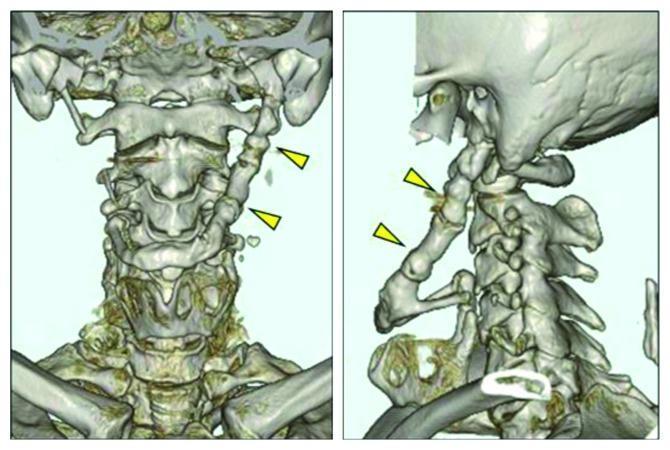
This diagnostic image consists of two panels showing a three-dimensional (3D) CT reconstruction of the human cervical region and skull base in posterior and oblique views. The primary finding is a significantly elongated and thickened left styloid process (indicated by yellow arrowheads) that exhibits complete bony fusion with the hyoid bone. This ossified stylohyoid complex measures approximately 70 mm in length and maintains a uniform thickness of roughly 10 mm. The surrounding anatomical structures, including the cervical vertebrae (C1-C7), mandible, and upper thoracic cage (clavicles and ribs), appear normally aligned. The right styloid process is visible but significantly shorter and disconnected from the hyoid. This presentation is characteristic of a severe anatomical variant of Eagle Syndrome, demonstrating the progressive ossification of the stylohyoid ligament and its impact on the anatomical relationship between the temporal bone and the hyoid apparatus, which can lead to symptoms like dysphagia and orofacial pain.
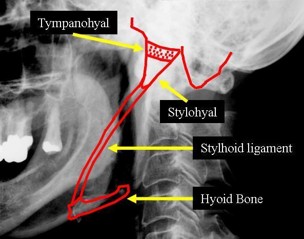
This lateral neck radiograph features a diagnostic overlay illustrating Eagle syndrome or stylohyoid ligament ossification. A red outline traces the pathological ossification of the stylohyoid chain, extending from the skull base to the hyoid bone. Key anatomical landmarks are labeled with yellow arrows: the Tympanohyal region (superiorly near the temporal bone), the Stylohyal (the proximal portion of the styloid process), the elongated and ossified Stylohyoid ligament, and its distal attachment at the Hyoid Bone. The image demonstrates the relationship of this ossified structure to adjacent anatomy, including the posterior border of the mandible and the cervical spine. The visual focus is the continuous, elongated bony projection that can cause clinical symptoms such as dysphagia or facial pain. This material is suitable for dental, ENT, and radiology students studying craniofacial anatomy and calcification disorders of the head and neck.
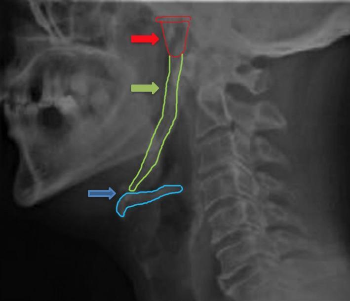
A lateral neck X-ray (radiography) demonstrating extensive ossification of the stylohyoid complex. The image features color-coded outlines and arrows highlighting key anatomical structures: a red arrow and outline identify the styloid process of the temporal bone; a green arrow and outline delineate the continuous, elongated radiopaque ossification of the stylohyoid ligament; and a blue arrow and outline mark the hyoid bone. The ossification forms a near-continuous bony chain extending from the skull base to the lesser cornua of the hyoid bone, showing non-uniform thickness and density along its length. This radiographic finding is characteristic of Eagle syndrome or asymptomatic stylohyoid ligament mineralization. Other visible landmarks include the cervical vertebrae (C1-C7) and the mandible. This diagnostic image is intended for educational use in oral and maxillofacial radiology or otolaryngology to illustrate pathological mineralization of soft tissues in the neck.
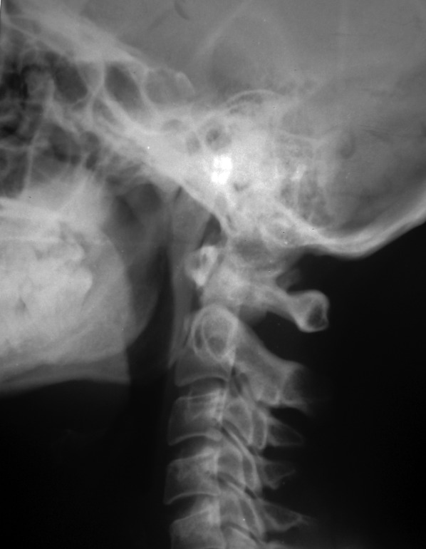
**Modality:** Lateral projection plain radiograph (X-ray). **Anatomic Region:** Upper cervical spine and skull base, including the craniovertebral junction and oropharyngeal soft tissue area. **Observations:** The image demonstrates a prominent, elongated, and heavily ossified structure originating from the styloid process at the skull base. This radiopaque structure extends anteroinferiorly and caudally toward the level of the hyoid bone. The ossification appears continuous and robust, occupying the lateral cervical space. The cervical vertebrae (C1-C5 visible) show preserved alignment and disc heights, though there is superimposed radiopacity from the ossified ligament over the upper cervical segments. **Pathology/Clinical Entity:** The findings are highly characteristic of **Eagle syndrome** (stylohyoid syndrome), specifically demonstrating complete or near-complete **ossification of the stylohyoid ligament**. **Key Diagnostic Features:** - Significant elongation and mineralization of the styloid process. - Continuous radiopaque band extending to the hyoid bone. - Differentiation from vascular calcification based on the linear, anatomical trajectory of the stylohyoid apparatus.
Styloid Apparatus
Components (from above downward)
| Component | Description |
|---|---|
| Styloid process | Slender bone projection from the inferior surface of the petrous temporal bone; normally ~2.5 cm long |
| Stylohyoid ligament | Fibrous band connecting the tip of the styloid process to the lesser horn (cornu minus) of the hyoid bone |
| Lesser horn (cornu minus) | Small bony projection forming the upper part of the hyoid bone |
| Body of the hyoid bone | (Superior portion derived from 2nd arch; inferior portion from 3rd arch) |
Embryological Origin: Second Pharyngeal Arch (Reichert's Cartilage)
- Proximally: styloid process, manubrium of the malleus, long process of the incus, stapes suprastructure
- Centrally: withers → forms the stylohyoid ligament
- Distally: superior body and lesser cornu of the hyoid bone
Muscles Attached to the Styloid Process ("Stylo-Trio")
| Muscle | Nerve | Action |
|---|---|---|
| Stylohyoid | Facial nerve (CN VII) | Elevates and retracts hyoid |
| Styloglossus | Hypoglossal nerve (CN XII) | Retracts and elevates tongue |
| Stylopharyngeus | Glossopharyngeal nerve (CN IX) | Elevates pharynx and larynx |
Relations of the Styloid Process
- The stylohyoid ligament forms the second attachment line of the lateral pharyngeal wall - running from the tip of the styloid to the lesser horn of the hyoid, then along the greater horn. - Gray's Anatomy for Students
- The internal carotid artery lies medial to the styloid process
- The external carotid artery lies lateral to stylopharyngeus
- CN IX (glossopharyngeal) winds around the lateral aspect of the styloid
Clinical Relevance: Eagle's Syndrome
- Normal styloid length: ~2.5 cm; elongated if >4 cm
- Elongated styloid present in ~4% of the population, but only ~4% of those develop symptoms
- Symptoms: dull pharyngeal pain (often in the tonsillar fossa), radiation to the ipsilateral ear, odynophagia, foreign body sensation in the throat, dysphagia, increased salivation, and potentially carotidynia (from impingement on the carotid vessels)
- Nerve involvement: trigeminal, facial, glossopharyngeal, and vagus nerves can be irritated
- Originally described by Eagle (1937) following tonsillectomy, though tonsillectomy is not always an aetiological factor
- Calcified stylohyoid ligament is an incidental finding in ~4% of the normal population
-
Conservative: steroids, local anaesthetic injection, carbamazepine
-
Surgical (preferred): reduction/shortening of the styloid process via transoral (through tonsillar fossa) or external transcervical approach
-
Scott-Brown's Otorhinolaryngology, KJ Lee's Essential Otolaryngology
Radiological Images of the Styloid Apparatus
Summary
Piriform fossa
piriform fossa recess laryngopharynx anatomy diagram
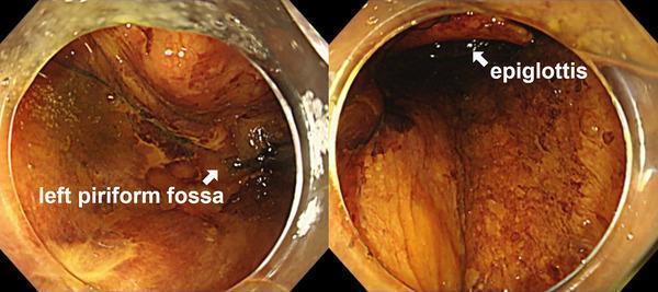
This diagnostic endoscopic clinical photograph captures two views of the laryngopharynx following an accidental reflux of Lugol's iodine solution during an endoscopic submucosal dissection (ESD) procedure. The mucosa of the pharyngeal region, including the epiglottis and the left piriform fossa, is prominently stained with a dark brown, mahogany-colored solution, which contrast significantly with the normal pale pink appearance of healthy pharyngeal tissue. On the left side of the composite image, the left piriform fossa is labeled, showing the deep recess of the laryngopharynx. The right side of the image highlights the superior portion of the epiglottis. The visualization demonstrates the extent of iodine dispersion across the laryngeal inlet and surrounding structures. This image is clinically significant for illustrating potential complications of chromoendoscopy, specifically the inadvertent exposure of sensitive laryngeal mucosa to caustic staining agents like Lugol's solution, which can lead to chemical-induced laryngitis and subsequent edema or erosion.
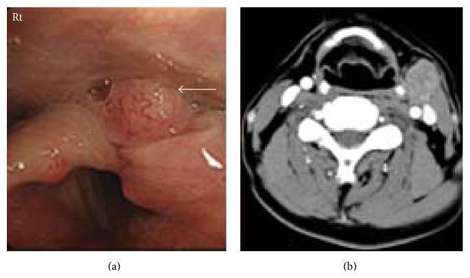
This dual-panel image displays diagnostic findings for hypopharyngeal squamous cell carcinoma. Panel (a) is an endoscopic clinical photograph of the laryngopharynx, showing a pinkish-red, irregular mass lesion located in the left piriform recess (indicated by a white arrow). The lesion exhibits a nodular surface texture characteristic of malignancy. Panel (b) is an axial contrast-enhanced computed tomography (CT) scan of the neck at the level of the larynx. It demonstrates a primary soft-tissue mass in the left hypopharyngeal region and a corresponding enlarged, contrast-enhancing metastatic cervical lymph node on the left side (Level II/III). The lymph node appears oval with increased density compared to adjacent muscular structures. Together, these images illustrate the primary site and regional lymphatic spread of Stage IVa hypopharyngeal cancer, highlighting the role of endoscopy and cross-sectional imaging in oncological staging and diagnosis within otorhinolaryngology.
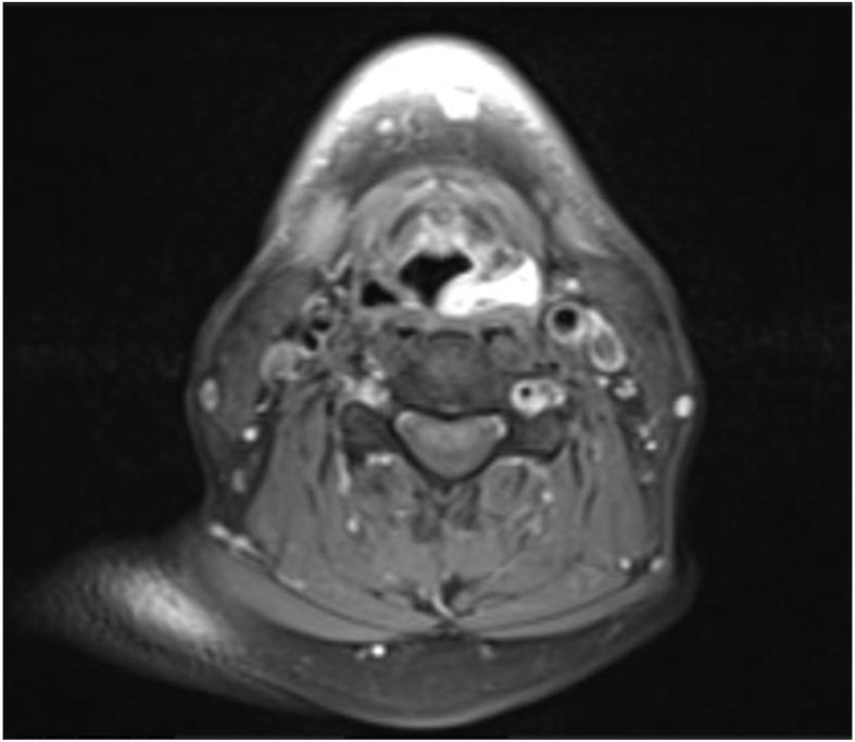
This diagnostic image is an axial cross-section of the upper neck obtained via Magnetic Resonance Imaging (MRI). The scan focuses on the hypopharynx and larynx at the level of the piriform fossae. A prominent, well-defined pathological lesion is visualized within the left piriform fossa. On this post-contrast T1-weighted sequence, the lesion demonstrates intense, bright enhancement compared to the surrounding soft tissues, indicating high vascularity or contrast uptake. The lesion occupies the left piriform sinus space, causing partial obliteration of the anatomical recess. Surrounding anatomical structures include the cervical vertebrae posteriorly, the laryngeal airway centrally, and the major neurovascular bundles of the neck laterally. The clinical significance of these findings relates to the identification of a soft tissue mass in the hypopharynx, which in this context corresponds to a lipomatous hamartoma. The image is intended for intermediate to advanced medical learners focusing on radiology, otorhinolaryngology, and head and neck pathology.
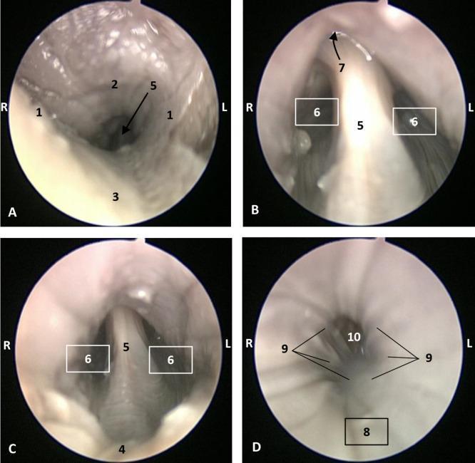
A series of four endoscopic images (A–D) detailing the anatomy of the pharyngeal cavity. Image A illustrates the oropharynx and the isthmus of the fauces, showing the arcus palatoglossus (1), soft palate (2), and tongue root (3). Image B provides a view of the laryngopharynx, highlighting the lingual surface of the epiglottis (5), the bilateral piriform recesses (6), and the superiorly located intrapharyngeal orifice (7). Image C further demonstrates the laryngopharyngeal structures, including the epiglottic vallecula (4), epiglottis (5), and piriform recesses (6). Image D captures the transition toward the esophagus, showing the oesophageal vestibule (8), the pharyngoesophageal limit (9), and the longitudinally folded oesophageal mucosa (10). The images showcase the healthy mucosal appearance, transitions in tissue texture between the pharyngeal and esophageal regions, and the spatial relationships between key landmarks such as the epiglottis and the piriform recesses, which are critical for understanding deglutition and airway protection.
Piriform Fossa (Piriform Recess / Pyriform Sinus)
Location Within the Hypopharynx
- Paired pyriform sinuses/fossae (most lateral - the subject here)
- Postcricoid area
- Posterior pharyngeal wall
Boundaries
| Wall | Structure |
|---|---|
| Medial wall | Aryepiglottic fold (superiorly), arytenoid and cricoid cartilages (inferiorly) |
| Lateral wall | Thyrohyoid membrane (superiorly), inner surface of thyroid cartilage (inferiorly); this wall is contiguous with the posterior pharyngeal wall |
| Apex | The pointed inferior tip - at the junction of the medial and lateral walls, deep within the fossa |
| Opening | Posteriorly into the pharyngeal lumen |
Key Nerve Relation - The Internal Branch of the Superior Laryngeal Nerve (iBSLN)
- The internal branch of the superior laryngeal nerve (CN X branch) descends in the medial wall of the piriform fossa, running beneath the mucous membrane
- It pierces the thyrohyoid membrane and then divides:
- Upper branch: supplies mucosa of lower pharynx, epiglottis, vallecula, vestibule of larynx
- Lower branch: descends in the medial wall of the piriform fossa beneath the mucosa → supplies the aryepiglottic fold and laryngeal mucosa down to the level of the vocal folds
- Because it lies directly under the piriform fossa mucosa, the nerve is accessible for local anaesthetic injection here, producing excellent anaesthesia for most of the piriform fossa and supraglottis - Scott-Brown's Otorhinolaryngology
Blood Supply
- Superior thyroid artery (primary supply)
- Branches from lingual and ascending pharyngeal arteries also contribute collaterals
Lymphatic Drainage
- Piriform sinus lymphatics pass through the thyrohyoid membrane primarily to the jugulodigastric (level II/III) lymph nodes
- The apex of the piriform sinus also drains to paratracheal and precricoid (Delphian) lymph nodes
- This explains why piriform sinus carcinomas frequently present with early cervical node metastases - Cummings Otolaryngology
Histology / Wall Layers
- Mucosal lining - stratified squamous epithelium over loose stroma
- Fibrous layer - pharyngeal aponeurosis
- Muscular layer - posterior cricoarytenoid (anteriorly), middle and inferior constrictor (posteriorly)
- Fascial layer - buccopharyngeal fascia
The Pyriform Apex - Special Surgical Significance
- Related laterally to the thyroid cartilage, medially to the cricoid cartilage, and lies inferior to the paraglottic space
- Tumours at the apex can spread superiorly into the paraglottic space → causing vocal cord fixity
- Close proximity to cricoid cartilage means oncological removal often requires cricoid cartilage removal
- Tumours can also escape via the cricothyroid membrane early, bypassing the laryngeal framework
- Scott-Brown's Otorhinolaryngology
Clinical Importance
1. Foreign Body Impaction
2. Carcinoma of the Piriform Sinus
- The commonest subsite of hypopharyngeal carcinoma (~70% of hypopharyngeal cancers)
- Higher incidence in northern France (14.8 per 100,000)
- Presents late with dysphagia, hoarseness, or referred otalgia
- Clinical sign: unilateral pooling of saliva in the piriform fossa on flexible endoscopy
- Tumours spread through the thyrohyoid membrane laterally without cartilage destruction, keeping laryngeal function partially intact - useful for treatment planning
- Treatment: chemoradiotherapy for organ preservation; major surgery (total laryngectomy + pharyngectomy with reconstruction) for advanced or recurrent disease
- Scott-Brown's Otorhinolaryngology, Bailey & Love's
3. Third Branchial Arch Fistula (Pyriform Sinus Fistula)
- An anomaly where a fistulous tract opens internally at the apex of the pyriform sinus, passing along the course of the third branchial arch
- Presents as recurrent neck abscess or suppurative thyroiditis (left side more common)
- Confirmed by laryngoscopy showing the internal opening
- Treatment: endoscopic cautery of the pyriform sinus tract, or surgical excision (often with hemithyroidectomy)
Endoscopic Appearance
Summary
- The internal branch of the superior laryngeal nerve runs under its medial wall mucosa (accessible for anaesthetic injection, and responsible for referred otalgia when diseased)
- Lymphatics drain through the thyrohyoid membrane to jugulodigastric nodes
- It is the commonest site of hypopharyngeal carcinoma (~70%)
- Foreign bodies pool here
- Third branchial fistulae open internally at its apex
Horners syndrome
Horner syndrome ptosis miosis anhidrosis oculosympathetic pathway
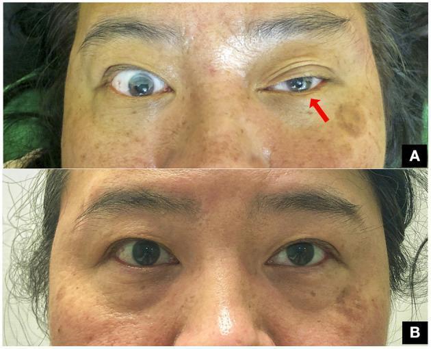
This clinical comparison photograph shows a 47-year-old woman presenting with classic oculofacial features of Horner’s syndrome following a clavicle fracture. Image A demonstrates the acute presentation of the oculosympathetic defect in the left eye, characterized by significant blepharoptosis (drooping of the upper eyelid) and ipsilateral miosis (constriction of the pupil) compared to the normal right eye. A red arrow points to the affected left eye to emphasize these findings. Image B shows the same patient at a three-month follow-up after surgical fixation of the fracture. It illustrates complete resolution of the syndrome, with symmetric eyelid positions and equal pupillary diameters (isocoria) in both eyes. This visual progression highlights the classic clinical triad (ptosis and miosis, with anhidrosis typically associated) and the potential for recovery following the management of the underlying traumatic compression of the preganglionic sympathetic pathway. The image is a valuable educational resource for neurology, ophthalmology, and trauma surgery.
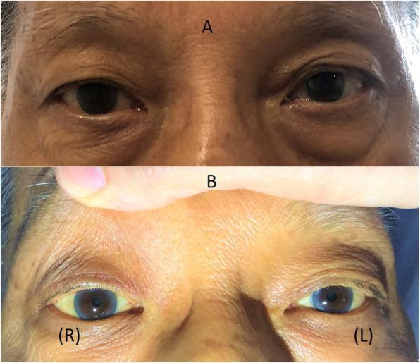
Two-panel clinical photograph demonstrating Horner’s syndrome in a patient. Panel A shows the patient's eyes at rest, highlighting a mild right-sided ptosis (drooping of the upper eyelid) and a subtle 'upside-down ptosis' (elevation of the right lower eyelid). Panel B provides a close-up view of the eyes with the upper eyelids manually retracted to reveal anisocoria; the right pupil (labeled R) is significantly smaller (miotic) compared to the left pupil (labeled L). The clinical findings illustrate the classic triad of sympathetic denervation: ptosis, miosis, and enophthalmos appearance due to narrowing of the palpebral fissure. This visual evidence supports a diagnosis of Horner’s syndrome, often associated with neurological or vascular pathology affecting the oculosympathetic pathway. The image serves as an educational tool for identifying autonomic ocular dysfunction in neurology and ophthalmology.
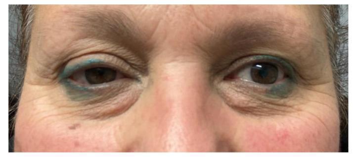
This clinical photograph shows a close-up, frontal view of a patient's periorbital region, demonstrating the classic triad of Horner syndrome on the left side. Key diagnostic features include visible ptosis (drooping) of the left upper eyelid and mild elevation of the left lower eyelid (inverse ptosis), resulting in a narrowed palpebral fissure. Comparison of the pupils reveals miosis (constriction) of the left pupil compared to the right, indicating an anisocoria that is typically more pronounced in dim light. The surrounding skin exhibits signs of aging, including rhytids (wrinkles) and mild dermatochalasis. The patient has blue cosmetic tattooing or permanent eyeliner on both the upper and lower eyelids. This presentation is characteristic of a disruption in the oculosympathetic pathway, which in this clinical context followed the surgical resection of a parapharyngeal space schwannoma involving the cervical sympathetic chain.
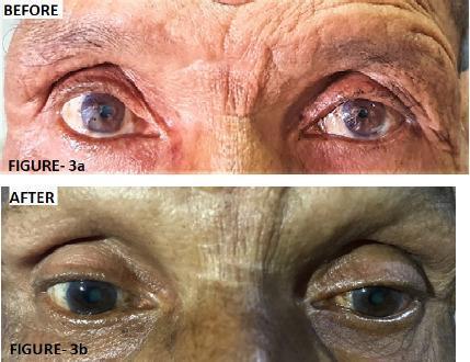
Two-panel clinical photograph displaying the 'Before' and 'After' ophthalmic findings in a patient with left-sided Horner's syndrome. The 'Before' image (Figure 3a) demonstrates classic clinical signs including left upper eyelid ptosis (drooping) and left-sided miosis (constricted pupil) compared to the right eye. The 'After' image (Figure 3b) illustrates the clinical response following three cycles of chemotherapy for an underlying superior sulcus tumor (Pancoast tumor). There is visible improvement in the left ocular presentation, characterized by the elevation of the left upper eyelid, reduction in ptosis severity, and a comparative increase in left pupil diameter (resolution of miosis). The image highlights the ocular manifestations of sympathetic nerve pathway disruption and its subsequent recovery during systemic treatment of the primary thoracic malignancy. Educational focus includes the recognition of Horner's syndrome triad (ptosis, miosis, and anhidrosis) and its clinical association with apical lung lesions.
Horner's Syndrome (Oculosympathetic Palsy)
The Three-Neuron Pathway
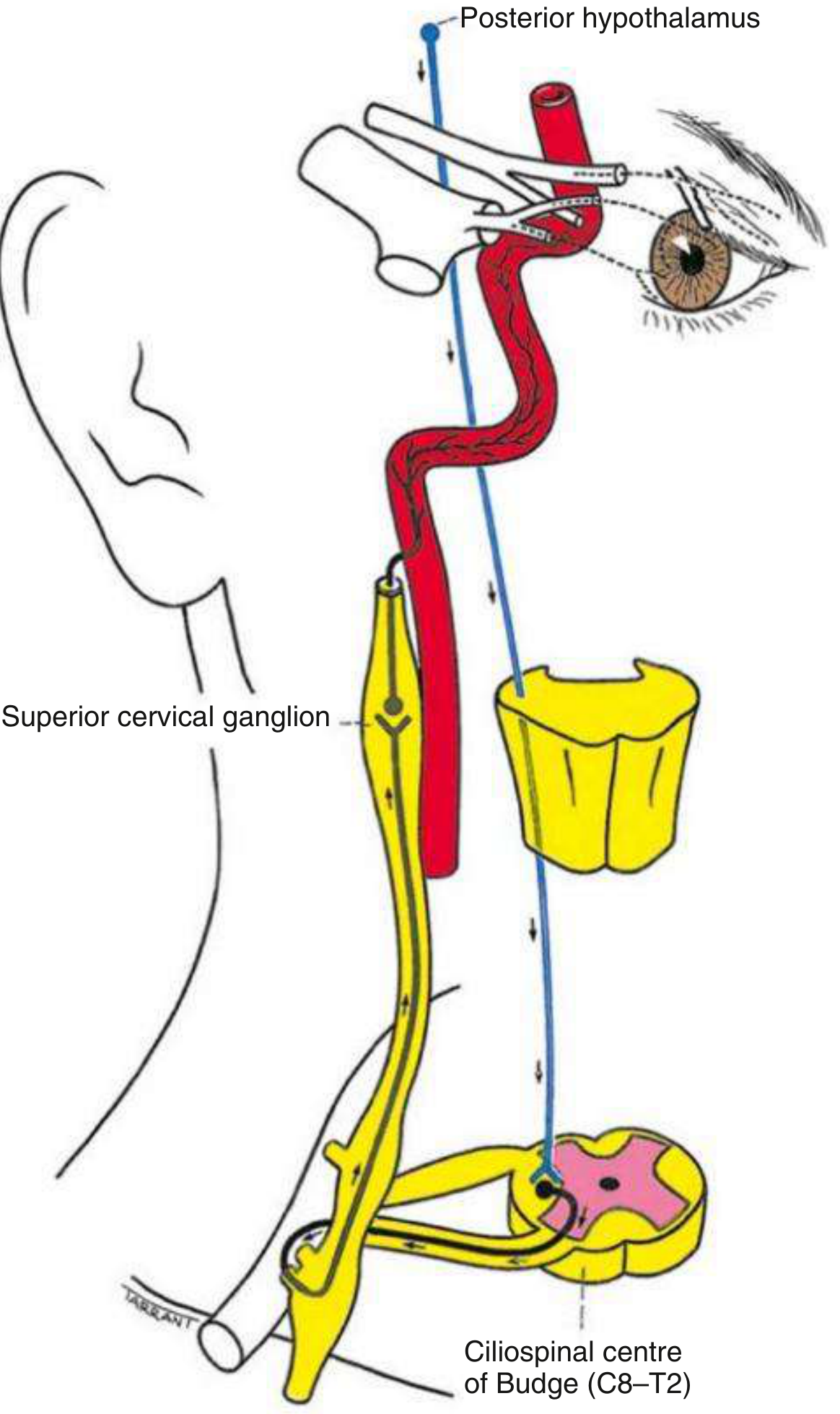
| Neuron | Order | Course | Key Relations |
|---|---|---|---|
| First-order (central) | 1st | Posterior hypothalamus → descends uncrossed through brainstem → ciliospinal centre of Budge (C8-T2 intermediolateral horn) | Brainstem, lateral tegmentum, cervical cord |
| Second-order (preganglionic) | 2nd | Ciliospinal centre → exits ventral root → over apex of lung → along subclavian artery → up cervical sympathetic chain → synapses at superior cervical ganglion | Closely related to apical pleura; vulnerable to Pancoast tumour and neck surgery |
| Third-order (postganglionic) | 3rd | Superior cervical ganglion → ascends along internal carotid artery → cavernous sinus → joins ophthalmic division of CN V → nasociliary nerve → long ciliary nerves → dilator pupillae and Müller's muscle | Internal carotid artery, cavernous sinus |
- Kanski's Clinical Ophthalmology, Neuroanatomy through Clinical Cases
Clinical Features
Classic Triad
- Ptosis (1-2 mm) - loss of innervation to Müller's smooth muscle (superior tarsal muscle) in the upper lid
- Miosis - loss of sympathetic innervation to the pupillary dilator muscle → unopposed sphincter pupillae action
- Anhidrosis - loss of sympathetic sudomotor innervation to ipsilateral face/neck
Additional Features
- Lower lid elevation ("upside-down ptosis" / inverse ptosis) - weakness of the inferior tarsal muscle
- Apparent enophthalmos - an illusion created by narrowing of the palpebral fissure (true enophthalmos is debated)
- Anisocoria accentuated in dim light (Horner pupil fails to dilate; dark-induced anisocoria also diminishes over time)
- Dilation lag - the Horner pupil dilates more slowly than the normal pupil when light is removed
- Normal light reflex - pupillary constriction to light and near stimuli is preserved (unlike CN III palsy)
- Heterochromia iridis - the Horner iris is lighter (hypochromic) in congenital or long-standing cases; due to failure of melanin deposition
- Ciliospinal reflex absent - painful pinch to the neck normally causes pupillary dilation via cervical sympathetics; absent on the affected side
Anhidrosis Localisation
-
Lesion at or below common carotid artery: loss of sweating involves the entire face
-
Lesion distal to carotid bifurcation: no anhidrosis, or confined to medial forehead and side of nose (sudomotor fibres travel along the external carotid, not internal)
-
Postganglionic lesions above the bifurcation generally have no anhidrosis
-
Adams & Victor's Neurology, Kanski's Clinical Ophthalmology
Clinical Photo
Causes by Neuron Level
First-Order (Central) Neuron Lesions
- Brainstem stroke (especially lateral medullary / Wallenberg syndrome - PICA territory infarct)
- Brainstem tumour or demyelination
- Syringomyelia
- Cervical spinal cord lesion
- Diabetic autonomic neuropathy
Second-Order (Preganglionic) Neuron Lesions
- Pancoast (apical lung) tumour - the classic cause
- Carotid and aortic aneurysm or dissection
- Thoracic spinal cord lesion
- Neck lesions: thyroid tumour, enlarged lymph nodes, trauma, post-surgical
Third-Order (Postganglionic) Neuron Lesions
- Internal carotid artery dissection (painful, acute-onset Horner - emergency)
- Cavernous sinus mass (thrombosis, infection, aneurysm, neoplasm)
- Nasopharyngeal tumour
- Cluster headache (migrainous neuralgia) - transient Horner during attacks
- Otitis media
- Orbit infection or neoplasm
Key clinical rule: Acute painful Horner's syndrome = carotid dissection until proven otherwise. Treat as an emergency. - Kanski's Clinical Ophthalmology
Special Forms
- Congenital Horner: often from perinatal injury to the sympathetic chain; associated with heterochromia (lighter iris on affected side)
- Hereditary Horner: autosomal dominant; usually with heterochromia
- Bilateral Horner: rare; seen in autonomic neuropathies and high cervical cord transection
- Wallenberg syndrome: lateral medullary infarction - ipsilateral Horner plus crossed hemianaesthesia, ataxia, dysphagia
Pharmacological Tests
| Drug | Mechanism | Normal Pupil | Horner Pupil | Use |
|---|---|---|---|---|
| Apraclonidine 0.5-1% | Alpha-1 agonist; acts on upregulated receptors in denervated dilator | No dilation | Dilates (reversal of anisocoria + ptosis improves) | Confirm diagnosis (sensitivity ~90%, specificity ~100%) |
| Cocaine 4-10% | Blocks noradrenaline reuptake | Dilates | No dilation (no NA being released) | Confirm diagnosis (less used now) |
| Hydroxyamphetamine 1% | Releases NA from postganglionic terminals | Dilates | Dilates if pre-ganglionic; no dilation if postganglionic | Localise: pre vs postganglionic |
| Phenylephrine 1% | Direct alpha-1 agonist (denervation hypersensitivity) | Minimal dilation | Dilates if postganglionic (>10 days established) | Localise postganglionic |
-
Apraclonidine note: has a latent period of ~7 days before the test becomes positive; avoid in infants (crosses blood-brain barrier)
-
Hydroxyamphetamine/Phenylephrine logic: postganglionic lesion → no NA release possible / denervation hypersensitivity. Preganglionic lesion → 3rd neuron intact → still releases NA
-
Kanski's Clinical Ophthalmology
Investigation
- CT or MR angiography from the aortic arch to circle of Willis - first-line to exclude carotid dissection, Pancoast tumour, thyroid/skull base lesions
- MRI for brainstem stroke
- CXR / CT chest if Pancoast suspected
- Acute onset = emergency imaging required immediately
Differential Diagnosis of Anisocoria
| Condition | Small pupil side | Anisocoria worse in | Light reflex |
|---|---|---|---|
| Horner's syndrome | Affected side (miosis) | Dark | Normal |
| CN III palsy | Unaffected side (mydriasis affected) | Light | Absent (affected eye) |
| Physiological anisocoria | Either | Same in all light | Normal |
| Adie pupil | Affected (large, sluggish) | Light | Absent/sluggish; tonic near response |