
What is Ventilation/perfusion mismatch 10 mark answer
Reading File
Now let me search the medical library for V/Q mismatch content in parallel.
Finding Sources
Finding Sources
Excellent. Let me now read the most comprehensive sources in parallel - Ganong's physiology, Murray & Nadel's respiratory medicine, and Morgan & Mikhail's clinical anesthesiology sections on V/Q mismatch.
Reading File
Reading File
Reading File
Now let me read the Murray & Nadel section on V/Q relationships more thoroughly and also read the Fishman's Pulmonary section on hypoxemia/V-Q mismatch:
Reading File
Reading File
Searching Images
ventilation perfusion ratio diagram lung zones dead space shunt
Let me fetch the V/Q diagram image from Ganong's and also read more from Murray & Nadel's section on V/Q distribution and disease:
Loading Image
Finding Sources
Excellent! The V/Q diagram is perfect. Now let me read the Costanzo Physiology section on V/Q defects and the Murray & Nadel section on dead space for a thorough answer:
Reading File
Reading File
Reading File
Let me fetch the Costanzo V/Q defects diagram and also read the continuation of the Costanzo section:
Loading Image
Reading File
I now have comprehensive material from multiple authoritative textbooks. Let me compile a thorough 10-mark answer.
Ventilation/Perfusion (V/Q) Mismatch
Definition and Normal V/Q Ratio
Ventilation/perfusion (V/Q) mismatch refers to an imbalance between alveolar ventilation (V) and pulmonary capillary blood flow (Q) in the lung. Effective gas exchange requires that ventilated alveoli be closely matched to perfused pulmonary capillaries. The normal overall V/Q ratio is approximately 0.8, derived from a resting alveolar ventilation of ~4 L/min and a cardiac output (pulmonary blood flow) of ~5 L/min. V/Q mismatch is, by far, the most common cause of hypoxemia in clinical practice.
- Ganong's Review of Medical Physiology, 26th ed.
- Costanzo Physiology, 7th ed.
Regional Variation in the Normal Lung
Even in healthy lungs, perfect matching does not exist uniformly. In the upright position:
- Base of the lung: Both ventilation and perfusion are greater than at the apex. However, the relative increase in blood flow exceeds the increase in ventilation, resulting in a low V/Q ratio (~0.6) at the base.
- Apex of the lung: Blood flow is reduced more than ventilation, giving a high V/Q ratio (~3.3) at the apex.
- This gradient is largely attributed to gravity, but persists even in microgravity, suggesting additional structural factors.
At the apex: alveolar PO2 is high (~130 mmHg) and PCO2 is low. At the base: PO2 is lower and PCO2 is higher. Overall, the lung operates as if it contains approximately 500 million alveoli with a distribution of V/Q ratios around the mean.
- Ganong's Review of Medical Physiology, 26th ed.
Spectrum of V/Q Defects
The V/Q ratio can range from zero to infinity, and any value in between can exist:
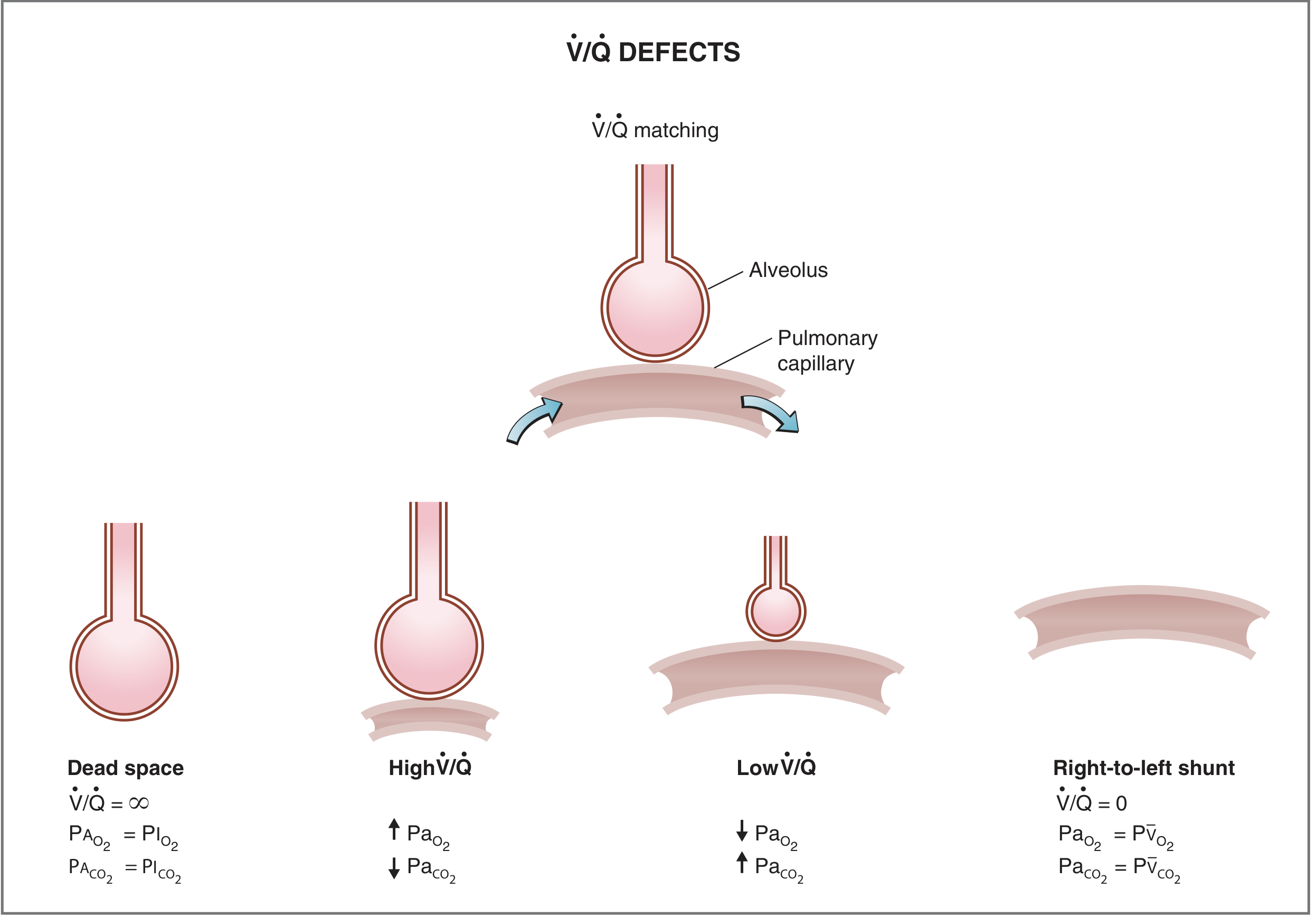
1. Dead Space (V/Q = ∞)
- Alveoli are ventilated but not perfused
- No gas exchange occurs - ventilation is "wasted"
- Alveolar gas equilibrates with inspired air: PA,O2 = 150 mmHg, PA,CO2 = 0 mmHg
- Physiological dead space = anatomic dead space + alveolar dead space
- Clinical example: Pulmonary embolism (blood flow to a lung region is blocked)
- Anatomic dead space (~150 mL in a 150-lb adult; ~1 mL/lb body weight) is the gas in conducting airways; alveolar dead space is ventilated but unperfused alveoli. Together they form physiologic dead space.
2. High V/Q
- High ventilation relative to reduced (but not absent) perfusion
- Blood leaving these alveoli has high PO2 and low PCO2
- Represents a partial step toward dead space
3. Low V/Q
- Reduced ventilation relative to maintained (or relatively increased) perfusion
- Blood leaving these alveoli has low PO2 and high PCO2
- The most common contributor to clinical hypoxemia
- Clinical examples: COPD, asthma, interstitial edema
4. Right-to-Left Shunt (V/Q = 0)
-
Alveoli are perfused but not ventilated - blood bypasses gas exchange entirely
-
Pulmonary capillary blood has the same composition as mixed venous blood: PaO2 ~40 mmHg, PaCO2 ~46 mmHg
-
Clinical examples: Pneumonia, atelectasis, alveolar flooding (pulmonary edema), right-to-left cardiac shunts (ASD, VSD)
-
Costanzo Physiology, 7th ed.
-
Murray & Nadel's Textbook of Respiratory Medicine
Mechanism of Hypoxemia in V/Q Mismatch
The key diagram below compares ideal vs. uncompensated V/Q mismatch:
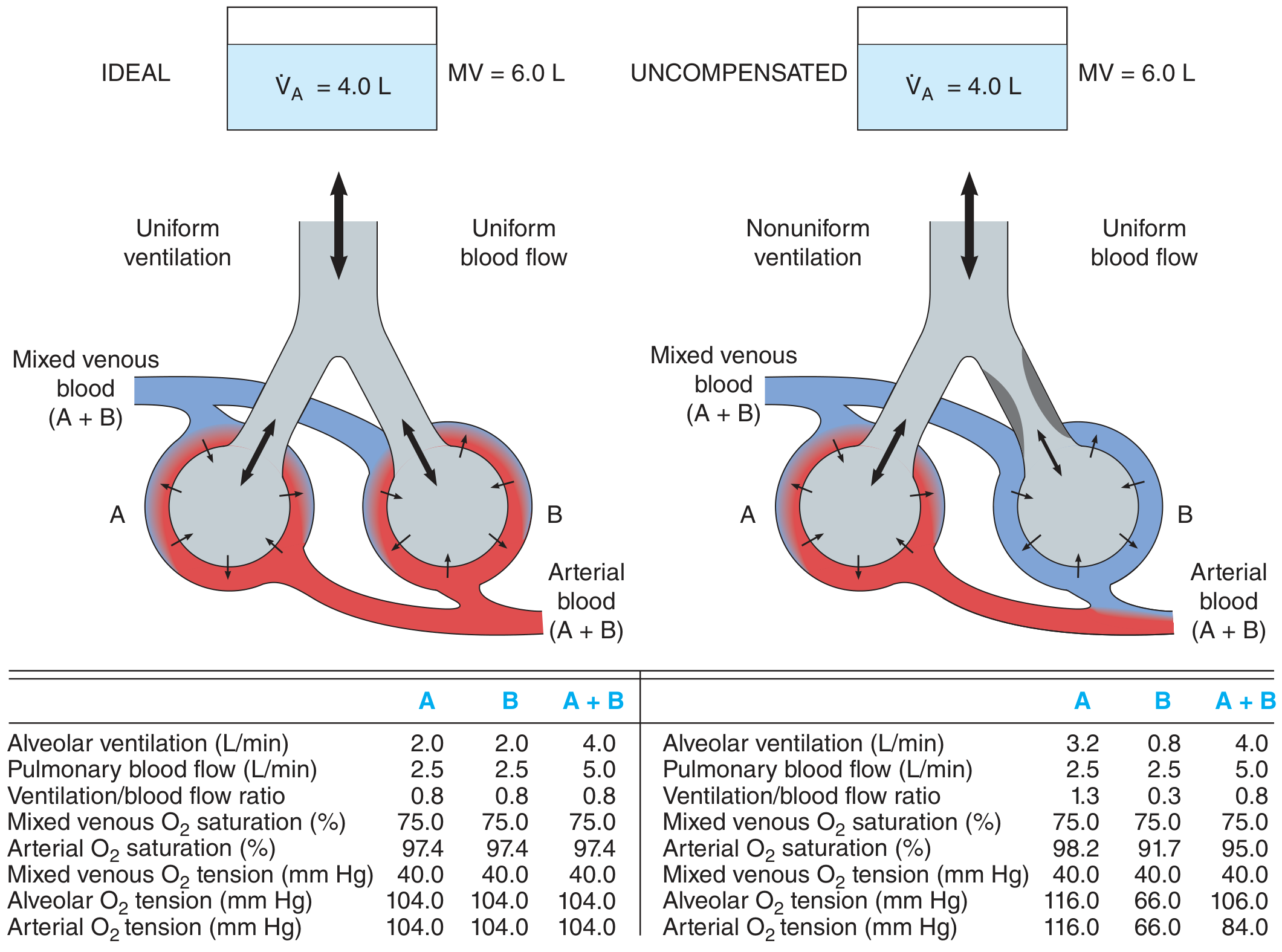
- In the ideal lung (left): both alveoli A and B have V/Q = 0.8; arterial O2 saturation = 97.4%
- In the uncompensated lung (right): alveolus A is overventilated (V/Q = 1.3), alveolus B is underventilated (V/Q = 0.3). The mixed arterial O2 saturation falls to 95% and arterial PO2 drops to 84 mmHg.
Why can't the overventilated alveolus compensate? Because hemoglobin is nearly fully saturated at normal PO2; raising alveolar PO2 further adds very little extra O2 to already-saturated hemoglobin. The underventilated alveolus produces significantly desaturated blood, and this cannot be fully corrected by the overventilated unit. This is the oxyhemoglobin dissociation curve asymmetry at work.
Effect on CO2: CO2 is generally maintained at normal levels in pure V/Q mismatch because the overventilated units can "blow off" extra CO2 (CO2 dissociation is nearly linear), compensating for CO2 retention by underventilated units. However, in severe mismatch, CO2 retention (hypercapnia) may occur.
- Ganong's Review of Medical Physiology, 26th ed.
Alveolar-Arterial (A-a) Gradient
V/Q mismatch produces an elevated alveolar-arterial PO2 difference (A-a gradient):
- Normal A-a gradient: ~5-15 mmHg (increases with age)
- Formula: A-a gradient = PAO2 - PaO2
- PAO2 = PIO2 - (PaCO2/RQ) = [FiO2 × (Patm - PH2O)] - (PaCO2/0.8)
- V/Q mismatch causes elevated A-a gradient with hypoxemia that responds to supplemental oxygen (distinguishes it from shunt, which does not)
Compensatory Mechanisms
-
Hypoxic Pulmonary Vasoconstriction (HPV): When alveolar PO2 falls (low V/Q regions), local pulmonary arterioles constrict, diverting blood flow away from poorly ventilated areas to better-ventilated ones. This helps reduce mismatch. If extensive and prolonged, HPV can cause pulmonary hypertension and cor pulmonale.
-
Increased minute ventilation: Chemoreceptors detecting hypoxemia or hypercapnia stimulate increased ventilation. This increases V/Q in underventilated areas and reduces CO2 retention.
-
Response to supplemental O2: Increasing FiO2 raises alveolar PO2 in low-V/Q units, improving oxygenation. This is why V/Q mismatch (unlike pure shunt) responds well to supplemental oxygen.
- Fishman's Pulmonary Diseases and Disorders
Clinical Conditions Causing V/Q Mismatch
| Condition | Mechanism |
|---|---|
| COPD | Airflow obstruction → low V/Q; emphysema causes dead space |
| Asthma | Bronchospasm → low V/Q |
| Pulmonary embolism | Blocked perfusion → high V/Q / dead space |
| Pneumonia | Alveolar filling → low V/Q / shunt |
| Pulmonary edema | Interstitial/alveolar fluid → low V/Q |
| ARDS | Widespread low V/Q and shunt |
| Atelectasis | No ventilation → shunt (V/Q = 0) |
| COVID-19 | Increased V/Q mismatch from alveolar/interstitial damage and prothrombotic effects |
Measurement of V/Q Distribution
The Multiple Inert Gas Elimination Technique (MIGET) is the gold standard for assessing V/Q distribution. A mixture of 6 inert gases (e.g., SF6, ethane, cyclopropane, isoflurane, ether, acetone) is infused intravenously. Arterial and mixed-expired samples are analyzed; the retention-solubility and excretion-solubility curves reveal the full distribution of V/Q ratios in diseased lungs.
V/Q scanning (nuclear medicine) using inhaled radioactive gas (ventilation) and injected radiolabeled particles (perfusion) provides clinical assessment, especially in pulmonary embolism diagnosis.
- Murray & Nadel's Textbook of Respiratory Medicine
Summary Table
| Parameter | Dead Space (V/Q = ∞) | Low V/Q | Shunt (V/Q = 0) |
|---|---|---|---|
| Ventilation | Present | Reduced | Absent |
| Perfusion | Absent | Present | Present |
| PaO2 | = PI,O2 (~150 mmHg) | Decreased | = Mixed venous (~40 mmHg) |
| PaCO2 | = 0 | Increased | = Mixed venous (~46 mmHg) |
| Response to O2 | - | Improves | Minimal improvement |
| Example | PE | COPD, asthma | Pneumonia, atelectasis |
Key takeaways for 10-mark answer:
- V/Q mismatch is the most common cause of hypoxemia in clinical medicine
- Normal V/Q = 0.8; ranges from 0 (shunt) to ∞ (dead space)
- Low V/Q units cause hypoxemia because of the sigmoid shape of the oxyhemoglobin dissociation curve (high V/Q units cannot compensate)
- CO2 is less affected than O2 because of the linear CO2 dissociation curve
- Hypoxic pulmonary vasoconstriction is the main compensatory mechanism
- V/Q mismatch-related hypoxemia corrects with supplemental O2; true shunt does not
This is a shared conversation. Sign in to Orris to start your own chat.