
Where is ecg
The ECG (electrocardiogram) is recorded by placing electrodes on the skin of the chest, limbs, and sometimes other areas. Here is a quick overview:
Standard 12-lead ECG electrode placement:
- Limb leads (4 electrodes): Right arm (RA), Left arm (LA), Right leg (RL - ground), Left leg (LL)
- Precordial (chest) leads (6 electrodes):
- V1: 4th intercostal space, right sternal border
- V2: 4th intercostal space, left sternal border
- V3: Between V2 and V4
- V4: 5th intercostal space, midclavicular line
- V5: Anterior axillary line (same level as V4)
- V6: Midaxillary line (same level as V4 and V5)
Searching sources...
Let me pull the authoritative details from the medical library.
Reading File
Finding Sources
Searching the Web
ECG electrode placement 12 lead position anatomy
Finding Sources
Finding Sources
Reading File
Finding Sources
Finding Sources
Reading File
Reading File
Loading Image
Loading Image
Here is a complete, textbook-sourced answer on where the ECG electrodes are placed:
ECG Electrode Placement - Standard 12-Lead
Precordial (Chest) Leads - V1 to V6
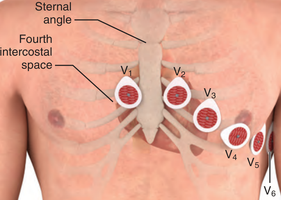
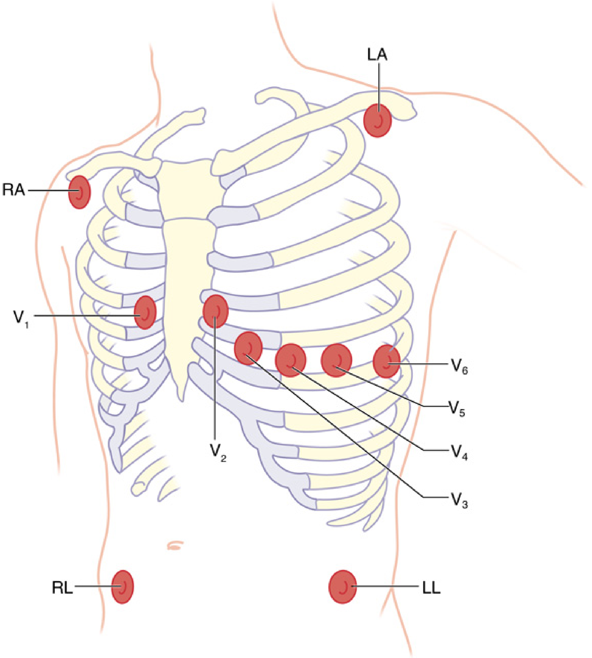
| Lead | Position |
|---|---|
| V1 | 4th intercostal space, right sternal border |
| V2 | 4th intercostal space, left sternal border |
| V3 | Between V2 and V4 (diagonal) |
| V4 | 5th intercostal space, midclavicular line |
| V5 | Same horizontal level as V4, anterior axillary line |
| V6 | Same horizontal level as V4/V5, midaxillary line |
Key landmark tip: Start at the sternal angle of Louis (manubriosternal junction) - the rib space immediately below it is the 2nd intercostal space. Count down to the 4th for V1/V2, and to the 5th for V4.
Limb Leads (4 electrodes)
| Electrode | Standard placement |
|---|---|
| RA (Right Arm) | Right wrist (distal to shoulder) |
| LA (Left Arm) | Left wrist (distal to shoulder) |
| RL (Right Leg) | Right ankle (ground electrode) |
| LL (Left Leg) | Left ankle |
Electrodes may be moved more proximally (e.g., to shoulders/thighs) if necessary due to amputation or injury, with a notation made on the ECG.
Important notes
- V5 is the most sensitive single lead for detecting myocardial ischemia during anesthesia and exercise testing.
- V3/V4 may be preferred for detecting postoperative ischemia in high-risk vascular surgery patients.
- Misplacement is common - studies show up to 50% of clinicians misplace V1/V2. Always count ribs from the sternal angle, never guess.
- Mason-Likar modification (used by paramedics/monitors): limb leads are moved to the torso (infraclavicular fossae for arms, iliac crests for legs), but precordial placement stays the same. This can cause a rightward axis shift and reduce inferior Q waves.
Sources: Roberts and Hedges' Clinical Procedures in Emergency Medicine, Fig. 14.4 | Miller's Anesthesia 10e, Fig. 32.4 | Pfenninger and Fowler's Procedures for Primary Care
This is a shared conversation. Sign in to Orris to start your own chat.