
Recent treatment modalities in malignant otitis media and malignant otitis media in detail as a long question answer according to standard ENT textbook in simplified manner
"malignant otitis externa" AND treatment
malignant otitis externa granulation tissue external auditory canal
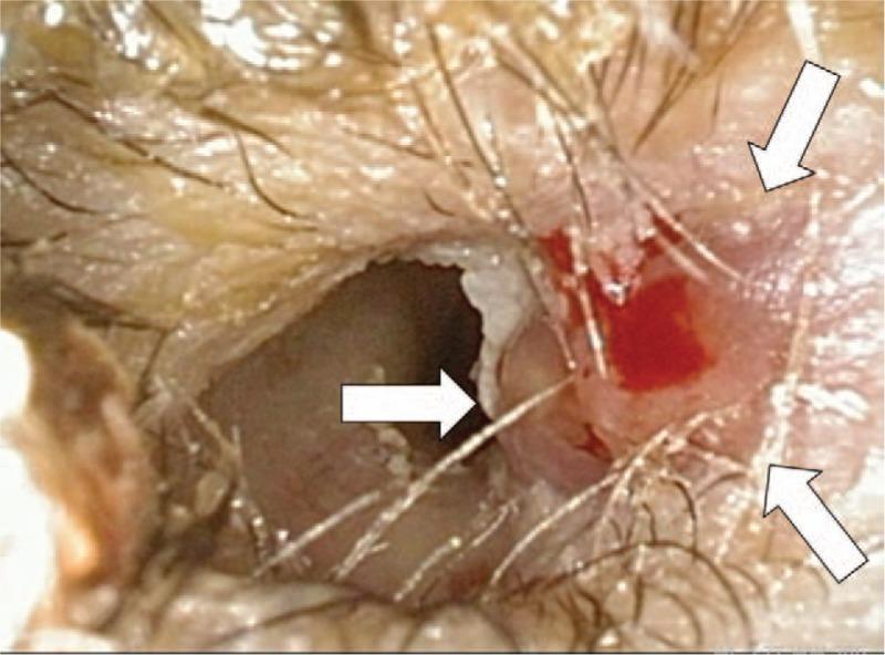
Clinical photograph of an otoscopic view showing the right external auditory canal. The image demonstrates significant pathological changes consistent with malignant external otitis. White arrows highlight the presence of friable, erythematous granulation tissue and focal ulceration on the floor and posterior wall of the canal. There is evidence of active inflammation with associated serosanguinous discharge and minor blood spotting. The canal lumen appears narrowed due to soft tissue edema. This visual evidence illustrates a hallmark clinical sign of skull base osteomyelitis (malignant otitis externa), particularly in the context of immunocompromised states such as diabetes mellitus or post-radiation therapy for head and neck squamous cell carcinoma. The surrounding skin of the meatus shows sparse hairs and mild maceration.
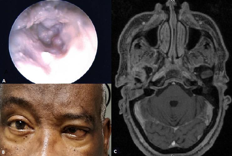
This composite figure illustrates a clinical case of fungal malignant external otitis (necrotizing otitis externa) through three modalities: endoscopic photography, clinical photography, and diagnostic imaging. Image A is an otoscopic view of the external auditory canal showing extensive pale, fleshy granulation tissue with irregular, bumpy morphology and inflammatory exudate obstructing the lumen. Image B is a clinical photograph of a patient's mid-face demonstrating cranial nerve involvement, specifically characterized by facial nerve palsy manifesting as right-sided facial asymmetry and ptosis. Image C is an axial MRI scan (T1-weighted) at the level of the skull base, showing soft tissue infiltration and inflammatory changes in the infratemporal fossa and the vicinity of the stylomastoid foramen, consistent with the spread of infection. Collectively, these images represent the diagnostic triad of malignant external otitis: visible otoscopic pathology, neurological complications such as facial nerve paralysis, and radiological evidence of skull base osteomyelitis or soft tissue extension.
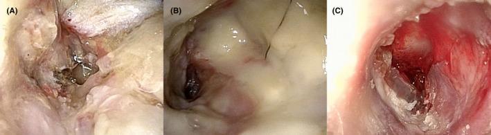
Three-panel endoscopic clinical photograph illustrating the progression of malignant otitis externa (MOE) and postoperative healing in the left external auditory canal. Panel (A), taken two weeks post-canal wall down mastoidectomy, shows a healing surgical cavity with pale granulation tissue and early epithelialization. Panel (B), at two weeks post-discharge, demonstrates clinical worsening characterized by a pale, edematous mass and purulent-appearing discharge obscuring the surgical site, associated with the emergence of MRSA. Panel (C), following readmission and targeted antibiotic treatment (vancomycin and cefepime/ceftazidime), shows intense erythema, raw inflammatory tissue, and active drainage (otorrhea) indicative of severe inflammation and skull base osteomyelitis. The sequence provides an educational visual timeline of surgical management, complication by antibiotic-resistant pathogens, and subsequent inflammatory response during systemic treatment in a high-risk patient with diabetes and chronic renal failure.
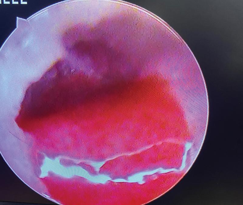
This clinical photograph is an endoscopic view of the external auditory canal (EAC), illustrating a granulation polyp. The primary feature is a large, erythematous, and fleshy mass that partially or completely occupies the canal lumen. The mass displays a heterogeneous red-to-purple hue, suggesting significant vascularity and inflammation. Its surface texture is irregular and granular, characteristic of granulation tissue. Near the inferior aspect of the lesion, light-colored, winding bands are visible, representing serous or purulent fluid-filled tracks or mucopurulent discharge commonly associated with chronic suppurative otitis media (CSOM). This visual provides a clinical example of an aural polyp, which is a significant finding in otolaryngology as it may obscure the view of the tympanic membrane and can be associated with underlying conditions such as cholesteatoma or malignant otitis externa. The image is intended for medical students and clinicians to recognize inflammatory middle ear disease complications.
Malignant Otitis Media (Malignant Otitis Externa / Necrotizing Otitis Externa)
INTRODUCTION AND TERMINOLOGY
Note on Nomenclature: The term "malignant" does not imply a neoplastic process. It was coined by Chandler in 1968 to reflect the aggressive, often fatal course. The condition was first described by Meltzer and Kelemen in 1959. The term "Necrotizing Otitis Externa" or "Skull Base Osteomyelitis" better describes its pathophysiology.
EPIDEMIOLOGY
- Predominantly affects elderly diabetic patients - glucose intolerance may be present in up to 90% of cases
- Also seen in other immunocompromised states: HIV/AIDS, myeloid malignancies, pharmacological immunosuppression, organ transplant recipients
- Rarely reported in immunocompetent individuals
- Mortality: 5-20% even in the antibiotic era; higher with intracranial extension
ETIOLOGY AND PATHOGENESIS
Why Diabetics Are Affected
- Microangiopathy of the ear canal vessels - impairs local blood supply
- Increased pH of cerumen in diabetics - promotes bacterial growth
- Impaired polymorphonuclear leukocyte function - reduces ability to fight infection
- Diabetic microangiopathy causes vascular thrombosis → coagulative necrosis of surrounding tissue
Causative Organism
- Pseudomonas aeruginosa accounts for >90% of cases
- Other organisms (rare): Staphylococcus aureus, S. epidermidis, Proteus mirabilis, Klebsiella oxytoca, fungal species
- Fungal MOE (rare): most commonly Aspergillus fumigatus, associated with HIV-positive patients; 84% of fungal MOE cases occur in immunocompromised individuals
- Fluoroquinolone-resistant Pseudomonas is an increasing and concerning problem
SPREAD AND PATHOLOGY
- Starts in soft tissues of the EAC (skin and subcutaneous tissue of the cartilaginous canal)
- Spreads to the tympanic bone via:
- Fissures of Santorini (gaps in the cartilaginous floor of the EAC)
- Tympanomastoid suture → stylomastoid foramen → jugular foramen
- Hallmark: Does NOT spread through pneumatized tracts like typical temporal bone infections; the middle ear is rarely involved until late
- Spreads via Haversian system of compact bone, forming multiple abscesses and bony sequestra
- Extension pathways:
- Petrotympanic fissure → temporomandibular joint (TMJ) and infratemporal fossa
- Inferiorly → soft tissue spaces below skull base (periparotid, cervical)
- Medially → carotid canal, jugular foramen
- Intracranially → via venous channels and facial planes → dural sinuses
CLINICAL FEATURES
Symptoms
- Severe, deep-seated otalgia - typically worse at night, out of proportion to examination findings
- Otorrhoea (ear discharge)
- Aural fullness
- Fever (may be absent even in active disease)
Signs
| Finding | Significance |
|---|---|
| Granulation tissue at bony-cartilaginous junction of EAC | Pathognomonic / Cardinal sign |
| Ear canal edema and skin necrosis | Active disease |
| Proptosis of the auricle | Extension into periauricular tissue |
| Cranial nerve VII palsy (facial palsy) | Most common cranial nerve affected - stylomastoid foramen involvement |
| CNs IX, X, XI, XII palsy | Disease extending to jugular foramen |
| CNs V and VI palsy | Extension to the petrous apex |
| Meningeal signs (headache, neck stiffness, fever, altered consciousness) | Intracranial extension |
| "Picket fence" spiking fevers | Septic thrombophlebitis of sigmoid sinus |
Key point: Multiple cranial neuropathies indicate a worse prognosis. The facial nerve is most commonly involved due to proximity of the stylomastoid foramen to the floor of the EAC.
INVESTIGATIONS
1. Biopsy of Granulation Tissue
- Mandatory - both to rule out malignancy and for culture purposes
- Bacterial and fungal cultures must be sent simultaneously
2. Laboratory Tests
- ESR (Erythrocyte Sedimentation Rate): typically elevated; nonspecific but useful to follow treatment response and detect possible recurrence
- White blood cell count: may be normal even in active disease
- Blood glucose
3. Imaging
A. Nuclear Medicine (Most Important)
| Scan | Role |
|---|---|
| Technetium-99m (Tc-99) Scintigraphy | Imaging of choice to CONFIRM diagnosis; concentrates in areas of osteoblastic activity; high sensitivity for bone infection; turns positive BEFORE CT shows changes; SPECT more sensitive than planar imaging; however, remains positive for months even after treatment - NOT useful to follow response |
| Gallium-67 (Ga-67) scan | Shows areas of inflammatory cell activity; used to MONITOR treatment response - normalizes as infection resolves; scan every 4 weeks during treatment |
| Indium-111 labeled leukocyte scan | Shows inflammatory cell activity; sensitivity increased when combined with Tc-99 scan |
Memory tip: Tc-99 = Diagnosis; Ga-67 = Follow-up/treatment monitoring
B. CT Scan (High-Resolution Temporal Bone CT)
- Demonstrates bony erosion and soft tissue abnormalities around EAC
- Cannot distinguish infection from malignancy
- Significant bony erosion must occur before findings appear - early changes may be missed
- Not useful for following treatment response
- Early sign: subtle soft tissue thickening in EAC; later: cortical bone erosions
C. MRI
- Better than CT for detecting soft tissue changes and dural/intracranial involvement
- Imaging of choice when infection spreads beyond temporal bone (facial nerve or intracranially)
- MR Angiography can evaluate patency of dural sinuses
DIAGNOSIS - COHEN AND FRIEDMAN CRITERIA
- Refractory otitis externa
- Severe nocturnal otalgia
- Purulent otorrhoea
- Granulation tissue in the EAC
- Positive Tc-99 bone scan
- Immunocompromised state (especially diabetes)
TREATMENT
A. General / Supportive Measures
- Aggressive blood glucose control in diabetic patients - essential and cannot be overemphasized
- Correct other causes of immunodeficiency where possible (e.g., adjust immunosuppressive medications, treat HIV)
- Pain management - analgesics (NSAIDs, opioids as needed)
- Aural toilet / EAC debridement - regular cleaning of the ear canal (daily debridement)
B. Antibiotic Therapy (Cornerstone of Treatment)
First-Line: Oral Fluoroquinolone
- Ciprofloxacin 750 mg orally twice daily is the antibiotic of choice for MOE
- Fluoroquinolones are the only oral antibiotics with reliable antipseudomonal activity
- Monotherapy with oral ciprofloxacin is effective in patients without intracranial complications or cranial neuropathies
- Dose recommended by Scott-Brown's: 1.5 g/day over 6-12 weeks
Duration of Treatment
- Minimum 6-8 weeks (Cummings, Scott-Brown's)
- Treatment should continue until clinical examination normalizes
- Can treat until Gallium-67 scan normalizes (most reliable endpoint, though may prolong treatment)
When to Escalate to IV/Parenteral Antibiotics
- Advanced cases (intracranial extension, cranial neuropathy)
- Failure of outpatient oral therapy (failure rate up to 33% in some series)
- When the patient cannot take oral medications
- In these cases: IV fluoroquinolone via PICC line, or parenteral antipseudomonal aminopenicillins
- Patients stable on IV can be discharged home with frequent outpatient debridement
Fluoroquinolone-Resistant Pseudomonas
- An increasing problem; may require:
- 3rd or 4th generation cephalosporins (e.g., ceftazidime, cefepime) ± aminoglycoside
- Culture-directed therapy is strongly preferred whenever cultures are available
Double Coverage
- Empiric double coverage directed against Pseudomonas is begun after cultures are obtained in advanced cases (Shambaugh)
C. Antifungal Therapy (for Fungal MOE)
- Parenteral antifungal therapy for culture-positive fungal MOE
- Amphotericin B: traditionally used, but has significant side effects (nephrotoxicity, etc.)
- Voriconazole: now the preferred first-line therapy for invasive Aspergillosis; more favourable side-effect profile than Amphotericin B
- Other azoles (fluconazole, itraconazole) used depending on species and sensitivity
D. Surgical Treatment
| Indication | Procedure |
|---|---|
| Abscess formation | Drainage |
| Necrotic bone (sequestrum) | Debridement of bony sequestrum |
| Recalcitrant/progressive disease | More extensive debridement |
| Granulation tissue | Local removal / debridement |
Important: Surgical intervention is NOT indicated for facial nerve paralysis in MOE - decompression of the facial nerve fails to address the extratemporal location of nerve involvement (Shambaugh). Heroic surgical measures are reserved for recalcitrant disease only.
E. Hyperbaric Oxygen Therapy (HBO)
- The role of HBO in MOE is unproven (KJ Lee's)
- Theoretically benefits anaerobic wound healing and enhances neutrophil killing
- Used as an adjunct in refractory cases in some centers
- Not part of routine standard management
MONITORING RESPONSE TO TREATMENT
| Parameter | Use |
|---|---|
| Gallium-67 scan (every 4 weeks) | Best modality to monitor treatment response |
| ESR | Nonspecific, but can follow trends |
| Clinical examination | Resolution of granulation tissue, otalgia, discharge |
| Tc-99 scan | NOT useful for monitoring (stays positive too long) |
COMPLICATIONS
| Complication | Mechanism |
|---|---|
| Facial nerve palsy (CN VII) | Stylomastoid foramen involvement - most common |
| Cranial nerve IX, X, XI, XII palsy | Jugular foramen extension |
| CN V, VI palsy | Petrous apex extension |
| Sigmoid sinus thrombosis | Spread via venous channels |
| Meningitis | Intracranial extension |
| Temporal lobe / cerebellar abscess | Intracranial extension |
| Cavernous sinus thrombosis | Medial spread |
| Death | End-stage untreated disease |
PROGNOSIS
- Mortality: 5-20% in the antibiotic era
- Extension to skull base, cranial nerve involvement, and intracranial extension at presentation all correlate with higher mortality
- Good prognostic factors: early diagnosis, good diabetic control, culture-directed antibiotic therapy
- Relapse can occur; Gallium-67 monitoring helps detect recurrence
CLINICAL IMAGES
RECENT UPDATES IN TREATMENT (2021-2026)
- Fluoroquinolone resistance in Pseudomonas is a growing challenge - culture-directed therapy is increasingly important
- Fungal MOE is now well-recognized, especially in HIV patients (PMID 39534824 - Sideris et al., 2024 systematic review); voriconazole has largely replaced amphotericin B for Aspergillus
- Diagnostic conundrum: NOE can be difficult to distinguish from other skull base pathologies - biopsy + imaging + nuclear medicine remain the diagnostic triad (PMID 38560038 - Ahmed et al., 2024)
- Mortality data: A 2025 meta-analysis (PMID 40765727) confirms significant mortality, particularly with intracranial extension
- Oral ciprofloxacin remains effective for uncomplicated MOE; intravenous options are needed for resistant or complicated disease
SUMMARY TABLE
| Feature | Detail |
|---|---|
| Definition | Aggressive osteomyelitis of EAC and skull base |
| Old names | Malignant OE, Necrotizing OE, Skull Base Osteomyelitis |
| Commonest in | Elderly diabetics; immunocompromised |
| Organism | Pseudomonas aeruginosa (>90%) |
| Hallmark sign | Granulation tissue at bony-cartilaginous junction |
| Pathognomonic imaging | Tc-99 bone scan (confirm); Ga-67 (monitor) |
| Antibiotic of choice | Ciprofloxacin 750 mg BD × 6-8 weeks |
| Surgery role | Limited - only abscess/necrotic bone debridement |
| CN most affected | VII (facial nerve) |
| Monitoring | Gallium-67 scan every 4 weeks; ESR |
| Mortality | 5-20% even in antibiotic era |