
differentiate submandibular gland and lymph node clinically
Reading File
Now let me search the medical library and web in parallel.
Finding Sources
Finding Sources
Searching the Web
clinical differentiation submandibular gland vs lymph node examination
The S Das Manual on Clinical Surgery result looks very relevant. Let me read those sections in detail.
Reading File
Reading File
Reading File
I now have comprehensive content from S Das Manual on Clinical Surgery. Let me also fetch the key image showing bimanual palpation to include in the answer.
Loading Image
I now have comprehensive information. Here is the complete, well-structured answer:
Clinical Differentiation: Submandibular Gland vs. Lymph Node
The submandibular triangle contains both the submandibular salivary gland and level IB lymph nodes in close proximity, making clinical differentiation one of the classic challenges in head and neck examination.
1. History
| Feature | Submandibular Gland | Lymph Node |
|---|---|---|
| Meal-related symptoms | Swelling increases at mealtimes (colicky pain, tense swelling) - strongly suggests ductal calculus | No relationship to meals |
| Associated symptoms | Dry mouth, foul-tasting saliva, pain on eating | Fever, sore throat, dental pain (reactive), weight loss/night sweats (lymphoma/malignancy) |
| Duration | Often intermittent (calculus) or slowly progressive (tumour) | Acute (infection), chronic (TB, lymphoma) |
| Source of primary disease | Oral cavity, floor of mouth | Search for primary in oral cavity, oropharynx, scalp, skin |
A history of colicky submandibular swelling that appears during meals and subsides afterwards is essentially diagnostic of a submandibular duct calculus. - S Das Manual on Clinical Surgery, 13th Ed., p. 362
2. Inspection
| Feature | Submandibular Gland | Lymph Node |
|---|---|---|
| Number of swellings | Single, smooth, diffuse swelling | Often multiple (discrete or matted) |
| Response to lemon juice | Swelling appears/enlarges promptly when patient sucks lemon (stimulates salivary flow against obstruction) | No change |
| Wharton's duct orifice | May show redness, swelling, pus, or a visible stone at the orifice near the frenulum linguae | Normal orifice |
3. Palpation - The Key Differentiating Step
Number and character of swellings:
- Submandibular gland enlargement = one swelling, uniform
- Lymph node enlargement = nodular, either discrete or matted
Bimanual palpation (the most important manoeuvre):
One finger is placed on the floor of the mouth (intraoral), medial to the alveolus and lateral to the tongue, pressed as far back as possible. The fingers of the other hand are placed externally just medial to the inferior border of the mandible and pushed upward.
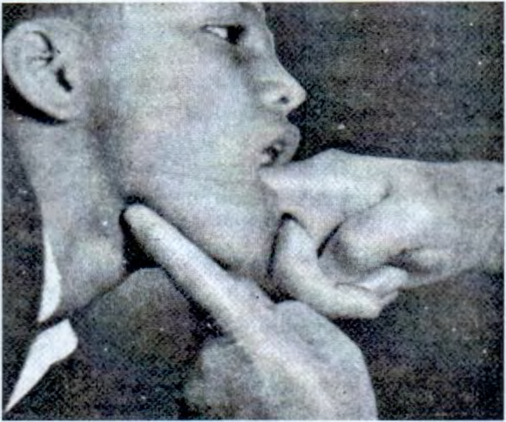
Bimanual palpation - S Das Manual on Clinical Surgery, Fig. 25.14
The anatomical key: The finger inside the mouth can feel the deep part of the salivary gland (which lies above the mylohyoid muscle) but cannot feel lymph nodes (which lie below the mylohyoid muscle). This is the single most reliable clinical sign.
| Palpation Feature | Submandibular Gland | Lymph Node |
|---|---|---|
| Deep lobe palpable intraorally | Yes - felt above mylohyoid | No - lies below mylohyoid |
| Consistency | Firm, uniform, possibly a hard calculus | Rubbery (lymphoma), firm-hard (metastasis), soft/fluctuant (abscess) |
| Surface | Smooth | Nodular, discrete, or matted |
| Mobility | Moves with the gland | Mobile (reactive/lymphoma), fixed (metastatic) |
| Tenderness | Tender in infection/calculus | Tender in acute infection, non-tender in lymphoma/metastasis |
| Calculus palpable | Sometimes felt along Wharton's duct | Never |
4. Additional Oral Examination
If a lymph node is suspected, a thorough search for the primary source is mandatory:
- Floor of mouth, tongue (especially posterior 1/3), buccal mucosa, tonsils, lower lip, teeth/gums
- This is because submandibular (level IB) nodes drain the floor of mouth, tongue, lower lip, and anterior oral cavity
"For the latter case one must examine thoroughly the inside of the mouth including the upper lip, the lower lip, the cheeks, the tongue and the floor of the mouth." - S Das Manual on Clinical Surgery, p. 363
5. Special Tests
| Test | Relevance |
|---|---|
| Lemon/citric acid stimulation | Enlarges gland swelling if calculus present; no effect on lymph node |
| Swab test (Wharton's duct) | Dry swab on affected side = stone impacted in duct |
| Milking the gland | Pus from orifice = gland infection; normal saliva or nothing in lymph node pathology |
| X-ray floor of mouth | Radio-opaque calculus seen in ~80% of submandibular stones (most are radio-opaque unlike parotid) |
Summary: The 3 Most Reliable Differentiating Points
- Bimanual palpation - deep lobe palpable intraorally = salivary gland (above mylohyoid); not palpable = lymph node (below mylohyoid)
- Meal-related fluctuation - increases with meals/citric stimulation = gland (especially calculus)
- Number - single swelling = more likely gland; multiple nodular swellings = lymph nodes
Source: S Das Manual on Clinical Surgery, 13th Edition, pp. 362-363
This is a shared conversation. Sign in to Orris to start your own chat.