
What is purpura physiology mbbs 1st year
Reading File
Now let me search the medical library for purpura physiology content:
Finding Sources
Finding Sources
Let me read the most relevant sections - Harrison's comprehensive purpura section and the Andrews' dermatology section:
Reading File
Reading File
Finding Sources
Now let me get the physiology-specific content from Ganong's and Robbins, plus the Harrison's full text:
Reading File
Reading File
Reading File
Let me fetch the petechiae image from Robbins and also get more content from Harrison's on palpable vs non-palpable purpura:
Loading Image
Reading File
I now have comprehensive information from multiple authoritative textbooks. Let me compile the complete answer.
Purpura - Physiology (MBBS 1st Year)
Definition
Purpura is the term for extravasation (leakage) of red blood cells (RBCs) into the skin, mucous membranes, or serosal surfaces, producing lesions that do not blanch with pressure (diascopy). This non-blanching nature is the key clinical differentiator from erythema or vasodilation, which DO blanch.
- Andrews' Diseases of the Skin, p. 947
- Robbins & Kumar Basic Pathology
Types by Size
| Term | Size | Description |
|---|---|---|
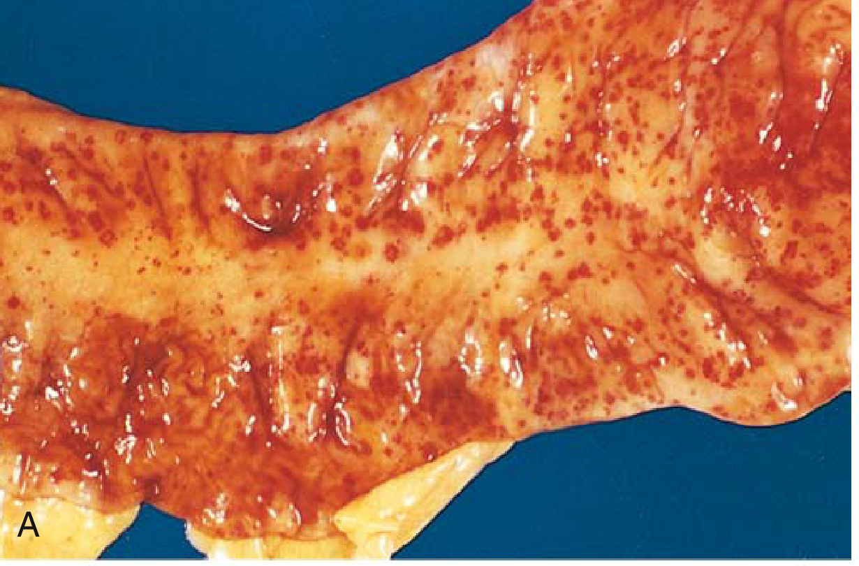
| Petechiae | < 2-3 mm | Pinhead-sized, round, superficial hemorrhagic macules; bright red initially, then brownish/rust-colored; occur in crops on dependent areas; classically suggest platelet disorders |
| Purpura | 3-5 mm | Slightly larger hemorrhages; same mechanisms as petechiae |
| Ecchymoses | 1-2 cm | "Bruises" - deeper, irregularly shaped blue-purple patches; turn yellow as hemoglobin is degraded; suggest coagulation factor defects |
| Vibices | Linear | Linear purpuric streaks |
| Hematoma | Large | A palpable, fluctuant pool of extravasated blood in a tissue dead space |
Physiological Basis - Why Purpura Occurs
Normal hemostasis prevents RBC leakage via three mechanisms. Failure of any one causes purpura:
1. Platelet Disorders (most common cause of petechiae/purpura)
- Normally, platelets adhere to exposed subendothelial collagen and von Willebrand factor (vWF) after vessel injury
- Activated platelets release ADP, thromboxane A2, and PAF, triggering platelet aggregation and plug formation
- When platelet count is LOW (thrombocytopenia) or platelet function is ABNORMAL:
- Clot retraction is deficient
- Ruptured vessels fail to constrict adequately
- Result: Thrombocytopenic purpura - easy bruisability and multiple subcutaneous hemorrhages
- Purpura can also occur with a normal platelet count if platelets are functionally abnormal: Thrombasthenic purpura
- Ganong's Review of Medical Physiology, 26e
2. Vascular Wall Defects (increased fragility)
- The dermal connective tissue provides physical support to blood vessels
- When this support is lost, minor trauma easily ruptures vessels:
- Scurvy (Vitamin C deficiency): Vit C is a cofactor for lysyl hydroxylase, needed for collagen cross-linking. Loss of collagen support = perifollicular hemorrhages + purpura
- Senile/Solar (actinic) purpura: UV damage and aging degrade supporting connective tissue, especially on extensor forearms
- Steroid purpura: Glucocorticoids (topical or systemic) cause dermal atrophy
- Ehlers-Danlos syndrome: Defective collagen structure
- Amyloidosis: Amyloid deposits weaken vessel walls
- Harrison's Principles of Internal Medicine 22e
3. Coagulation Factor Defects
- Failure of the coagulation cascade (hemophilia, warfarin, Vitamin K deficiency, liver disease) leads to larger ecchymoses rather than petechiae
- There is often a component of trauma
- Andrews' Diseases of the Skin, p. 947
4. Intravascular Thrombi / Vascular Occlusion
- Paradoxically, excessive clotting can cause purpura by occluding small dermal vessels
- This produces retiform (net-like) purpura
- Causes: DIC, TTP, antiphospholipid antibody syndrome, cryoglobulinemia, warfarin/heparin reactions
- These thrombi are demonstrable on skin biopsy
5. Vascular Inflammation (Vasculitis)
- Inflammatory destruction of vessel walls allows RBC leakage
- Produces palpable purpura (raised, palpable elevation) - the classic sign of vasculitis
- Seen in: IgA vasculitis (Henoch-Schonlein), meningococcemia, Rocky Mountain spotted fever
- Tintinalli's Emergency Medicine
Classification Summary
PURPURA
├── NONPALPABLE (flat/macular)
│   ├── Platelet disorders (thrombocytopenia, thrombasthenia)
│   ├── Coagulation defects (hemophilia, anticoagulants)
│   ├── Vascular fragility (scurvy, Ehlers-Danlos, amyloidosis)
│   ├── Increased venous pressure (stasis purpura)
│   └── Intravascular thrombi (DIC, TTP) → retiform pattern
│
└── PALPABLE (raised, elevated)
    └── Vasculitis (vessel wall inflammation)
Key Points for MBBS 1st Year
- Purpura = RBCs leaked into dermis = does NOT blanch (vs. erythema/hyperemia which blanches because blood is still in vessels)
- Petechiae → platelet problem | Ecchymoses → coagulation factor problem (clinical rule)
- The color change of bruises (red → blue-green → yellow) reflects sequential enzymatic degradation: hemoglobin → biliverdin → bilirubin → hemosiderin
- Palpable purpura = vasculitis until proven otherwise
- In scurvy, purpura occurs because Vitamin C deficiency impairs collagen synthesis (specifically cross-linking) - weakening vascular support tissue
- Thrombocytopenic purpura: platelets < 50,000/µL usually needed for spontaneous purpura; < 20,000/µL for serious hemorrhage
Sources:
- Ganong's Review of Medical Physiology 26e - Chapter on Platelets, p. 93
- Robbins & Kumar Basic Pathology - Chapter on Hemorrhage, p. 82
- Harrison's Principles of Internal Medicine 22e (2025) - Chapter 61, p. 450
- Andrews' Diseases of the Skin - Purpura, p. 947
- Tintinalli's Emergency Medicine - Chapter 253
Create a study guide comparing platelet vs coagulation purpura
Finding Sources
Finding Sources
Reading File
Reading File
Finding Sources
Finding Sources
Reading File
Reading File
Finding Sources
I now have all the information needed. Let me build the comprehensive study guide.
📚 Study Guide: Platelet Purpura vs. Coagulation Purpura
Part 1 - The Foundation: Primary vs. Secondary Hemostasis
Understanding purpura requires understanding the two-stage system that normally keeps blood inside vessels.
| Primary Hemostasis | Secondary Hemostasis | |
|---|---|---|
| What it is | Platelet plug formation | Coagulation cascade (clotting factors) |
| Speed | Fast (seconds to minutes) | Slower (minutes) |
| Key players | Platelets, vWF, GpIb, GpIIb-IIIa | Factors VII, IX, X, V, VIII, prothrombin, fibrinogen |
| Trigger | Exposed subendothelial vWF and collagen | Exposed tissue factor (TF) |
| End product | Soft platelet plug | Hard fibrin mesh (stabilizes plug) |
| What it seals | Small vessels, capillaries, mucosa | Larger vessels |
"Primary hemostasis occurs even in patients with hemophilia." - K.J. Lee's Essential Otolaryngology
Memory hook: Think of it as a two-lock door - platelet plug = the first lock (quick), coagulation = the second lock (permanent). Both must work for normal hemostasis.
Part 2 - The Big Comparison Table
| Feature | Platelet (Primary) Purpura | Coagulation (Secondary) Purpura |
|---|---|---|
| Defect | ↓ Platelet number OR ↓ platelet function | Deficiency/dysfunction of clotting factors |
| Type of lesion | Petechiae (1-2 mm) and purpura (3-5 mm) | Ecchymoses (1-2 cm), hematomas |
| Skin appearance | Multiple small, superficial pinpoint spots | Large irregular bruises, deep bleeds |
| Distribution | Mucous membranes, skin (especially dependent areas) | Soft tissues, muscles, joints |
| Blanching? | No | No |
| Palpable? | No (flat, macular) | Can be palpable if hematoma forms |
| Joints involved? | Rarely | YES - hemarthrosis is characteristic |
| Mucosal bleeding | YES - epistaxis, gum bleeding, menorrhagia | Less common |
| Bleeding time | Prolonged | Normal |
| PT / PTT | Normal | Prolonged (PT in extrinsic/common defects; PTT in intrinsic pathway) |
| Platelet count | Low (in thrombocytopenia) or normal (in thrombasthenia) | Normal |
| Clot retraction | Abnormal | Normal |
| Onset after trauma | Immediate | Delayed (bleeds recur hours later) |
| Spontaneous bleeding threshold | Platelets < 20,000/µL | Severe factor deficiency (< 1% activity) |
Sources: Robbins, Cotran & Kumar Pathologic Basis of Disease; Robbins & Kumar Basic Pathology; Andrews' Diseases of the Skin
Part 3 - Causes at a Glance
🔴 Platelet Purpura - Causes
A. Thrombocytopenia (Low Platelet Count)
| Mechanism | Examples |
|---|---|
| Decreased production | Aplastic anemia, leukemia (marrow infiltration), megaloblastic anemia, alcohol, cytotoxic drugs |
| Increased destruction (immune) | ITP (Immune Thrombocytopenic Purpura), SLE, drug-induced (heparin, quinidine, sulfa) |
| Increased destruction (non-immune) | DIC, TTP, HUS, microangiopathic hemolytic anemia |
| Sequestration | Hypersplenism |
| Dilutional | Massive transfusions |
B. Platelet Dysfunction (Normal Count, Abnormal Function)
| Type | Examples |
|---|---|
| Congenital | Glanzmann's thrombasthenia (GpIIb-IIIa defect), Bernard-Soulier syndrome (GpIb defect) |
| Acquired | Aspirin (COX inhibitor), uremia, liver disease, gammopathy |
| vWF defect | von Willebrand disease |
🔵 Coagulation Purpura - Causes
| Factor Defect | Pathway | Disease |
|---|---|---|
| Factor VIII | Intrinsic | Hemophilia A |
| Factor IX | Intrinsic | Hemophilia B (Christmas disease) |
| Factor VII | Extrinsic | Rare congenital deficiency |
| Multiple factors | Extrinsic + Common | Liver disease, Warfarin (Vit K antagonist) |
| Multiple factors | All pathways | DIC (also causes thrombocytopenia - mixed picture) |
| Fibrinogen | Common | Afibrinogenemia |
Part 4 - Clinical Pattern Recognition
Platelet Purpura - The Classic Picture
- Tiny scattered pinpoint red spots (petechiae) on legs and oral mucosa
- Gum bleeding after brushing teeth
- Nosebleeds (epistaxis)
- Heavy periods (menorrhagia) in females
- Bleeding stops within minutes with pressure (primary plug forms quickly even with fewer platelets)
- No hemarthrosis
Coagulation Purpura - The Classic Picture
- Large spreading purple bruises in soft tissues
- Hemarthrosis (joint bleeds) - hallmark of hemophilia
- Bleeding recurs hours after apparent control (the platelet plug forms but dissolves without fibrin stabilization)
- Deep muscle hematomas after injections
- No petechiae
Part 5 - Lab Tests Decoded
Suspected bleeding disorder
         |
    Full blood count + Platelet count
         |
    ┌────┴────┐
Low platelets    Normal platelets
    |                |
Thrombocytopenia   ┌──────────┐
(platelet purpura) PT/PTT tests  Platelet function
                   |            tests (PFA-100)
               ┌───┴───┐           |
           PT long  PTT long   Abnormal →
           |        |          Thrombasthenia/
         Extrinsic Intrinsic   vWD
         pathway   pathway
         (VII)     (VIII,IX)
| Test | What it measures | Abnormal in |
|---|---|---|
| Platelet count | Number of platelets | Thrombocytopenia |
| Bleeding time / PFA-100 | Primary hemostasis | Platelet dysfunction, vWD |
| PT (Prothrombin Time) | Extrinsic pathway (Factor VII, X, V, II, fibrinogen) | Liver disease, warfarin, Factor VII deficiency |
| PTT (Partial Thromboplastin Time) | Intrinsic pathway (XII, XI, IX, VIII, X, V, II, fibrinogen) | Hemophilia A (Factor VIII), Hemophilia B (Factor IX) |
| Clot retraction | Platelet contraction ability | Thrombocytopenia, Glanzmann's thrombasthenia |
Source: Robbins, Cotran & Kumar Pathologic Basis of Disease, p. 619
Part 6 - Prototypical Diseases Side-by-Side
| ITP (Immune Thrombocytopenic Purpura) | Hemophilia A | |
|---|---|---|
| Category | Platelet (primary hemostasis defect) | Coagulation (secondary hemostasis defect) |
| Mechanism | Autoantibodies (IgG) against GpIIb-IIIa → platelet destruction by spleen | Deficiency of Factor VIII |
| Inheritance | Acquired (autoimmune) | X-linked recessive (males affected) |
| Platelet count | Low (<50,000/µL in bleeding) | Normal |
| PT | Normal | Normal |
| PTT | Normal | PROLONGED |
| Bleeding sites | Skin, mucous membranes | Joints (hemarthrosis), deep muscles |
| Lesion type | Petechiae, purpura | Ecchymoses, hematomas |
| Peripheral smear | Low platelets, large platelets (megaplatelets) | Normal |
| Treatment | Corticosteroids, IVIG, splenectomy, TPO agonists | Factor VIII replacement |
Part 7 - Quick Memory Tricks
"Platelets = Petechiae = Primary = Plug" (all P's) - Small vessels, skin, mucosa
"Coagulation = C = Cavities and deep tissues" - Joints, muscles, hematomas
"If it's tiny and many = platelet problem; If it's huge and one = clotting problem"
"Platelet bleeding stops quickly but comes back IMMEDIATELY; coagulation bleeding takes longer to start but is HARDER to stop"
Part 8 - Flowchart for Exams
Patient has abnormal bleeding / purpura
              |
    ┌─────────┴──────────┐
Petechiae / purpura    Ecchymoses / hematoma / hemarthrosis
(small, superficial)    (large, deep)
         |                          |
Platelet defect              Coagulation defect
    |                               |
Check platelet count          Check PT / PTT
    |                               |
Low → Thrombocytopenia        PT long → Factor VII, X, V, II, fibrinogen
Normal → Platelet dysfunction  PTT long → Factor VIII (Hemophilia A),
(check PFA-100)                           Factor IX (Hemophilia B)
Part 9 - Vascular Purpura (Third Category - Don't Forget!)
Both platelet count AND coagulation tests are normal but patient still bleeds.
| Cause | Mechanism |
|---|---|
| Scurvy (Vitamin C deficiency) | Defective collagen synthesis → weak vessel walls → perifollicular hemorrhages |
| Senile/Solar purpura | UV damage → dermal connective tissue atrophy → vessels unsupported |
| Ehlers-Danlos syndrome | Defective collagen → fragile vessels |
| Amyloidosis | Amyloid deposits weaken vessels |
| Vasculitis (e.g., IgA/HSP) | Inflammatory destruction of vessel wall → palpable purpura |
| Steroid purpura | Glucocorticoids cause skin atrophy → vessel fragility |
"If platelet count is normal AND PT/PTT are normal → think vessel wall"
Sources used:
- Robbins, Cotran & Kumar Pathologic Basis of Disease - Chapter on Hemorrhagic Disorders, p. 618-619
- Robbins & Kumar Basic Pathology - Thrombocytopenia, p. 3077
- Harrison's Principles of Internal Medicine 22e (2025) - Purpura classification
- Andrews' Diseases of the Skin - Pathogenesis of Purpura, p. 947
- Ganong's Review of Medical Physiology 26e - Platelets, p. 93
This is a shared conversation. Sign in to Orris to start your own chat.