


Was less 2 days back then it has gotten this extensive
- Legs/forearms: Extensive, intensely red, confluent erythematous plaques with dense vesicles/microvesicles, weeping/oozing, edema - covering a large area of both limbs
- Face: Unilateral erythematous patch over the right temporal region with a dermatomal distribution (following the trigeminal nerve - V1/V2 branches)
herpes zoster shingles vesicular rash skin
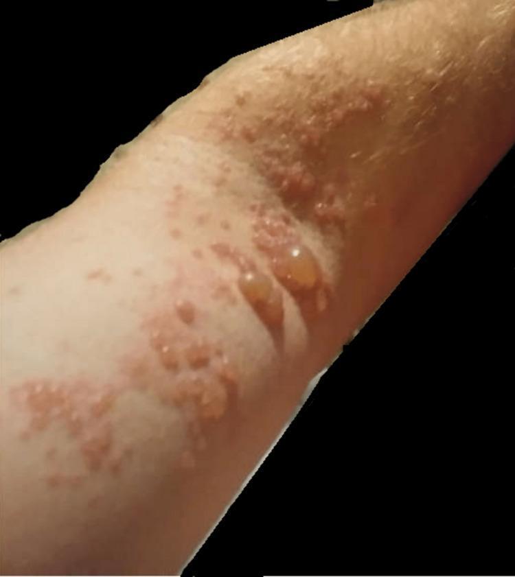
A clinical photograph of the left upper extremity demonstrating a vesicular and bullous eruption characteristic of Herpes Zoster (shingles). The rash exhibits a dermatomal distribution, featuring clusters of lesions on an erythematous base. Morphologically, the lesions range from small, grouped vesicles filled with clear fluid to larger, tense, dome-shaped bullae measuring over one centimeter. Several lesions appear ruptured or umbilicated, resulting in shallow erosions and crusting. The surrounding skin shows signs of inflammation and localized hyperemia. This visual presentation is indicative of Varicella-Zoster Virus (VZV) reactivation, illustrating the classic presentation of painful cutaneous lesions confined to a specific nerve root territory (C5-C6), often associated with neuropathic pain and potential motor weakness in the affected limb.
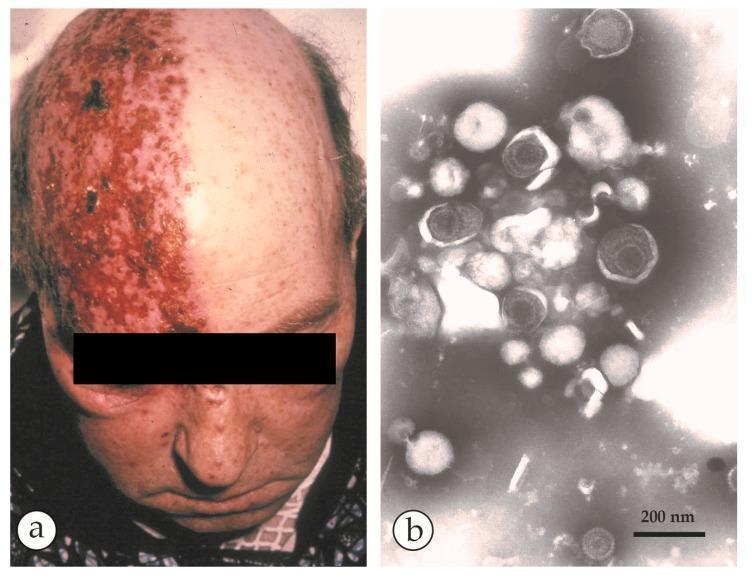
This composite educational image illustrates the clinical and microscopic features of shingles (Herpes Zoster). Panel (a) is a clinical photograph of an elderly patient demonstrating a classic unilateral vesicular rash. The lesions follow a specific dermatomal distribution on the right side of the scalp and forehead, sharply demarcated at the midline. The skin shows confluent erythematous plaques with clustered vesicles and areas of crusting, characteristic of a reactivated Varicella-Zoster Virus (VZV) infection. Panel (b) is a negative-stain electron micrograph (DEM) of fluid collected from a vesicle. It reveals an aggregate of herpesvirus particles. The virions exhibit typical morphology: a 110 nm icosahedral capsid core surrounded by a protein tegument and a loose outer envelope, totaling approximately 150-180 nm in diameter. Some capsids appear dark or 'empty' due to PTA-stain penetration. The presence of these spherical particles alongside cellular detritus confirms the diagnosis. A 200 nm scale bar is provided for size reference. This material is designed for dermatology and infectious disease studies.
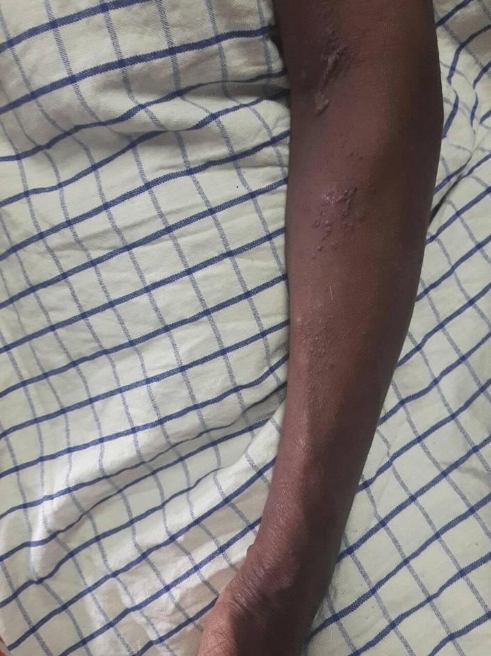
This clinical photograph displays a vesicular rash on the left upper limb of a patient with a darker skin tone. The lesions are primarily characterized as fluid-filled vesicles and small papules situated on an underlying erythematous base. The distribution is clustered and appears to follow a dermatomal pattern, typical of Herpes Zoster (shingles). Notable morphological features include areas of crusting and scabbing near the antecubital fossa, while more distal regions toward the forearm show active, raised vesicular clusters. The surrounding skin appears slightly dry with subtle pigmentary changes, but is otherwise intact. This visual serves as a primary educational example of the cutaneous manifestations of Varicella Zoster Virus reactivation, highlighting the progression from active vesicles to crusted lesions and the diagnostic importance of dermatomal distribution in clinical dermatology and infectious disease.
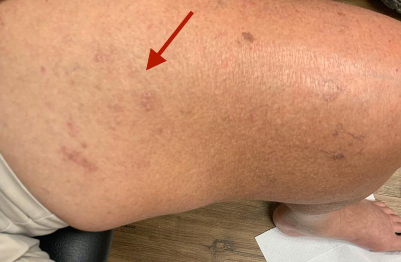
Clinical photograph of the left medial thigh demonstrating a resolving vesicular rash consistent with Herpes zoster (shingles). A red arrow indicates an area of post-inflammatory skin changes along the L2 and L3 dermatomes. The affected region shows scattered, erythematous to violaceous macules and flattened papules, suggesting a healing or crusting phase of the viral infection. The skin texture in the involved area appears slightly irregular compared to the adjacent healthy skin. Visible secondary features include mild localized edema, scattered benign-appearing nevi, and faint superficial venous markings. The image illustrates the typical dermatomal distribution and morphology of subacute Varicella-Zoster Virus reactivation in an adult patient. This visual is relevant for neurology and dermatology education, particularly in the context of post-herpetic neurological complications such as motor neuropathy or radiculopathy.
⚠️ This is a Medical Emergency - Please Read Carefully
What I'm Seeing in the Images
- Intense, confluent bright-red erythematous plaques covering large areas of both limbs
- Dense clusters of tiny vesicles (fluid-filled blisters) and larger bullae
- Weeping, oozing, and early serous crusting
- The rash is spreading rapidly along a dermatomal pattern
- Unilateral erythematous patch over the right temporal region, sharply confined to one side
- This follows the ophthalmic (V1) or maxillary (V2) branch of the trigeminal nerve
- This is called Herpes Zoster of the Face/Head
Why This is Urgent
-
Facial involvement near the eye (V1 trigeminal branch): This raises serious concern for Herpes Zoster Ophthalmicus (HZO) - the virus can spread to the eye and cause blindness. Per Tintinalli's Emergency Medicine, when the ophthalmic branch is involved, urgent ophthalmology evaluation is required. Ask if she has any eye pain, redness, or blurred vision.
-
Disseminated zoster: The extensive spread over multiple sites strongly suggests immune compromise (possible underlying diabetes, cancer, HIV, steroid use, or simply advanced age lowering immunity). This form requires IV antiviral therapy, not just oral medication.
What Needs to Happen RIGHT NOW
| Treatment | Details |
|---|---|
| IV Acyclovir | 500 mg/m² every 8 hours for 7 days - this is the standard for disseminated or severe zoster, per Fitzpatrick's Dermatology |
| Eye examination | Urgent ophthalmology consult for the facial involvement |
| Blood work | CBC, blood sugar (rule out diabetes), HIV test, and immune workup |
| Pain control | NSAIDs, paracetamol, or stronger analgesics for the nerve pain |
| Wound care | Cool compresses, calamine lotion to dry lesions; NO steroid creams on the blisters |
What NOT to Do at Home
- Do NOT apply any steroid cream (hydrocortisone, betamethasone, etc.) on the blistered areas - it will worsen the viral infection
- Do NOT burst or scratch the blisters - risk of secondary bacterial infection
- Do NOT let her near pregnant women, newborns, or anyone who has never had chickenpox - she is infectious until all blisters crust over
- Do NOT delay - the antiviral works best within 72 hours of rash onset. She is already 2+ days in, and the facial/eye involvement means she should receive antivirals regardless of timing, per Fitzpatrick's Dermatology
About Post-Herpetic Neuralgia (PHN)
- Fitzpatrick's Dermatology, Herpes Zoster Treatment section
- Tintinalli's Emergency Medicine, Chapter 250
And
- Did you want to share more photos of other affected areas?
- Are you asking about a specific symptom (like ear pain, eye redness, fever)?
- Do you want to know about home care while waiting to get to the hospital?
- Is there something else going on with her that you wanted to mention?
What else should be included in management
Complete Management of Disseminated Herpes Zoster (Elderly Patient)
1. Antiviral Therapy (Already Covered - Key Recap)
- IV Acyclovir 10 mg/kg every 8 hours for 7–10 days (for disseminated/severe disease)
- Switch to oral Valacyclovir 1g three times daily once new lesion formation stops and patient is stable
- Start even beyond 72 hours given facial/cranial nerve involvement and ongoing new vesicle formation
2. Ophthalmology Referral (URGENT)
- The right-sided facial rash in the V1/V2 trigeminal distribution puts the eye at risk
- Ask specifically about: eye pain, redness, tearing, blurred vision, light sensitivity
- Look for Hutchinson's sign - vesicles on the tip of the nose - which predicts ocular involvement
- Ophthalmologist will check for: keratitis, uveitis, retinal necrosis
- If eye is involved: topical antiviral eye drops + steroid eye drops under specialist supervision
3. Investigations / Workup
| Test | Reason |
|---|---|
| CBC with differential | Lymphopenia suggests immune compromise |
| Fasting blood glucose / HbA1c | Diabetes is a major trigger for zoster reactivation |
| HIV test | Must be done in any disseminated zoster case |
| Liver and kidney function | Baseline before IV acyclovir (acyclovir is nephrotoxic) |
| VZV PCR (vesicle swab) | Confirms diagnosis if uncertain |
| Chest X-ray | Rule out visceral dissemination (zoster pneumonitis) |
| Serum LDH | Marker of disseminated viral activity |
4. Pain Management - Stepwise
- Step 1: Paracetamol (acetaminophen) 500–1000 mg every 6–8 hours
- Step 2: Add NSAIDs (ibuprofen/naproxen) if no contraindication (check renal function first)
- Step 3: Weak opioids (tramadol) for moderate-severe pain
- Step 4: Gabapentin 300 mg at night, titrating up - addresses the neuropathic component and also reduces risk of post-herpetic neuralgia (PHN)
- Tricyclic antidepressants (amitriptyline 10–25 mg at night) can be added for neuropathic pain, but use with caution in elderly due to fall risk
5. Wound and Skin Care
- Cool saline compresses 3–4 times daily to weeping/oozing areas - reduces discomfort and promotes drying
- Calamine lotion over vesicular areas - soothing and drying
- Chlorhexidine wash (dilute) if there is suspicion of secondary bacterial infection
- If superinfection (increasing warmth, pus, spreading redness with fever): oral/IV antibiotics - flucloxacillin or clindamycin for staphylococcal cover
- Keep lesions loosely covered with non-adhesive dressings - avoid tight wrapping
- No steroid creams whatsoever on any lesion
6. Nutritional and Supportive Care
- Adequate hydration - especially important with IV acyclovir to prevent renal crystallization (ensure good urine output)
- High-protein diet to support skin healing
- Vitamin C and zinc supplementation may aid wound healing
- Monitor for dehydration in elderly patients who may reduce fluid intake due to pain
7. Isolation Precautions
- She is infectious until all lesions have completely crusted over (typically 7–10 days)
- Keep away from:
- Pregnant women (fetal varicella syndrome risk)
- Newborns and infants
- Immunocompromised individuals
- Anyone who has never had chickenpox or been vaccinated
- Healthcare workers should use contact + airborne precautions when caring for her
8. Monitoring for Complications
| Complication | Watch For |
|---|---|
| Zoster Ophthalmicus | Eye pain, redness, vision change |
| Ramsay Hunt Syndrome | Ear pain, vesicles in ear canal, facial paralysis, hearing loss - needs urgent ENT |
| Zoster Encephalitis | Confusion, headache, fever, neck stiffness |
| Bacterial superinfection | Increasing pain, pus, fever, spreading redness |
| Post-herpetic Neuralgia (PHN) | Persistent burning pain after rash heals - very common in elderly |
| Visceral dissemination | Respiratory symptoms, abdominal pain, hepatitis |
| Motor weakness | Rare - zoster paresis in affected limb |
9. Post-Herpetic Neuralgia (PHN) Prevention and Treatment
- PHN occurs in up to 50% of patients over 60 after zoster
- Early aggressive antiviral treatment is the best prevention
- If PHN develops after rash heals:
- Gabapentin or pregabalin (first-line)
- Tricyclic antidepressants (amitriptyline)
- Topical lidocaine 5% patches over affected area
- Capsaicin 8% patch (applied in clinic setting)
- Opioids (tramadol/oxycodone) as last resort
10. Immunization Counseling (After Recovery)
- Recombinant Zoster Vaccine (Shingrix) - 2 doses given 2–6 months apart
- Recommended for adults 50+ even after a zoster episode (reduces recurrence and PHN)
- Do not give during active infection - wait until fully recovered
- This is especially important given she already had severe disseminated disease
11. Address the Underlying Cause
- Screen for undiagnosed diabetes
- Screen for occult malignancy (lymphoma especially - zoster is a presenting feature)
- Review all medications for immunosuppressants (steroids, DMARDs)
- HIV serology if not previously done
There’s a blister formed on the hand which is 1cmX1cm in size. Should it be popped
Do NOT Pop the Blister
Why You Must NOT Pop It
-
The fluid inside is infectious - it contains live Varicella-Zoster Virus. Popping it releases the virus onto surrounding skin, causing the rash to spread further.
-
The blister roof is protecting the skin underneath - it acts as a natural sterile dressing. Once removed, the raw skin underneath is exposed and vulnerable.
-
High risk of bacterial infection - once the skin barrier is broken, bacteria (especially Staph and Strep) can enter and cause cellulitis or even sepsis - very dangerous in an elderly patient.
-
The fluid can infect others - anyone who touches the fluid and has not had chickenpox can catch chickenpox from it.
-
It increases scarring - intact blisters heal with less scarring than those that are opened.
What To Do With It Instead
| Action | How |
|---|---|
| Leave it intact | Do not touch, squeeze, or poke it |
| Cover it loosely | Use a clean, non-stick sterile dressing (e.g. Mepitel or non-adhesive gauze) - do not wrap tightly |
| Cool compress | Gently apply a cool, clean, damp cloth for 10-15 minutes to reduce discomfort |
| Calamine lotion | Apply gently around (not rupturing) the blister to soothe the area |
| Keep it dry | Avoid soaking the hand in water |
| Watch for signs of infection | Increasing redness spreading outward, warmth, pus, fever - if any of these appear, go to hospital immediately |
If It Bursts on Its Own
- Do not peel off the roof/skin - leave it in place as a cover
- Gently clean the area with normal saline or clean water only
- Pat dry carefully
- Cover with a clean non-stick dressing
- Apply a thin layer of mupirocin (Bactroban) ointment if available - to prevent bacterial infection
- Change dressing once or twice daily