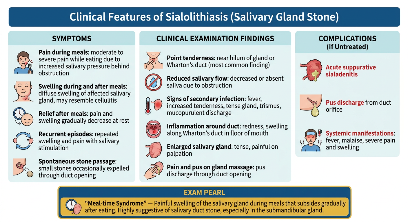
Clinical Features of Sialolithiasis (Salivary Gland Stone) - Simplified Symptoms Pain during meals Moderate to severe pain occurs while eating, especially when saliva production increases. Pain is caused by increased pressure behind the obstructed duct. Swelling during and after meals Swelling of the affected salivary gland appears during eating. It may be diffuse and sometimes resemble cellulitis. Relief after meals Pain and swelling gradually decrease during rest when saliva production falls. Recurrent episodes Patients often give a history of repeated swelling and pain whenever salivary flow is stimulated. Symptoms continue until the obstruction is relieved. Spontaneous passage of stone Occasionally, small salivary stones may be expelled spontaneously through the duct opening. Clinical Examination Findings Point tenderness Most common finding. Tenderness is usually present near: Hilum of the gland, or Wharton's duct (submandibular gland duct). Reduced salivary flow Saliva secretion is decreased or completely absent due to duct obstruction. Signs of secondary infection Fever Increased tenderness Tense gland Trismus (difficulty opening the mouth) Mucopurulent discharge Thick, cloudy, gelatinous, or pus-like material may be seen mixed with saliva. Results from inflammation and stagnation of saliva behind the obstruction. Inflammation around the duct Redness, swelling, and tenderness along the course of Wharton's duct. Most noticeable in the floor of the mouth. Enlarged salivary gland Gland becomes enlarged, tense, and painful on palpation. Pain and pus on gland massage Palpation or milking of the gland may produce pain and discharge of pus through the duct opening. Complications (If Untreated) Acute suppurative sialadenitis Severe infection develops due to prolonged obstruction. Pus discharge from duct orifice Frank pus may exude from the duct opening. Systemic manifestations Fever Malaise Severe pain and swelling Exam Pearl (OMFS) Classic presentation of sialolithiasis: "Meal-time syndrome" → Painful swelling of the salivary gland during meals that subsides gradually after eating. This is highly suggestive of a salivary duct stone, especially in the submandibular gland. give it as image
Medical education infographic titled "Clinical Features of Sialolithiasis (Salivary Gland Stone)" with a clean clinical layout. Include the following sections clearly organized: SYMPTOMS section with icons: - Pain during meals: moderate to severe pain while eating due to increased salivary pressure behind obstruction - Swelling during and after meals: diffuse swelling of affected salivary gland, may resemble cellulitis - Relief after meals: pain and swelling gradually decrease at rest - Recurrent episodes: repeated swelling and pain with salivary stimulation - Spontaneous stone passage: small stones occasionally expelled through duct opening CLINICAL EXAMINATION FINDINGS section: - Point tenderness: near hilum of gland or Wharton's duct (most common finding) - Reduced salivary flow: decreased or absent saliva due to obstruction - Signs of secondary infection: fever, increased tenderness, tense gland, trismus, mucopurulent discharge - Inflammation around duct: redness, swelling along Wharton's duct in floor of mouth - Enlarged salivary gland: tense, painful on palpation - Pain and pus on gland massage: pus discharge through duct opening COMPLICATIONS (If Untreated) section: - Acute suppurative sialadenitis - Pus discharge from duct orifice - Systemic manifestations: fever, malaise, severe pain and swelling EXAM PEARL box at bottom (highlighted in gold/yellow): "Meal-time Syndrome" — Painful swelling of the salivary gland during meals that subsides gradually after eating. Highly suggestive of salivary duct stone, especially in the submandibular gland. Use a professional medical color scheme: deep blue headers, white background, light blue/teal section backgrounds, red highlights for complications, gold for exam pearl box. Include small anatomical icons. Clean modern typography.